Volume: 16 Issue: 4 August 2018
FULL TEXT
Generalized pustular psoriasis appears as an uncommon variant form of psoriasis consisting of widespread pustules on an erythematous background (von Zumbusch). A 39-year-old male patient with a history of plaque psoriasis since the age of 9 who had an acute relapse of generalized pustular psoriasis 12 days following a successful renal transplantation is presented. Despite administered immunosuppression for transplantation, the addition of cyclosporine and methotrexate did not reverse the ongoing process of disease and the patient died on the 57th posttransplant day due to multiorgan failure subsequent to severe bone marrow suppression.
Key words : Renal transplant, Pustular psoriasis, Cyclosporine, Methotrexate
Introduction
Generalized pustular psoriasis is an uncommon skin disease usually resistant to medical treatment. Patients who have the disease present with an extensive area of erythematous background and multiple small pustules. The activation of T-lymphocytes and tumour necrosis factor alpha (TNF-α) determines the type and severity of psoriasis.1 Acute generalized type (von Zumbusch variant) is associated with fever and toxicity. Both genders are affected equally. Cause has not been completely clarified. Although many factors can be considered as triggering for a disease flare-up, the most important contributors are considered the abrupt withdrawal of steroids after systematic use and the administration of drugs including salicylates, lithium, and penicillin.1 Generalized pustular psoriasis rarely appears in patients after organ transplantation, with only 2 cases presented so far in the literature.2,3
Case Report
A 39-year-old male patient with a 20-year history of chronic kidney disease of unknown cause on regular hemodialysis for the last 9 years is presented. A history of allergy to nonsteroidal anti-inflammatory drugs and vancomycin was reported. Since childhood, he had mild psoriasis, receiving local corticosteroids and moisturizers. Skin lesions had seasonal accentuation, with aggravation in spring and remission during summer.
The patient received a renal allograft from a deceased donor with 2 HLA matches (A2 and DR14). He received induction therapy with daclizumab 1 mg/kg/day and a triple immunosuppressive regimen with tacrolimus 0.1 mg/kg/day, mycophenolate mofetil (MMF) 2 g/day and methylprednisolone (1 g intravenously during surgery followed by prednisolone 200 mg on the 1st postoperative day, which was tapered to oral prednisolone 20 mg/day within 2 weeks and to 10 mg/day within 1 month), according to protocol.
Due to delayed graft function, he remained on hemodialysis. On the 6th postoperative day, he developed trunk erythematous lesions and local corticoid treatment (fluticasone) was applied (Figure 1). Twelve days postoperatively, skin lesions became aggravated, expanding to the whole trunk and extremities while multiple clusters of nonfollicular, superficial 2- to 3-mm pustules appeared. Pus culture from pustules was negative. After prednisolone was reduced to 10 mg daily, on the 30th postoperative day, the patient exhibited a dramatic deterioration of skin lesions consisting of a widespread and generalized body pustular exanthema. A skin biopsy revealed pustular psoriasis of the von Zumbusch variant, with eosinophilic infiltrate indicative of drug toxicity (Figure 2). On the 37th postoperative day, the patient became febrile, and urine output was reduced with gradual deterioration of renal graft function. MMF and daclizumab were discontinued, tacrolimus was substituted with cyclosporine (3.0 mg/kg/day), and steroids were increased to 30 mg/day. As skin lesions were extended to the whole body surface, becoming life threatening, methotrexate (50 mg intravenously) was administered on days 39 and 46 postoperatively. The patient developed sepsis, with increased body temperature to 39.5oC, pancytopenia (white blood cell count: 0.320 103/mm3, hematocrit level: 25.1%, hemoglobin: 8.4 g/dL, platelets: 29 103/ mm3), signs of disseminated intravascular coagulation, oliguria (400 mL/day), and respiratory distress. He was supported with transfusions of red blood cells and platelets as well as granulocyte colony stimulating factor. On postoperative day 57, the patient died from multiorgan failure.
Discussion
Psoriasis is a chronic, inflammatory skin disease affecting approximately 2% of white individuals. The cause for these variations is likely to be both genetic and environmental.1 Pustular psoriasis is an uncommon variant of psoriasis and may be classified into several types depending on the clinical course (acute, subacute, or chronic). The acute type (von Zumbusch variant) occurs with erythematous lesions producing burning sensation and may be associated with upper respiratory system or other systemic infections. High temperature appears suddenly with shiver and chills. Formation of multiple small pustules on an erythematous background in a generalized pattern occurs. In the acute type, the pustules often become confluent to form lakes of pus.4
Cyclosporine has been reported to be effective in controlling disease activity in pustular psoriasis. Cyclosporine inhibits T lymphocytes and affects the function of phospholipase A and the activation of interleukins 1 and 2, via its action against calcineurin, factors determinant for psoriasis.5 Furthermore retinoids including natural retinoids, vitamin A, and its active derivatives have been successfully employed in treating psoriasis. Possibly, retinoids contribute to improvement of symptoms by normalizing keratinization and proliferation of epidermal cells that control the production of cytokines (TNF-α, IL-1, and IL-6). Methotrexate has been also proposed as a treatment of severe psoriasis. It inhibits the differentiation and proliferation of epidermis.6 Due to numerous adverse effects and toxicity, methotrexate should be used cautiously especially in patients with renal failure. In our patient, methotrexate administration was necessary, despite delayed graft function, due to deterioration of skin disease and because the substitution of tacrolimus with cyclosporine was ineffective. Whether sepsis and multiorgan failure were related to pustular psoriasis per se or to overimmunosuppression was not elucidated. A novel concept in the treatment of pustular psoriasis is the use of biologic therapies, using anti-TNF-α, designed to selectively interfere with the immune mechanisms that induce psoriasis.7 Other anti-inflammatory and immunosuppressive drugs such as corticosteroids are used for pustular psoriasis, particularly early in the acute stage. Reduction or withdrawal of corticosteroids may lead to relapse of disease. Local treatment includes corticosteroid compounds and products of vitamin D3. While standard systemic therapy is effective, in the short term, unpredictable toxicity and gradual decline in efficacy could complicate a long-term use.
The case presented is the third case reported in the literature. However the other 2 reported patients responded well to combination therapy with cyclosporine, retinoids, and methotrexate.2,3 It is noteworthy that, in our patient, cyclosporine, an immunosuppressive agent, drug of choice for transplant, and main treatment for psoriasis was not able to prevent rapid deterioration. In addition, supplementation of treatment regimen with methotrexate did not control psoriasis activity, but it is possible that it led to overimmunosuppression, complicating our patient’s recovery. In contrast to already reported cases describing flare-up of psoriasis in stable renal transplant recipients, our patient presented an acute relapse of psoriasis a few days after renal transplant surgery while on delayed renal allograft function and undoubtedly during a period of intense immunosuppression.
Pustular psoriasis is a rare disease among patients undergoing transplant procedures, with only 2 reports in the literature. With only 2 cases presented so far as well as ours with a dismal prognosis, it is difficult to extract definite conclusions whether patients with history of psoriasis and chronic renal failure should be listed for transplantation.
References:
- Ohkawara A, Yasuda H, Kobayashi H, et al. Generalized pustular psoriasis in
Japan: two distinct groups formed by differences in symptoms and genetic
background. Acta Derm Venereol. 1996;76(1):68-71.
PubMed - Coulson IH, Evans CD, Holden CA. Generalized pustular psoriasis after renal
transplantation--failure to suppress with cyclosporin A. Clin Exp Dermatol.
1988;13(6):416-417.
CrossRef - PubMed - Kaaroud H, Beji S, Jebali A, et al. Pustular psoriasis after renal
transplantation. Transplant Proc. 2007;39(4):1130-1131.
CrossRef - PubMed - Zelickson BD, Muller SA. Generalized pustular psoriasis. A review of 63
cases. Arch Dermatol. 1991;127(9):1339-1345.
CrossRef - PubMed - Sarzi-Puttini P, Cazzola M, Panni B, et al. Long-term safety and efficacy of
low-dose cyclosporin A in severe psoriatic arthritis. Rheumatol Int.
2002;21(6):234-238.
CrossRef - PubMed - Boffa MJ, Chalmers RJ. Methotrexate for psoriasis. Clin Exp Dermatol.
1996;21(6):399-408.
CrossRef - PubMed - Gisondi P, Girolomoni G. Biologic therapies in psoriasis: a new therapeutic
approach. Autoimmun Rev. 2007;6(8):515-519.
CrossRef - PubMed

Volume : 16
Issue : 4
Pages : 488 - 490
DOI : 10.6002/ect.2016.0036
From the 1First Department of Surgery, Transplant Unit; the 2Department of
Dermatology; and the 3Department of Pathology, “Evangelismos” General Hospital
of Athens, Greece
Acknowledgements: The authors declare that they have no sources of funding for
this study, and they have no conflicts of interest to declare.
Corresponding author: Spiros Drakopoulos, Transplant Unit, Evangelismos General
Hospital of Athens, Greece; 45-47 Ipsilantou Street, Athens 10676, Greece
Phone: +30 210 720 1032
E-mail: spdrakopoulos@gmail.com

Figure 1. Psoriasiform Epidermal Hyperplasia With Spongiform Pustules of Kogoj
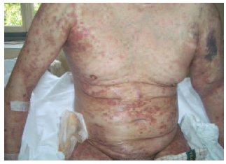
Figure 2. Generalized Pustular Psoriasis of the Body and Upper Extremities