Volume: 12 Issue: 1 March 2014 - Supplement - 1
FULL TEXT
Objectives: This study sought to analyze the incidence and risk factors of skin complications in liver transplant recipients.
Materials and Methods: Patients with skin biopsies were selected from 408 liver transplant recipients from January 1990 to December 2012 at Baskent University in Ankara, Turkey. Demographic and clinical findings including age, sex, primary liver disease, immunosuppressive therapy, and the time between transplant and cutaneous lesions were examined.
Results: There were 38 patients who had skin biopsy after liver transplantation. The mean age at transplantation was 30.5 years. The primary liver disease was hepatocellular carcinoma seconder to viral hepatitis in 15 patients, Wilson disease in 7 patients, criptogenic cirrhosis in 3, familial hypercholesterolemia in 3 patients, and 1 each patients of Byler disease, Caroli disease, tyrosinemia, Budd-Chiari sendrom, congenital hepatic fibrosis, alcholic cirrhosis, and biliar atresia. The primary disease in 3 patients were unknown. The histologic diagnosis included precancerous and cancerous lesions (n=4), infectious diseases (n=4); xantho-matous lesions (n=4); vasculopathic lesions (n=4); melanocytic lesions (n=4); benign adnexial tumors (n=2); skin atrophy (n=2); calcinosis cutis (n=2), keratinous cyst (n=2), neutrophilic dermatosis (n=2), perivascular dermatitis (n=2) and miscellaneous lesions (n=6).
Conclusions: We presented the cutaneous manifestations in liver transplant recipients. The most common skin lesion in these patients were precancerous and cancerous lesions, infectious, xanthomatous, and melanocytic lesions. The findings emphasize the importance of dermatologic screening in these patients that can provide early diagnosis and a better quality of life.
Key words : Liver, Skin malignancies, Infections
Introduction
Liver transplant is often the only remaining therapy for patients with end-stage liver cirrhosis, acute liver failure, and some metabolic and congenital hepatic diseases.1,2 Recipients are usually susceptible to skin lesions, mostly because of immunosuppressive treatment.1-4 Age, sex, sun exposure, and viral infections also are possible factors that can predict skin complications.1-5 Although several studies concerning skin manifestations in kidney transplant recipients have been reported, few concern liver transplants.1-7 This study sought to report our experience regarding incidence and risk factors of skin complications in liver transplant recipients.
Materials and Methods
Patients with skin biopsies were selected from 408 liver transplant recipients (25 men [65.7%] 13 women [34.3%]; mean age at the time of transplant, 30.5 y; graft source: living-related donor in 20 patients [52.6%], deceased donor in 18 patients [47.4%]) who were operated on between January 1990 and December 2012 at Başkent University in Ankara, Turkey. The study was approved by local Ethics Committee of the university. All protocols conformed with the ethical guidelines of the 1975 Helsinki Declaration. Informed consent was obtained from all subjects. Demographic and clinical findings including age, sex, immunosuppressive therapy, age at transplant, and (for malignant lesions) the time between transplant and tumor development were examined.
Statistical analyses were performed with SPSS software (SPSS: An IBM Company, version 16.0, IBM Corporation, Armonk, NY, USA). All values were presented as means ± SE (standard error) and were analyzed with Kruskal-Wallis test followed by the Mann-Whitney U test. Value of P < .05 was considered statistically significant.
Results
Among 408 liver transplant recipients, 38 patients (9.3%) had skin biopsies. Several regimens of immunosuppression were used, but all 38 recipients received a corticosteroid. Additionally, 14 of them (37%) were receiving cyclosporine, 6 of them (15.7%) were receiving both cyclosporine and tacrolimus, and 18 of them (47.3%) were receiving tacrolimus. Among 38 patients studied, 25 (65.7%) never had acute rejection episodes, 8 (21%) had at least 1 rejection episode, and 5 (13.1%) had more than 1. Eight patients died during follow-up.
The indications for liver transplant were 15 (39.4%) hepatocellular carcinoma secondary to viral hepatitis, 7 (18.4%) Wilson disease, 3 (7.8%) cryptogenic cirrhosis, 3 (7.8%) familial hypercholesterolemia, and 1 (2.6%), each, patients of Byler disease, Caroli disease, tyrosinemia, Budd-Chiari syndrome, congenital hepatic fibrosis, alcoholic cirrhosis, and biliary atresia. The primary disease in 3 patients (7.8%) were unknown.
The cutaneous manifestations included pre-cancerous or cancerous lesions (n=4), infectious diseases (n=4), xanthomatous lesions (n=4), vasculopathic lesions (n=4), melanocytic lesions (n=4), benign adnexal tumors (n=2), skin atrophy (n=2), calcinosis cutis (n=2), keratinous cyst (n=2), neutrophilic dermatosis (n=2), perivascular dermatitis (n=2), and miscellaneous lesions (n=6). The clinical characteristics of 38 patients are shown in Table 1.
There were 4 premalignant/malignant lesions in our study. These lesions were squamous cell carcinoma (n=1), basal cell carcinoma (n=1), and in situ squamous cell carcinoma (n=2). Histologically, in squamous cell carcinoma, irregular masses of atypical epidermal cells proliferate downward into the dermis. In in situ squamous cell carcinoma, there is no stromal invasion, but atypical squamous cells infiltrate all the epidermis (Figure 1A). Basal cell carcinoma contains atypical basaloid cells that are arranged in a palisading fashion. All of our precancerous/cancerous lesions occurred on sun-exposed sites. The mean age was 58.2 years and all the patients were men. Two of these patients (50%) were being administered cyclosporine and the other 2 (50%) were receiving tacrolimus-based immuno-suppression regimen. The cause of the liver disease were hepatocellular carcinoma in all of the precancerous, and cancerous lesions in our study, and all these patients are alive at the time of this writing. The mean interval to malignancy after transplant was 38.5 months (range, 6-74 mo). There were no significant differences in the mean interval to malignancy after transplant between the treatment modalities, tumor types, or between the patients who have rejection.
Four cutaneous infections were detected including verruca vulgaris (n=2), condylomata acuminatum (n=1), and leishmaniasis (n=1). Leishmaniasis was detected on the scalp of a 6-year-old boy with, Wilson disease. Histologically, the biopsy of verruca vulgaris showed hyperkeratosis, acanthosis, and papillomatosis. Groups of large, vacuolated cells lay in the upper stratum malpighii and in the granular layer of epidermis (Figure 1B). In leishmaniasis, diffuse dermal infiltrates of histiocytes were detected in the cytoplasm of these histiocytes, numerous dull blue-grey, round to oval microorganisms were seen (Figure 1C).
Melanocytic lesions were intradermal naevus (n=2), compound naevus (n=1), and dysplastic naevus (n=1). Three of them, including dysplastic naevus, were detected before transplant, 1 of intradermal naevus was seen after liver transplant.
Xanthomatous lesions were xanthoma in 3 patients and juvenile xanthogranuloma in 1 patient; the primary disease of these patients was familial hypercholesterolemia in 3 and biliary atresia in 1. All of the lesions were located on the distal extremities. Histologically, the lesion was composed of lipid-laden histiocytes, called foamy cells (Figure 1D).
Discussion
Organ transplant recipients are predisposed to several cutaneous complications, especially because of immunosuppressive therapy.1-5 Skin lesions related to the use of immunosuppressive drugs directly affect the patient’s quality of life. This study evaluated the spectrum of cutaneous complications seen in liver transplant recipients. The most common cutaneous manifestations in our study were precancerous/cancerous lesions, infectious diseases, xanthomatous, and vasculopathic lesions.
Skin cancers are the most common malignant conditions in organ transplant recipients, with the incidence as high as 35% to 70% at 20 years in parts of the world where sun exposure is more common.1-4 The incidence of skin cancer is lower in liver transplant recipients than in other solid-organ transplant recipients, because the requirement of immunosuppression is lower after a liver transplant.2 The incidence varies between 1.6% and 22.5% in different studies.1,2,3,6 This variability may be because the differences in the length of follow-up, immunosuppressive regimen, or geographic area.1-6 Squamous cell carcinoma and basal cell carcinoma account for more than 90% of all skin cancers in transplant recipients in almost all the studies.1-6 While basal cell carcinoma is the most common skin cancer in nontransplant population, in organ transplant recipients, squamous cell carcinoma is the most frequent form of skin cancer. And because squamous cell carcinoma is more aggressive than basal cell carcinoma and the tendency toward recurrence and metastases is greater, early detection is important, especially in transplant recipients.
In our study, the incidence of premalignant and malignant lesions was 10.5%, and most of them (75%) were of squamous origin. In some studies, age or male sex was found to be an independent risk factor for skin cancer.2,6 Although there have been some studies showing increased rates of skin cancer in patients who were receiving cyclosporine,5,7 similar to our study, some studies found no significant differences between patients receiving cyclosporine or tacrolimus-based protocols.8 Herrero and associates suggest that transplant for hepatocellular carcinoma is a risk factor for development of more than 1 skin cancer.2 Similarly, the cause of hepatocellular carcinoma has been reported to be predictive for skin cancer in the Spanish series by Xiol and associates.9 In our study, all patients that had premalignant or malignant lesions underwent transplant for hepatocellular carcinoma. We observed that older age, male sex, and the cause of hepatocellular carcinoma were related to the development of skin cancer.
The role of immunosuppressive agents also increased the risk of cutaneous infections in organ transplant recipients. In some articles, fungal infections,10 and in others,1,7 viral infections are reported to be the most frequent cause of skin infections. However, in all studies, viral warts were the most common reason of viral infections.10-12 Infection with human papillomavirus is responsible for developing viral warts that may cause significant morbidity in individuals unable to mount an adequate T-helper-cell–mediated immune response.11 Because some of this viral warts may present with atypical features and may progress into squamous cell carcinoma, early detection of these lesions is essential for transplanted patients.11 In the literature, the incidence of viral warts in organ transplant recipients varies from 24% to 53%, depending on the level of immunosuppression.12-14 However, lower rates had been reported in some studies.13 In our study, the incidence of viral warts was 7.8%. Any of them had atypical features, and all of them were detected during the first year of transplant.
Leishmaniasis is an infection caused by a protozoan parasite. Clinical presentations of infection include visceral, cutaneous, and mucocutaneous forms. Leishmaniasis is endemic in Africa, Asia, Europe, and South America. Cases of cutaneous and visceral leishmaniasis have been reported in organ transplant recipients in endemic areas.15,16 We had 1 patient who had cutaneous leishmaniasis in a 6-year-old boy.
Calcinosis cutis is depositing of insoluble calcium salts in the cutaneous tissues. Four different types of calcinosis cutis can be identified: dystrophic, metastatic, idiopathic, and iatrogenic.17,18 Dystrophic calcinosis is the most common type and occurs in previously damaged tissue. Metastatic calcinosis occurs in normal tissues as a result of a disturbance in systemic calcium homeostasis, such as renal failure. The pathogenesis of calcinosis in liver transplant recipients is uncertain, with previous reports suggesting that these calcifications could be either dystrophic or metastatic.18,19 Liver transplant recipients also are open to severe metabolic problems, including calcium homeostasis and renal failure around the time of surgery and during the postoperative period. Therefore, calcification can be metastatic, or local trauma because of subcutaneous injections can be precipitate calcification (dystrophic).17-19 In our report, we had 2 cases with calcinosis cutis, and they both occurred on the hands of these patients. In both cases, serum calcium levels were within normal limits. It is probably because the pathogenesis of calcium deposition is multifactorial.
Vasculopathic reactions are defined by the damage of vascular structures. Histologically, vascular dilatation, erythrosis extravasation, inflammatory infiltration around vascular channels, endothelial swelling, and reactive nuclear changes in the endothelium are defined as vasculopathic reactions.20,21 Its difference from vasculitis is the absence of fibrin deposition in the vessel wall. Vasculopathic reactions are mainly because of infectious complications or drug-related adverse events, and they also may indicate the recurrence of the original disease.20 In the literature, few case reports of cutaneous vasculitis have been reported after transplant.20,22 In our study, we had 4 skin biopsies including vasculopathic lesions, and in all of them, additional to histologic findings of vasculopathy, eosinophil-leukocyte infiltration was seen, and this finding let us think that these reactions were drug-related adverse events. All these patients were receiving cyclosporine-based regimen.
Xanthomatous lesions are often seen in patients with high levels of serum cholesterol.21 Also primary biliary cirrhosis, is well-known for its presentation. We had 4 patients who had xanthomatous lesions and in 3 of them primary disease was familial hyper-cholesterolemia. All had high cholesterol levels. Similarly, in the literature, a few cases have been mentioned about xanthomatous lesion, which were primary biliary cirrhosis or familial hyper-cholesterolemia in transplanted patients.23,24
In conclusion, liver transplant recipients show a high risk of cutaneous complications, compared with the general population, with a particular predisposition to infections and skin cancers. Periodic dermatologic evaluation is essential for detecting early lesions and to provide a better quality of life for the patients.
References:
- Perera GK, Child FJ, Heaton N, O'Grady J, Higgins EM. Skin lesions in
adult liver transplant recipients: a study of 100 consecutive patients. Br J
Dermatol. 2006;154(5):868-872.
CrossRef - PubMed - Herrero JI, España A, D'Avola D, et al. Subsequent nonmelanoma skin
cancer after liver transplantation. Transplant Proc. 2012;44(6):1568-1570.
CrossRef - PubMed - Herrero JI, España A, Quiroga J, et al. Nonmelanoma skin cancer after
liver transplantation. Study of risk factors. Liver Transpl.
2005;11(9):1100-1106.
CrossRef - PubMed - Euvrard S, Kanitakis J, Claudy A. Skin cancers after organ
transplantation. N Engl J Med. 2003;348(17):1681-1691.
CrossRef - PubMed - Bouwes Bavinck JN, Hardie DR, Green A, et al. The risk of skin cancer in
renal transplant recipients in Queensland, Australia. A follow-up study.
Transplantation. 1996;61(5):715-721.
CrossRef - PubMed - Mithoefer AB, Supran S, Freeman RB. Risk factors associated with the
development of skin cancer after liver transplantation. Liver Transpl.
2002;8(10):939-944.
CrossRef - PubMed - Jensen P, Hansen S, Møller B, et al. Skin cancer in kidney and heart
transplant recipients and different long-term immunosuppressive therapy
regimens. J Am Acad Dermatol. 1999;40:177-786.
CrossRef - PubMed - Jonas S, Rayes N, Neumann U, et al. De novo malignancies after liver
transplantation using tacrolimus-based protocols or cyclosporine-based
quadruple immunosuppression with an interleukin-2 receptor antibody or
antithymocyte globulin. Cancer. 1997;80(6):1141-1150.
CrossRef - PubMed - Xiol X, Guardiola J, Menendez S, et al. Risk factors for development of
de novo neoplasia after liver transplantation. Liver Transpl.
2001;7(11):971-975.
CrossRef - PubMed - Belloni-Fortina A, Piaserico S, Bordignon M, et al. Skin cancer and
other cutaneous disorders in liver transplant recipients. Acta Derm
Venereol. 2012;92(4):411-415. doi: 10.2340/00015555-1316.
CrossRef - PubMed - Schmook T, Nindl I, Ulrich C, Meyer T, Sterry W, Stockfleth E. Viral
warts in organ transplant recipients: new aspects in therapy. Br J Dermatol.
2003;149(suppl 66):20-24.
CrossRef - PubMed - Salard D, Parriaux N, Derancourt C, et al. Cutaneous complications
following liver transplantation: epidemiologic and clinical study in 86
patients [in French]. Ann Dermatol Venereol. 2002;129(10 Pt 1):1134-1138.
PubMed - Rüdlinger R, Smith IW, Bunney MH, Hunter JA. Human papillomavirus
infections in a group of renal transplant recipients. Br J Dermatol.
1986;115(6):681-692.
CrossRef - PubMed - Ramsay HM, Fryer AA, Reece S, Smith AG, Harden PN. Clinical risk factors
associated with nonmelanoma skin cancer in renal transplant recipients. Am J
Kidney Dis. 2000;36(1):167-176.
CrossRef - PubMed - Mirzabeigi M, Farooq U, Baraniak S, Dowdy L, Ciancio G, Vincek V.
Reactivation of dormant cutaneous Leishmania infection in a kidney
transplant patient. J Cutan Pathol. 2006;33(10):701-704.
CrossRef - PubMed - Gontijo CM, Pacheco RS, Orefice F, Lasmar E, Silva ES, Melo MN.
Concurrent cutaneous, visceral and ocular leishmaniasis caused by Leishmania
(Viannia) braziliensis in a kidney transplant patient. Mem Inst Oswaldo
Cruz. 2002;97(5):751-753.
CrossRef - PubMed - Jucglà A, Sais G, Curco N, Marcoval J, Moreno A, Peyri J. Calcinosis
cutis following liver transplantation: a complication of intravenous calcium
administration. Br J Dermatol. 1995;132(2):275-278.
CrossRef - PubMed - Larralde M, Giachetti A, Kowalczuk A, D'Agostino D, Galimberti R.
Calcinosis cutis following liver transplantation in a pediatric patient.
Pediatr Dermatol. 2003 May-Jun;20(3):225-228.
CrossRef - PubMed - Lateo S, Charlton F, Hudson M, Lawrence CM. Cutaneous calcification
following liver transplantation. Clin Exp Dermatol. 2005;30(5):484-486.
CrossRef - PubMed - Rau S, Wollenberg A, Weiss M, Fischereder M, Schönermarck U. Cutaneous
vasculitis after renal transplantation: a case report. Transplant Proc.
2012;44(10):3048-3050. doi: 10.1016/j.transproceed.2012.04.037.
CrossRef - PubMed - Elder DE. Vascular Diseases. In: Lever WF, Elder DE, Elenitsas R. Lever’s Histopathology of the Skin. 9th ed. Philadelphia, PA: Lippincott Williams & Wilkins; 2005:215-220.
- Hassan G, Khalaf H, Mourad W. Dermatologic complications after liver
transplantation: a single-center experience. Transplant Proc.
2007;39(4):1190-1194.
CrossRef - PubMed - Onaca N, Goldstein RM, Klintmalm GB. Xanthomas disappear rapidly after
liver transplantation for familial hypercholesterolemia. Liver Transpl.
2003;9(12):1334-1335.
CrossRef - PubMed - Hsu JC, Su TC, Chen MF, Liau CS, Lee YT. Xanthoma striatum palmare in a
patient with primary biliary cirrhosis and hypercholesterolemia. J
Gastroenterol Hepatol. 2005;20(11):1799-1800.
CrossRef - PubMed

Volume : 12
Issue : 1
Pages : 101 - 105
DOI : 10.6002/ect.25Liver.P11
From the Division of 1Pathology and 2Transplantation Surgery, Başkent University
Faculty of Medicine, Ankara, Turkey
Acknowledgements: The authors have no conflicts of interest to declare, and
there was no funding for this study.
Corresponding author: Merih Tepeoğlu, MD, Department of Pathology, Başkent
University Faculty of Medicine, 79. Sokak, No: 7/4, Bahcelievler, Ankara 06490,
Turkey
Phone: +90 312 212 6591
Fax: +90 312 212 7572
E-mail: merihdemirel@yahoo.com.tr
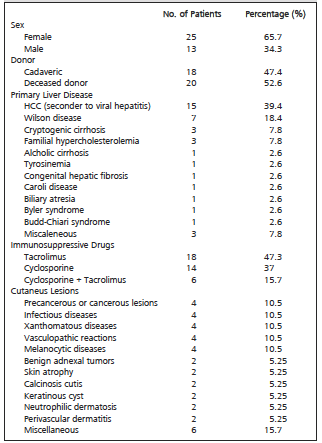
Table 1. Clinical Characteristics of 38 Liver Allograft Recipients With Cutaneus Manifestations
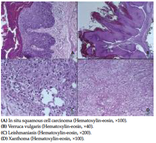
Figure 1. Cutaneous Lesions in Liver Transplant Recipients