Volume: 24 Issue: 8 August 2026
FULL TEXT
Clinical and Laboratory Outcomes in Biliary Atresia: Insights from the Shiraz Pediatric Liver Cirrhosis Cohort
Objectives: Biliary atresia is a progressive cholan-giopathy in neonates leading to liver failure and often requiring liver transplant. Although surgical inter-ventions like the Kasai portoenterostomy offer temporary relief, the disease continues to be the leading cause of liver transplant. This study aimed to evaluate the demographic, clinical, and laboratory characteristics of these patients and the effects of these characteristics on outcomes.
Materials and Methods: This cohort study included 167 pediatric patients (aged <18 years) with confirmed biliary atresia, selected from the Shiraz Pediatric Liver Cirrhosis Cohort Study between 2018 and March 2024. We analyzed data collected from the pediatric liver cirrhosis registry and assessed patient outcomes by comparing survivors versus those who did not survive.
Results: Among the 167 patients, 86 (51.5%) died and 81 (48.5%) survived. Those who died were significantly younger at enrollment (15.47 vs 36.31 months old) and had higher internal normalized ratio, prolonged partial thromboplastin time, and elevated liver enzymes compared with survivors. The Kasai portoenterostomy was performed in 77.8% of patients, with no significant difference in its distribution between survivors and patients who did not survive.
Conclusions: Although most children had a Kasai surgery and over one-third had a liver transplant, a significant number did not survive. To improve outcomes for children with biliary atresia, better perioperative and postoperative care, aggressive treatment of liver dysfunction, and robust nutritional support are essential.
Key words : Cholangiopathy, Middle Eastern population, Pediatric liver cirrhosis, Survival
Introduction
Biliary atresia (BA) is a cholangiopathy of unknown etiology in neonates that progresses through inflam-mation and fibrosis. Biliary atresia obliterates both intra- and extra-hepatic bile ducts and eventually leads to cholestasis and subsequent liver failure. Despite surgical intervention, BA continues to be the predominant reason for liver transplant (LT) and end-stage liver disease in the pediatric population.1
Hepatomegaly, clay-colored stool, urine discolo-ration, and concomitant congenital disorders, spec-ifically cardiac malformations, are among the clinical findings in patients with BA. Furthermore, an eleva-ted level of conjugated bilirubin and variations in the levels of liver enzymes are routine laboratory findings. Biopsy of the liver tissue remains the gold-standard diagnostic modality of choice in BA evaluation. However, making an accurate final diag-nosis requires a holistic approach encompassing the patient’s signs and symptoms together with paraclinical findings.2,3
Although BA is considered an uncommon disorder, BA has a wide rate of incidence worldwide, from 5 to 32 cases in every 100 000 live births, and is more prevalent in Asia and the Pacific region.4-6
The timing of LT in patients with BA is also of clinical significance.7 Liver transplant is expected in about 50% of patients with BA by the age of 2 years; in addition, before early adulthood, most patients with BA must undergo LT. According to the Society of Pediatric Liver Transplantation, the rate of graft and patient survival has been reported as 90% and 97%, respectively.8 Moreover, preoperative liver volume estimation, usually by computed tomography volumetry, is a standard step in the evaluation of donors and recipients before living donor LT since adequate graft size relative to recipient body size is crucial to a successful outcome after living donor LT.9 Postponing LT can result in increased risk of complications due to the progressive nature of cirrhosis. Thus, many centers favor early transplant.7 Conversely, early operation is associated with a greater likelihood of acquiring postoperative infections and biliary complications.10 Because of a shortage of deceased donors, especially for infants, transplant centers have used living related partial liver donations to expand access to transplant for children with BA; whole organ pediatric donors are scarce.11
Data on BA in the Middle East and specifically in Iran are scarce; therefore, in this study, we inves-tigated the individual and collective effects of demographic, clinical, and laboratory characteristics of patients with BA and evaluated the overall outcomes experienced by patients registered in the Shiraz Pediatric Liver Cohort Study (SPLCCS). Our ultimate goal was to incorporate the findings of current research into clinical practices, enhance diagnostic precision, refine therapeutic approaches for BA, and develop a deeper comprehension of the complexities surroun-ding this particular liver disorder.
Materials and Methods
Population and design
For our study cohort, we reviewed the Shiraz, Iran, registry database SPLCCS from 2018 to March 2024. The SPLCCS initiative commenced in September 2018 after approval was obtained from the ethics committee at Shiraz University of Medical Sciences (IR.SUMS.REC.1398.142). Parents or guardians of children provided informed consent before they were enrolled in SPLCCS.12
Patient selection
A total of167 patients with BA who were registered in SPLCCS were eligible for inclusion. The diagnosis was based on a comprehensive assessment that incorporated clinical presentation, biochemical markers, intraoperative cholangiography, and pat-hologic examination. Inclusion criteria consisted of patients younger than 18 years at enrollment, with documented follow-up data and no evidence of alternative liver diseases. Exclusion criteria included insufficient medical records or those who had undergone LT prior to enrollment. The main outcome assessed in this study was death. The main analysis was based on this factor, with comparisons made between survivors and those who did not survive.
Data collection
We used the Shiraz Pediatric Cirrhosis database (IR.SUMS.REC.1399.530) to prospectively gather demographic, clinical, and laboratory data. Among the significant clinical characteristics, we recorded comprehensive demographic data, anthropometric data, laboratory data, mean Pediatric End-Stage Liver Disease (PELD) and Model for End-Stage Liver Disease (MELD) scores, complications, operations (transplant-retransplant Kasai portoenterostomy [KPE] procedure), donor information, interval to death, and location.
For those who had undergone LT, additional information on transplant outcomes was retrieved from the Abu-Ali Sina Hospital Health Information System.
Ethical considerations
The study was conducted in accordance with the Declaration of Helsinki guidelines and approved by the ethics committee of Shiraz University of Medical Sciences, Shiraz, Iran (IR.SUMS.REC.1402.613).
Statistical analyses
We presented continuous and categorical variables as mean ± SD or as counts (%). We assessed normality of the data with the Shapiro-Wilk test, along with skewness and kurtosis measures. To compare the 2 groups (survivors vs those who did not survive), we used the t test (or Mann-Whitney test) and the χ2 tests. Multiple Cox proportional hazards regression analysis was used for the multivariate analysis to identify the most important variables, after model assumptions were verified. Variable selection was conducted by including variables that demonstrated significance at the 5% level in the bivariate analysis, and multicollinearity was also assessed. We calculated overall survival as the time from study enrollment to death from any cause.
We used SPSS software (version 16; SPSS Inc) to perform statistical analysis; P < .05 was considered statistically significant.
Results
Among the 167 patients included, 86 (51.5%) died and 81 (48.5%) survived, with sex distribution showing 44.9% male and 55.1% female patients. Mean age of included patients at time of death was 24.76 ± 28.10 months. Mean age at enrollment was 36.31 months in the group of patients who survived versus 15.47 months in the group of patients who died (P = .001). Of the total study population, 130 patients (77.8%) underwent the KPE procedure and 61 patients (36.5%) underwent LT, which was distributed evenly between those who survived and those who did not survive.
In terms of anthropometric measurements, pati-ents who survived showed significantly higher values across all parameters, including weight (13.04 vs 8.09 kg; P < .001) and height (85.19 vs 69.69 cm; P < .001); however, these significant differences did not remain when patient age and sex were considered. Z-scores for height and weight were equally dis-tributed among groups (P = .789 and P = .925, respectively). The mean PELD/MELD score was 18.41, 22.06, and 14.48 in the total cohort, in patients who died, and in patients who survived, respectively.
Patients who survived had a significantly greater mean red blood cell count (3.897 vs 3.626; P = .006) and lower mean white blood cell count (9.67 vs 11.88; P = .022). The mean hemoglobin level (10.62 vs 9.91 g/dL; P = .013) and hematocrit level (32.14% vs 30.4%; P = .028) were also higher in patients who survived, although the difference was not statistically significant.
Coagulation profile analysis showed that patients who died had significantly prolonged partial throm-boplastin time (45.74 vs 36.58; P = .011) and higher international normalized ratio for prothrombin time (INR) (1.60 vs 1.33; P = .017). In addition, liver function tests indicated that alanine aminotransferase (135.31 vs 194.93 U/L; P = .009), aspartate amino-transferase (195.67 vs 343.75 U/L; P < .001), and both total and direct bilirubin levels were significantly lower in patients who survived versus those who died (P < .001 for both parameters). No significant differences were observed for alkaline phosphatase, gamma-glutamyl transferase, albumin, and total protein. Table 1 lists other baseline characteristics of the included patients.
Among patients, 126 required hospital readmission, 75 required blood product transfusions, 5 had variceal bleeding, and 85 had ascites; however, the occurrence of these events was the same in those who survived versus those who did not, except for ascites, which was more prevalent in patients who did not survive (61.6% vs 39.5%; P = .004).
Among our patient group, 61 children underwent LT. Mean age at enrollment was 28.57 ± 45.32 months, with 42% (n = 26) of them being male (Table 2). Mean age at time of LT was 37.82 ± 49.01 months, with 73.8% (n = 45) having previously undergone a KPE. Retransplant was required in 4 patients (6.5%). Donor characteristics showed that 30 (54.4%) were male with mean age of 27.14 ± 11.50 years. Most organs were procured from living donors (62.3%, n = 38), with 37.7% (n = 23) from deceased donors. Among living donors, 34.4% were mothers, 24.6% were fathers, and 3.2% were other relatives.
The overall mortality rate in those who under-went LT was 41%. All deaths occurred in a hospital setting. The mean age at the time of death was 24.76 ± 28.10 months, and the average time from transplant to death was 3.86 ± 8.33 months.
Overall survival rates at 1, 2, 3, and 4 years were 63.7%, 49.1%, 43.5%, and 42%. Time to follow up was 11 days to 5.36 years. The median survival time was 1.85 years (Figure 1).
A multivariate analysis was performed to identify factors associated with increased mortality. Liver transplant significantly decreased the risk of mor-tality (odds ratio [OR] = 0.581; P = .031), indicating a higher risk of death in patients who could not undergo the procedure. Figure 2 shows the positive impact of LT on survival. In addition, age at enrollment was reversely associated with mortality risk (OR = 1.09; P = .013). Other variables did not show significant associations with mortality (Table 3).
Discussion
This study highlights several critical findings that hold important clinical implications for the man-agement of BA. First, the overall mortality rate of 51.5% underscores the severe nature of this disease, even in settings where both KPE and LT are available. Notably, LT proved to be a crucial intervention; however, 41% of LT recipients died, mostly in the initial months following surgery. These outcomes emphasize that, although LT is lifesaving for many, LT is not without substantial risks, particularly in the early postoperative period. In addition, patients who did not survive had significantly higher INR, prolonged partial thromboplastin time, and elevated liver enzymes and bilirubin levels at baseline, suggesting that coagulopathy and liver dysfunction are key prognostic indicators that may require more aggressive management to improve survival chances.
Study patients who survived and did not survive had significantly different ages at enrollment, with those who did not survive being younger at enrollment. This finding does not, however, neces-sarily mean that a lower prognosis is directly correla-ted with a younger age at disease onset. Rather, it might indicate that individuals who were included and presented earlier had more severe BA symptoms from the start, necessitating earlier medical care. The more critical initial status of these patients contri-buted to the higher mortality rate despite inter-ventions like KPE. This interpretation highlights the need for immediate recognition of severe cases to provide intensive management and potentially improve outcomes.
Most patients underwent KPE (77.8%), with a subsequent need for LT in approximately one-third of cases. Although KPE remains a cornerstone in the management of BA, its success in delaying or preventing LT varies. In our cohort, the number of KPEs performed was not significantly different between those who survived and those who did not survive, suggesting that factors beyond the initial surgery, such as liver fibrosis progression and other complications, may play a more critical role in determining long-term outcomes. For example, in a study of children with BA who did not undergo LT following KPE, survival rates were 63%, 54%, and 44% for their native livers at 5, 10, and 20 years, respectively.13 The survival rates between groups were significantly influenced by age at which the initial KPE was conducted, the surgical era, and the methods employed. By the age of 20 years, nearly half of the adult survivors with BA were shown to develop liver cirrhosis along with its related complications.14 Given these outcomes, early consideration for LT should be emphasized in BA patients.
Just like most centers, BA is the leading reason for LT in pediatric patients with liver diseases in our center.15 Two large-scale studies have investigated the long-term outcomes of LT for BA, finding that graft survival rates at 5 and 10 years varied from 68.0% to 98.0% and from 71.0% to 90.0%, respectively.16,17 Moreover, overall survival at 1, 2, 3, and 4 years were 63.7%, 49.1%, 43.5%, and 42% over the median follow-up of around 5 years. A higher rate of mortality, especially in the early postoperative period, under-scores the challenges of managing patient with BA after LT, including the risks of liver failure, infection, and complications related to transplant in our center. The significant association between LT and decreased mortality risk (OR = 0.581, P = .031) underscores the need for early LT. In another study, survival rates at 1, 5, and 10 years were reported as 85%, 82%, and 82% for BA patients who received LTs.18
Previous childhood cholestatic diseases have followed similar development patterns. For patients with Alagille syndrome for example, the probability of surviving into adulthood without LT is appro-ximately 50% once chronic cholestasis develops beyond infancy; this prognosis of long-term survival has been shown to greatly improve to about 87% post-LT.19
Malnutrition and sarcopenia are prevalent among children with end-stage liver disease, including BA, and serve as significant risk factors for clinical outcomes both before and after LT.20,21 Poor nutri-tional status in young infants is primarily caused by fat malabsorption due to cholestasis, gastric comp-ression from organomegaly associated with portal hypertension, and the presence of ascites.22 Accurate nutritional assessment in children with BA requires anthropometric measurements and careful tracking of these measurements over time to monitor the trends and to detect the changes.23 In children with BA, changes in weight and height should be inter-preted with caution, as factors like fluctuating ascites, progressive organomegaly, and subclinical edema can lead to weight gain that may inaccurately suggest adequate nutritional status. Our findings first showed that patients who did not survive had worse anthropometric indices; however, when the indices were adjusted for age and sex, no significant difference was shown between the groups in terms of height-for-age and weight-for-age z-scores. This observation is consistent with a previous study on Iranian children with end-stage liver disease, in which neither sarcopenia nor height-for-age affected mortality.24 As a remarkable variable in PELD calculation, further research and reconsideration of these issues are required.
We have thoroughly and prospectively reviewed possible associations between mortality and laboratory findings. Significantly lower concentrations of red blood cells and hemoglobulin and higher concentra-tions of white blood cells, that is, profound levels of anemia and leukocytosis, were found to be associated with mortality in our study. Consistently, pre-KPE hemoglobin level of <10 g/dL was reported as an independent predictor of postoperative complications (OR = 3.44) in a recent Malaysian study.25 Moreover, higher levels of INR, total and direct bilirubin, aspartate aminotransferase, and alanine aminotrans-ferase were linked with death in our cohort; however, no associations were shown between albumin, gamma-glutamyl transferase, and alkaline phospha-tase and poor outcomes in our population; this is in line with a trend shown in a previous retrospective Egyptian study.26 Our study revealed 2 other clinically significant associations between PELD score and ascites and clinical outcomes. This finding was compatible with previous retrospective studies.27,28
To the best of our knowledge, our study provided important insights on clinical, laboratory, and de-mographic factors influencing survival and outcomes in BA patients for the first time in a Middle Eastern prospective cohort. Because patients did not return for in-person follow-up visits, we had to collect information via telephone calls. As a result, we encountered some biases inherent in this method. Future studies should consider incorporation of alternative follow-up methods to mitigate these biases and enhance the robustness of the findings, in addition to those that could optimize treatment strategies and improve long-term outcomes for children with BA.
Conclusions
Although most children in our study underwent a Kasai procedure and more than one-third received LT, mortality remained significant. To reduce the rate of death in patients with BA, it is crucial to ensure early intervention, promptly identify severe cases, continuously monitor patient clinical and nutritional health, and provide specialized care throughout the transplant process.
References:
- Bezerra JA, Wells RG, Mack CL, et al. Biliary atresia: clinical and research challenges for the twenty-first century. Hepatology. 2018;68(3):1163-1173. doi:10.1002/hep.29905
CrossRef - PubMed - Moreira RK, Cabral R, Cowles RA, Lobritto SJ. Biliary atresia: a multidisciplinary approach to diagnosis and management. Arch Pathol Laboratory Med. 2012;136(7):746-760. doi:10.5858/arpa.2011-0623-RA
CrossRef - PubMed - Karbasian F, Mashhadiagha A, Anbardar MH, et al. Questioning diagnostic value of serum matrix metalloproteinase 7 for biliary atresia. J Clin Exp Hepatol. 2023;13(2):265-272. doi:10.1016/j.jceh.2022.10.001
CrossRef - PubMed - Chardot C. Biliary atresia. Orphanet J Rare Dis. 2006;1:28. doi:10.1186/1750-1172-1-28
CrossRef - PubMed - Schreiber RA, Barker CC, Roberts EA, et al. Biliary atresia: the Canadian experience. J Pediatr. 2007;151(6):659-665. e1. doi:10.1016/j.jpeds.2007.05.051
CrossRef - PubMed - Lin YC, Chang MH, Liao SF, et al. Decreasing rate of biliary atresia in Taiwan: a survey, 2004–2009. Pediatrics. 2011;128(3):e530-e536. doi:10.1542/peds.2011-0742
CrossRef - PubMed - Chung PH, Chok KS, Wong KK, Tam PK, Lo CM. Determining the optimal timing of liver transplant for pediatric patients after Kasai portoenterostomy based on disease severity scores. J Pediatr Surg. 2020;55(9):1892-1896. doi:10.1016/j.jpedsurg.2019.10.016
CrossRef - PubMed - Antala S, Taylor SA. Biliary atresia in children: update on disease mechanism, therapies, and patient outcomes. Clin Liver Dis. 2022;26(3):341-354. doi:10.1016/j.cld.2022.03.001
CrossRef - PubMed - Haberal KM, Kural Rahatlı F, Turnaoğlu H, Özgün G, Coşkun M. Use of computed tomography volumetry to assess liver weight in patients with cirrhosis during evaluation before living-donor liver transplant. Exp Clin Transplant. 2021;19(2):149-153. doi:10.6002/ect.2018.0008
CrossRef - PubMed - Kasahara M, Sakamoto S, Sasaki K, et al. Living donor liver transplantation during the first 3 months of life. Liver Transpl. 2017;23(8):1051-1057. doi:10.1002/lt.24743
CrossRef - PubMed - Haberal M, Telatar H, Bilgin N, et al. Living-related liver transplantation in an adult and a child. Exp Clin Transplant. 2008;6(2):95-100.
CrossRef - PubMed - Motazedian N, Geramizadeh B, Dehghani SM, et al. Cohort profile: Shiraz Pediatric Liver Cirrhosis Cohort (SPLCCS). Arch Iran Med. 2023;26(4):229-233. doi:10.34172/aim.2023.35
CrossRef - PubMed - Shinkai M, Ohhama Y, Take H, et al. Long-term outcome of children with biliary atresia who were not transplanted after the Kasai operation: >20-year experience at a children's hospital. J Pediatr Gastroenterol Nutr. 2009;48(4):443-450. doi:10.1097/mpg.0b013e318189f2d5
CrossRef - PubMed - Bijl E, Bharwani K, Houwen R, De Man R. The long-term outcome of the Kasai operation in patients with biliary atresia: a systematic review. Neth J Med. 2013;71(4):170-173.
CrossRef - PubMed - Shamsaeefar A, Motazedian N, Sayadi M, et al. Medication non-adherence among pediatric liver transplant recipients. Iran J Pediatr. 2023;33(5):e134365. doi:10.5812/ijp-134365
CrossRef - PubMed - Kasahara M, Umeshita K, Sakamoto S, et al. Living donor liver transplantation for biliary atresia: An analysis of 2085 cases in the registry of the Japanese Liver Transplantation Society. Am J Transplant. 2018;18(3):659-668. doi:10.1111/ajt.14489
CrossRef - PubMed - Wan P, Xu D, Zhang J, et al. Liver transplantation for biliary atresia: a nationwide investigation from 1996 to 2013 in mainland China. Pediatr Transplant. 2016;20(8):1051-1059. doi:10.1111/petr.12750
CrossRef - PubMed - Fouquet V, Alves A, Branchereau S, et al. Long-term outcome of pediatric liver transplantation for biliary atresia: a 10-year follow-up in a single center. Liver Transpl. 2005;11(2):152-160. doi:10.1002/lt.20358
CrossRef - PubMed - Haberal M, Arda IS, Karakayali H, et al. Successful heterotopic segmental liver transplantation from a live donor to a patient with Alagille syndrome. J Pediatr Surg. 2001;36(4):667-671. doi:10.1053/jpsu.2001.22317
CrossRef - PubMed - Sokol RJ, Shepherd RW, Superina R, Bezerra JA, Robuck P, Hoofnagle JH. Screening and outcomes in biliary atresia: summary of a National Institutes of Health workshop. Hepatology. 2007;46(2):566-581. doi:10.1002/hep.21790
CrossRef - PubMed - Ekramzadeh M, Moosavi SA, Mashhadiagha A, et al. Comparing body composition measures in children with end stage liver disease using noninvasive bioimpedance analysis. BMC Pediatr. 2024;24(1):549. doi:10.1186/s12887-024-04974-x
CrossRef - PubMed - Roggero P, Cataliotti E, Ulla L, et al. Factors influencing malnutrition in children waiting for liver transplants. Am J Clin Nutr. 1997;65(6):1852-1857. doi:10.1093/ajcn/65.6.1852
CrossRef - PubMed - Boster JM, Feldman AG, Mack CL, Sokol RJ, Sundaram SS. Malnutrition in biliary atresia: assessment, management, and outcomes. Liver Transpl. 2022;28(3):483-492. doi:10.1002/lt.26339
CrossRef - PubMed - Pourjafar S, Motazedian N, Shamsaeefar A, et al. Impact of sarcopenia on clinical outcomes in pediatric chronic liver disease post-liver transplantation: prevalence and implications. Gastroenterol Hepatol Bed Bench. 2024;17(2):171-179. doi:10.22037/ghfbb.v17i2.2908
CrossRef - PubMed - Adrin OE, Widyastuti Y, Sari D. Risk factors of early complications in pediatric biliary atresia after kasai procedure in Dr. Sardjito Hospital. Mal J Med Health Sci. 2024;20(Suppl 6):11-16. doi:10.47836/mjmhs.20.s6.3
CrossRef - PubMed - Gad EH, Kamel Y, Salem TA-H, Ali MA-H, Sallam AN. Short-and long-term outcomes after Kasai operation for type III biliary atresia: twenty years of experience in a single tertiary Egyptian center--a retrospective cohort study. Ann Med Surg (Lond). 2021;62:302-314. doi:10.1016/j.amsu.2021.01.052
CrossRef - PubMed - Superina R, Magee JC, Brandt ML, et al. The anatomic pattern of biliary atresia identified at time of Kasai hepatoportoenterostomy and early postoperative clearance of jaundice are significant predictors of transplant-free survival. Ann Surg. 2011;254(4):577-585. doi:10.1097/SLA.0b013e3182300950
CrossRef - PubMed - Guedes RR, Kieling CO, Dos Santos JL, et al. Severity of ascites is associated with increased mortality in patients with cirrhosis secondary to biliary atresia. Dig Dis Sci. 2020;65(11):3369-3377. doi:10.1007/s10620-019-06029-z
CrossRef - PubMed

Volume : 24
Issue : 8
Pages : 640 - 646
DOI : 10.6002/ect.2025.0282
From the 1Shiraz Transplant Research Center, the 2Abu-Ali Sina Organ Transplant Center, the 3Cardiovascular Research Centre, the 4Research Center for Youth Population and Active Aging (RCYPAA), and the 5Student Research Committee, Shiraz University of Medical Sciences, Shiraz, Iran
Acknowledgements: The authors have not received any funding or grants in support of the presented research or for the preparation of this work and have no declarations of potential conflicts of interest.
Corresponding author: Alireza Shamsaeefar, Shiraz Transplant Center, Abu Ali Sina Hospital, Shiraz University of Medical Sciences, Shiraz, Iran
E-mail: shamsaeefar@yahoo.com
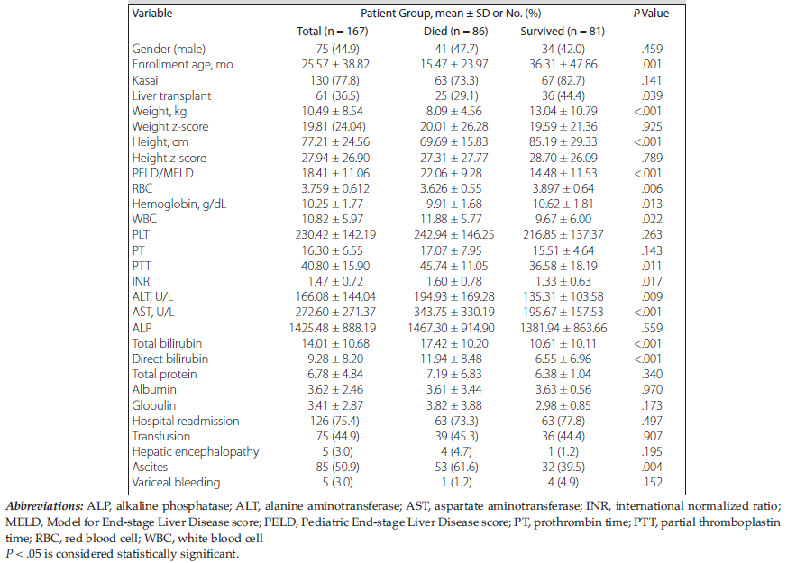
Table 1. Baseline Characteristics of Children With Biliary Atresia
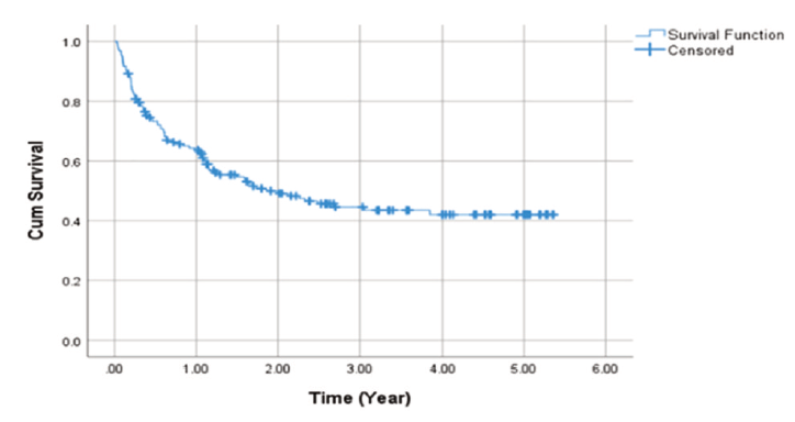
Figure 1. Survival Analysis of Children With Biliary Atresia
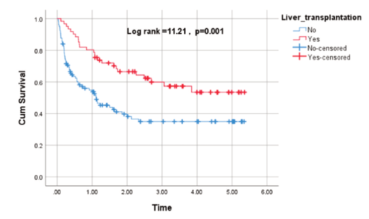
Figure 2. Differences in Survival Outcomes Between Liver Transplant Recipients and Non-Transplant Patients
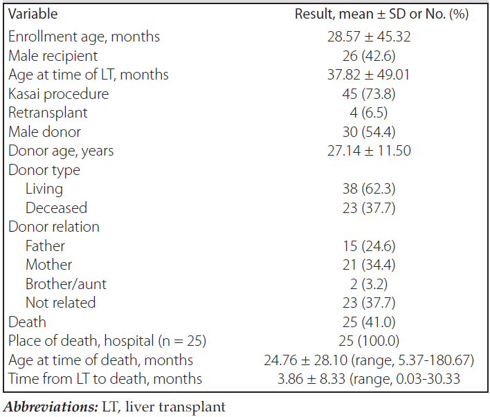
Table 2. Characteristics of Participants Who Underwent Liver Transplant (n = 61)
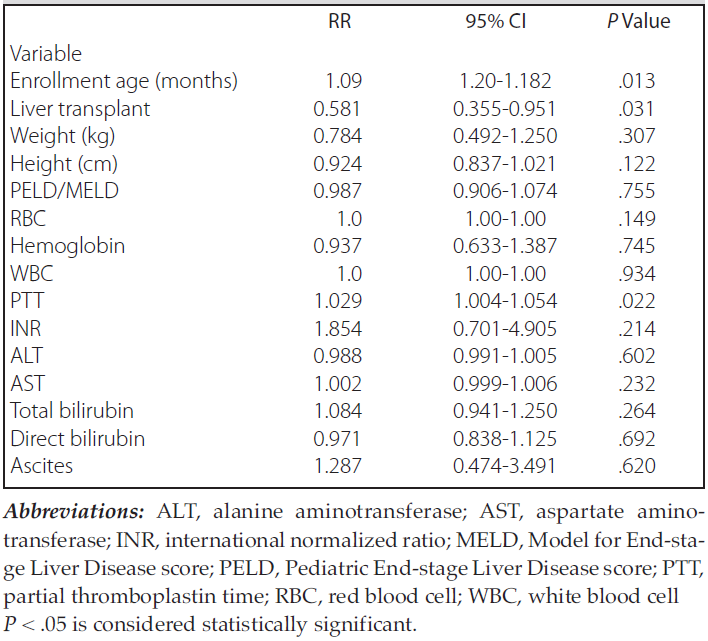
Table 3. Cox Regression Analysis Results of Children With Biliary Atresia