Volume: 24 Issue: 7 July 2026
FULL TEXT
Peripheral Blood Next-Generation RNA Sequencing as an Adjunctive Tool in the Evaluation of Borderline T-Cell-Mediated Rejection in BK Virus Nephropathy
Distinguishing BK virus nephropathy from T-cell-mediated rejection remains a clinicopathological chal-lenge in kidney transplants, because the 2 entities may share overlapping histological features while requiring opposite therapeutic approaches. We report a kidney transplant recipient with biopsy-proven BK virus nephropathy who later underwent repeat biopsy showing low-level BK virus nephropathy with borderline T-cell-mediated rejection. Despite histological concern for rejection, the broader clinical picture did not support clinically significant T-cell-mediated rejection; specifically, serum creatinine remained stable, BK viral load showed marked improvement, donor-specific antibodies were negative, and there were no vascular or microvascular features of active rejection. A peripheral blood next-generation RNA sequencing gene exp-ression profile yielded a low-risk acute rejection score of 6. Antirejection therapy was deferred, immuno-suppression was not escalated, and the patient remained clinically stable. This case highlights the potential adjunctive role of peripheral blood RNA profiling for resolution of histologically ambiguous cases of BK virus nephropathy and borderline T-cell-mediated rejection.
Key words : Gene expression profile, Kidney transplant, Tutivia prognostic assay
Introduction
A 56-year-old woman with end-stage kidney disease secondary to lupus nephritis underwent a second deceased donor kidney transplant after failure of a prior kidney allograft due to combined antibody-mediated rejection (AMR) and T-cell-mediated rejec-tion (TCMR). Induction immunosuppression consisted of thymoglobulin 4.5 mg/kg and methylprednisolone 1000 mg administered in 3 divided doses. Initial main-tenance immunosuppression included prednisone, mycophenolate mofetil (MMF), and tacrolimus.
The early posttransplant course was initially stable. However, by postoperative day (POD) 105, plasma BK virus DNA had become detectable by quantitative polymerase chain reaction at 3100 IU/mL, with serum creatinine of 1.15 mg/dL and donor-deri-ved cell-free DNA (dd-cfDNA) of 0.5%. The MMF dose was reduced to 750 mg twice daily, and tacrolimus levels were maintained between 8 and 10 ng/dL. Despite stepwise reduction in immunosuppression, plasma BK viral load continued to rise over the subsequent months, as follows: 5580 IU/mL on POD 132, 12600 IU/mL on POD 182, and 23200 IU/mL on POD 202. During this period, serum creatinine increased modestly from 1.38 to 1.59 mg/dL and then measured 1.38 mg/dL on POD 202. Parallel with worsening BK viremia, MMF was progressively reduced to 250 mg twice daily (Table 1, Figure 1).
Because of persistent BK viremia, a for-cause kidney allograft biopsy was performed. This biopsy demonstrated BK virus nephropathy (BKVN) class 2, with SV40 positivity in approximately 15% to 20% of tubular epithelial cells, Banff polyomavirus nephro-pathy class 3, and moderate tubulitis. These findings established biopsy-proven BKVN. By POD 224, plasma BK viral load had increased to 38300 IU/mL, serum creatinine was 1.68 mg/dL, and MMF was discontinued. Plasma BK viral load reached a peak of 103000 IU/mL on POD 245. Tacrolimus was switc-hed to cyclosporine, with target trough levels of 100 to 150 ng/dL. Serum creatinine subsequently reached 2.1 mg/dL on POD 253. Shortly thereafter, viral burden began to improve; specifically, plasma BK viral load decreased to 582 IU/mL by POD 279, and serum creatinine stabilized around 1.61 mg/dL by POD 330.
As BK viremia improved, immunosuppression was cautiously reintroduced. On POD 279, MMF was restarted at 250 mg twice daily and subsequently increased in a stepwise manner to 1000 mg twice daily oral dose. By POD 330, plasma BK viral load had fallen further to 100 IU/mL, but the level of dd-cfDNA had risen to 1.8%. At that time, the peripheral blood next-generation RNA sequencing gene expression profile (Tutivia prognostic assay) yielded a low-risk acute rejection score of 6. The contemporaneous score of an immune cell function assay that detects cell-mediated immunity in an immunosuppressed patients (ImmuKnow) was 282, which did not suggest an underimmunosuppressed state. Donor-specific antibodies were absent. By POD 360, tacrolimus was restarted with target trough levels of 8 to 10 ng/dL with cessation of cyclosporin.
Because of the increase in dd-cfDNA and concern for possible rejection, a repeat kidney allograft biopsy was performed on POD 356. This biopsy was inter-preted as concurrent low-level BKVN class 1 and suspicious for borderline TCMR, with Banff scores g0, ci1, ct1, i1, t3, ptc1, v0, and C4d0 (Figure 2). The biopsy showed interstitial inflammation (Banff score i1) in nonscarred cortex with associated severe tubulitis (t3), raising concern for borderline TCMR; however, there was no endarteritis or glomerulitis, and C4d staining was negative.
In light of the overall clinicopathological asses-sment, antirejection therapy was deferred. Immuno-suppression was not escalated, and the patient remained clinically stable. Subsequent follow-up continued to show stable kidney function, with serum creatinine 1.6 mg/dL on POD 372, whereas BKV quantitative polymerase chain reaction remained low at 61.6 IU/mL (Table 1, Figure 1).
Discussion
This case illustrates a classic but difficult problem in kidney transplants, in interpretation of borderline rejection-like histological findings in a patient with recent or resolving BKVN. The distinction is critical, because the therapeutic implications are opposite. It has been established that BKVN generally reflects excessive net immunosuppression and can be mana-ged by reduction of immunosuppression, whereas TCMR requires intensification of immunosuppression. In patients with overlapping biopsy findings, the risk of both undertreatment of rejection and exacerbation of viral injury must be carefully considered.1-3
The histological distinction between BKVN and TCMR rests on several features, summarized in Table 2. In BKVN, viral cytopathic change, SV40-positive tubular nuclei, and inflammation around infected tubules support virus-mediated injury. The inflammatory infiltrate generally consists of promi-nent plasma cells, neutrophils, CD20-positive cells, and tubulocentric macrophage clusters. In contrast, TCMR is favored when interstitial inflammation and tubulitis occur in nonscarred cortex away from virally infected tubules, particularly when accompanied by vascular inflammation, microvascular injury, or other supportive features. The inflammatory infiltrate often consists of T-cell-predominant infiltrate.1,4-7
Current biomarkers provide a noninvasive asses-sment of acute rejection risk but have limited ability to identify early rejection or distinguish TCMR from other causes of allograft injury. Such biomarkers often do not distinguish TCMR from AMR or BKVN and are generally more useful for their negative predictive value than for positive discrimination.8-10 Tutivia is a proprietary peripheral blood next-generation sequencing assay for evaluation of a 17-gene mRNA signature in combination with a prop-rietary artificial intelligence algorithm to categorize kidney transplant patients as low risk or high risk of acute rejection.11 In the validation study, Bestard and colleagues reported low Tutivia scores in patients with BKVN, suggesting potential utility to distinguish viral-related injury from acute rejection.11 Although RNA profiling may reflect general immune activation, it does not reflect a specific pathology.
The management of borderline TCMR remains controversial, because this Banff category is biolo-gically heterogeneous, its prognostic significance varies by clinical context, and the evidence suppor-ting uniform treatment is limited. The guidance from Kidney Disease: Improving Global Outcomes sug-gests treatment of subclinical and borderline acute rejection, although this recommendation is based on low-grade evidence.12 Banff updates have emphasized that not all tubulointerstitial inflammation above the Banff threshold for borderline TCMR necessarily reflects alloreactive T-cell injury, and thereby the present Banff perspective supports a more contextual interpretation of these biopsies.13 Zachariah and colleagues have reported that, for serial protocol biopsies performed after 1 year, borderline phenotypes were associated with adverse long-term graft function, supporting the view that at least some borderline lesions may represent clinically meaningful alloim-mune injury and may justify treatment or optimi-zation of immunosuppression.14 More recent data suggest that the value of treatment depends on biopsy context: Palmisano and colleagues have reported that treatment of acute borderline TCMR appeared justified mainly in biopsies performed for clinical indication,15 whereas Kirn and colleagues found that steroid pulse therapy for borderline TCMR detected on protocol biopsy was not associated with improved histological resolution, reduced fibrosis, or better graft function.16 Taken together, these data support a nuanced approach where borderline TCMR on for-cause biopsy is often treated because it may reflect active alloimmunity, whereas selected protocol-biopsy lesions may reasonably be observed with close follow-up when the broader clinical picture does not support clinically significant rejection.
In our present case, the first biopsy established BKVN, whereas the later biopsy raised concern for borderline TCMR in the setting of persistent low-level BKVN because interstitial inflammation with associated tubulitis was present in nonscarred cortex away from SV40-positive tubules. Despite this histo-logical concern, the broader clinical context did not support active rejection requiring treatment (Figure 2).
In this context, the peripheral blood biomarker data became clinically useful. The dd-cfDNA had increased to 1.8% on POD 330, raising concern for graft injury, but dd-cfDNA is not specific for rejection and may increase in other forms of allograft injury.10 In contrast, the peripheral blood next-generation RNA sequencing profile (Tutivia) yielded a low-risk acute rejection score of 6. The contemporaneous ImmuKnow assay value was 282. Although neither assay is independently diagnostic, together these did not suggest an underimmunosuppressed state that would have increased concern for clinically significant TCMR. When considered alongside stable serum creatinine, improving BK viral load, negative status for donor-specific antibodies, absence of proteinuria, and lack of other supportive histological features of active rejection, these results argued against clinically meaningful rejection requiring treatment escalation.
The subsequent clinical course supported the afore-mentioned decision against treatment escalation, with stable kidney function and persistently low-level BK viremia during follow-up (Table 1, Figure 1). Although one cannot conclude from a single case that perip-heral blood RNA profiling can reliably distinguish BKVN from borderline TCMR in all settings, our case suggests that it may provide useful adjunctive information when histology and clinical context are discordant.11 To the best of our knowledge, there are no prior published reports describing the use of peripheral blood next-generation RNA sequencing in this specific clinicopathology scenario of BKVN with biopsy findings suspicious for borderline TCMR. Additional studies and longer follow-up are needed to determine whether peripheral blood RNA pro-filing can reproducibly identify patients with borderline histology findings who may be safely observed rather than treated.
This report also underscores the importance of integrated clinicopathological interpretation in transplant biopsy. Borderline TCMR is, by definition, a low-grade histologic category, and its clinical signi-ficance may vary substantially depending on context. In a patient with recent biopsy-proven BKVN, impro-ving viremia, stable allograft function, and no sero-logical evidence of alloimmune activation, peripheral blood next-generation RNA sequencing may help support confidence in a conservative management approach.
This report had important limitations. This was a single case report and therefore could not establish diagnostic accuracy, clinical utility, or generalizability. There was no external validation of the biomarker findings, and follow-up was limited (372 days), so the long-term safety of deferred treatment in this setting remains unknown. In addition, biomarker specificity was uncertain because both dd-cfDNA and perip-heral blood gene expression assays may have reflec-ted nonspecific allograft injury or immune activation and could not definitively distinguish BKVN from borderline TCMR. Finally, publication bias must be considered, because cases with favorable short-term outcomes are more likely to be reported. For these reasons, the biomarker results should be interpreted only as supportive adjuncts within an integrated clinicopathological assessment.
Conclusions
Peripheral blood next-generation RNA sequencing may be a useful adjunct in the evaluation of kidney transplant recipients with BKVN and biopsy findings suspicious for borderline TCMR. In our case, the assay did not replace histology, but it helped contextualize a biopsy that suggested borderline TCMR despite a clinical picture arguing against clinically significant rejection. In patients with recent or resolving BKVN, this type of integrated assessment may help avoid unnecessary escalation of immunosuppression.
References:
- Hirsch HH, Randhawa P; AST Infectious Diseases Community of Practice. BK polyomavirus in solid organ transplantation. Am J Transplant. 2013;13 Suppl 4:179-188. doi:10.1111/ajt.12110
CrossRef - PubMed - Kotton CN, Kamar N, Wojciechowski D, et al; Transplantation Society International BK Polyomavirus Consensus Group. The Second International Consensus Guidelines on the Management of BK Polyomavirus in Kidney Transplantation. Transplantation. 2024;108(9):1834-1866. doi:10.1097/TP.0000000000004976
CrossRef - PubMed - Sharma R, Tzetzo S, Patel S, Zachariah M, Sharma S, Melendy T. BK virus in kidney transplant: current concepts, recent advances, and future directions. Exp Clin Transplant. 2016;14(4):377-384. doi:10.6002/ect.2016.0030
CrossRef - PubMed - Hirsch HH, Randhawa P; AST Infectious Diseases Community of Practice. BK virus in solid organ transplant recipients. Am J Transplant. 2009;9 Suppl 4:S136-146. doi:10.1111/j.1600-6143.2009.02904.x
CrossRef - PubMed - Masutani K, Shapiro R, Basu A, Tan H, Wijkstrom M, Randhawa P. The Banff 2009 Working Proposal for polyomavirus nephropathy: a critical evaluation of its utility as a determinant of clinical outcome. Am J Transplant. 2012;12(4):907-918. doi:10.1111/j.1600-6143.2012.03993.x
CrossRef - PubMed - Nickeleit V, Singh HK, Dadhania D, et al. The 2018 Banff Working Group classification of definitive polyomavirus nephropathy: a multicenter validation study in the modern era. Am J Transplant. 2021;21(2):669-680. doi:10.1111/ajt.16189
CrossRef - PubMed - Sar A, Worawichawong S, Benediktsson H, Zhang J, Yilmaz S, Trpkov K. Interobserver agreement for polyomavirus nephropathy grading in renal allografts using the working proposal from the 10th Banff Conference on Allograft Pathology. Hum Pathol. 2011;42(12):2018-2024. doi:10.1016/j.humpath.2011.03.008
CrossRef - PubMed - Edwards RL, Menteer J, Lestz RM, Baxter-Lowe LA. Cell-free DNA as a solid-organ transplant biomarker: technologies and approaches. Biomark Med. 2022;16(5):401-415. doi:10.2217/bmm-2021-0968
CrossRef - PubMed - Friedewald JJ, Kurian SM, Heilman RL, et al.; Clinical Trials in Organ Transplantation 08 (CTOT-08). Development and clinical validity of a novel blood-based molecular biomarker for subclinical acute rejection following kidney transplant. Am J Transplant. 2019;19(1):98-109. doi:10.1111/ajt.15011
CrossRef - PubMed - Park S, Guo K, Heilman RL, et al. Combining blood gene expression and cellfree DNA to diagnose subclinical rejection in kidney transplant recipients. Clin J Am Soc Nephrol. 2021;16(10):1539-1551. doi:10.2215/CJN.05530421
CrossRef - PubMed - Bestard O, Augustine J, Wee A, et al. Prospective observational study to validate a next-generation sequencing blood RNA signature to predict early kidney transplant rejection. Am J Transplant. 2024;24(3):436-447. doi:10.1016/j.ajt.2023.09.021
CrossRef - PubMed - Kasiske BL, Zeier MG, Chapman JR, et al; Kidney Disease: Improving Global Outcomes. KDIGO clinical practice guideline for the care of kidney transplant recipients: a summary. Kidney Int. 2010;77(4):299-311. doi:10.1038/ki.2009.377
CrossRef - PubMed - Naesens M, Roufosse C, Cornell LD, et al. The Banff 2024 Kidney Meeting Report: rejection as a spectrum of phenotypes and focus on differential diagnostic reasoning. Am J Transplant. 2026;26(5):922-936. doi:10.1016/j.ajt.2026.01.018
CrossRef - PubMed - Zachariah MS, Dwivedi AK, Yip CS, et al. Utility of serial protocol biopsies performed after 1 year in predicting long-term kidney allograft function according to histologic phenotype. Exp Clin Transplant. 2018;16(4):391-400. doi:10.6002/ect.2016.0323.
CrossRef - PubMed - Palmisano A, D'Angelo M, Gandolfini I, et al. Borderline rejection: to treat or not to treat? Transpl Immunol. 2024;84:102047. doi:10.1016/j.trim.2024.102047
CrossRef - PubMed - Kirn G, Chandran S, Rajalingam R, Feng S. Efficacy of steroid pulse to treat borderline T-cell mediated rejection on protocol kidney allograft biopsy. Clin Transplant. 2025;39(5):e70171. doi:10.1111/ctr.70171
CrossRef - PubMed

Volume : 24
Issue : 7
Pages : 569 - 574
DOI : 10.6002/ect.2026.0075
From the 1Department of Surgery, the 2Department of Pathology, and the 3Department of Nephrology, West Virginia University School of Medicine, Morgantown, West Virginia, United States
Acknowledgements: The authors have not received any funding or grants in support of the presented research or for the preparation of this work and have no declarations of potential conflicts of interest. The authors used ChatGPT (OpenAI) for language editing and proofreading during manuscript preparation. All content was reviewed and edited by the authors, who take full responsibility for the final manuscript.
Corresponding author: Rajeev Sharma, Department of Surgery, West Virginia University School of Medicine, 1 Medical Center Drive, Morgantown, WV 26506, USA
E-mail: Rajeev.Sharma@hsc.wvu.edu
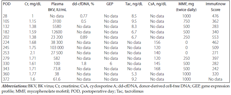
Table 1. Longitudinal Laboratory and Immunosuppression Data
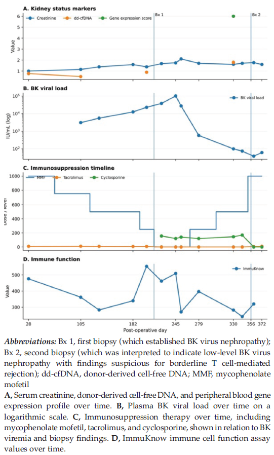
Figure 1. Longitudinal Clinical Course by Postoperative Day
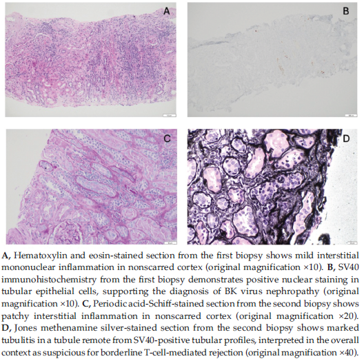
Figure 2. Histologic Findings in Sequential Kidney Allograft Biopsies
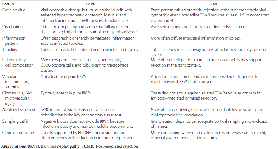
Table 2. Histologic Features Favoring BK Virus Nephropathy Versus T-Cell-Mediated Rejection in Kidney Allograft Biopsy