Volume: 24 Issue: 7 July 2026
FULL TEXT
Effect of Gene Expression Profiling Surveillance Compared With Endomyocardial Biopsy On Tricuspid Regurgitation in Patients with Heart Transplant
Objectives: Gene expression profiling is an emerging noninvasive method for rejection surveillance in heart transplants. Tricuspid regurgitation prevalence increases over time after heart transplant, and many prior studies have implicated invasive endomyocardial biopsy. The relationship between the use of gene expression profiling (AlloMap molecular expression test; CareDx) versus endomyocardial biopsy with regard to prevalence of tricuspid damage has never been studied.
Materials and Methods: Among 158 heart transplant patients with history of gene expression profiling and mean follow-up duration of 10.3 years that we reviewed, 114 patients were included in this study. Patients were divided into 4 groups based on mean endomyocardial biopsy and mean gene expression profiling sampling times. Tricuspid regurgitation grade change was compared between the group with less gene expression profiling and more endomyocardial biopsy versus the group with more gene expression profiling and less endomyocardial biopsy.
Results: Results from this single-center study showed that no statistically significant difference in tricuspid regurgitation for cardiac transplant patients who were monitored for rejection by endomyocardial biopsy versus gene expression profiling.
Conclusions: Preference of gene expression profiling instead of endomyocardial biopsy for rejection surveillance does not have a significant effect on tricuspid valve regurgitation.
Key words : Heart transplantation, Invasive biopsy, Rejection surveillance, Tricuspid valve disease
Introduction
Although endomyocardial biopsy (EMB) remains the clinical gold standard for the diagnosis of acute rejec-tion after heart transplant and is used as surveillance method,1 alternative approaches have been approved. The use of gene expression profiling (GEP), which is a noninvasive procedure that requires a peripheral blood draw for rejection monitoring, has increased since 2010. Many transplant centers have incorporated GEP methods (as approved by the US Food and Drug Administration) into their rejection surveillance protocols.2
Tricuspid regurgitation (TR) is the most common valvular dysfunction found after heart transplant.3 Tricuspid regurgitation events may be related to EMB performed for allograft rejection surveillance, and many previously published studies have stated that the prevalence of TR increases over time after heart transplant, from 5% at 1 year to 50% at 4 years posttransplant.4-8 Tricuspid regurgitation after heart transplant could also be related to pulmonary arterial hypertension, surgical technique, right ventricle anatomic distortion, allograft ischemia time, or acute cellular rejection (ACR).4,5,9
Although TR severity has been shown to increase over time after heart transplant, the association between the use of GEP as an alternative to EMB with regard to tricuspid damage has never been studied. We aimed to elucidate the lifetime severity of TR with regard to use of EMB versus GEP. Specifically, we investigated the effects of a noninvasive method such as GEP for rejection surveillance in heart transplant recipients versus the use of EMB with regard to TR events. Our hypothesis was that reduced use of in-vasive EMB in favor of increased use of a noninvasive method such as GEP can decrease tricuspid valve (TV) damage, with TR used as a marker of this damage.
Materials and Methods
This is a single-center retrospective observational study of heart transplant patients. This project was granted a full waiver of the typical authorization requirement (the Health Insurance Portability and Accountability Act of 1996) by a university hospital (institutional review board approval No. 202312252). The protocols and the study were approved by the ethics committee of the institution before the study began, and the protocols conformed to the ethical guidelines of the 1975 Declaration of Helsinki. Informed consent was obtained from patients or their guardians.
Patients were identified from electronic medical records via a self-service data reporting tool (SlicerDicer, Epic Systems). We reviewed charts and identified 158 patients (72% male) with heart transplant who were evaluated by both GEP and EMB for the period from 1993 to December 2023. Fourteen patients were excluded from the study because of technical difficulties in transthoracic echocardiogram or failure to visualize TV findings. One patient was excluded from the study because of technical difficulties in obtaining the echocardiogram report and images that were completed by an outside facility.
Given the retrospective nature of the study, a priori sample size calculation was not feasible. Therefore, a detectable-difference assessment was performed for the primary comparison of TR grade change between the group with less GEP and more EMB versus the group with more GEP and less EMB. With sample sizes of n = 34 and n = 36, we established that a 2-sided independent-sample t test at an α level of .05 provides approximately 80% power to detect a standardized mean difference (Cohen d) of ~0.68, which corresponds to an absolute difference of approximately 1.0 TR-grade unit based on the observed pooled standard deviation (~1.4) and indicates that our study was sufficiently powered to detect clinically meaningful differences of this magnitude.
The GEP was performed using a commercially available peripheral blood-based assay (AlloMap molecular expression test; CareDx) that quantifies the expression of a predefined panel of immune response and metabolic genes associated with ACR. The AlloMap assay generates a composite score that reflects the balance between immune activation and quiescence; lower scores indicate relative immune suppression, and higher scores suggest increased immune activity. The GEP method has been valida-ted for rejection surveillance in clinically stable heart transplant recipients beyond the early posttransplant period and has been shown to safely reduce the frequency of invasive EMB in selected populations.2
The biopsies were performed by either the transjugular approach or the transfemoral approach (5 patients). All echocardiograms were performed in the same center. The TR severity was compared by assigning scores according to the severity shown on echocardiograms. Absence of TR was accepted as a score of 0, and trace TR was accepted as a score of 1. Trace to mild TR was accepted as 2, mild TR was accepted as 3, mild to moderate TR was accepted as 4, moderate TR was accepted as 5, moderate to severe TR was accepted as 6, and severe TR was accepted as a score of 7. The difference between the TR severity at last follow-up was subtracted from the TR severity at 1 month after orthotopic heart transplant.
Patients with posttransplant mild to moderate TR or more severe cases (17 patients) were excluded from the study. Five patients were excluded due to long sheath use, which affected the bioptome passages across the tricuspid valve. We also excluded patients with at least grade 2 ACR (2 patients), patients with any antibody-mediated rejection (AMR) (1 patient), patients with history of both ACR and AMR at any time (2 patients), and patients with any multiple rejection episodes (1 patient). In addition to patients with biopsy-proven rejection, those with a decline in left ventricular ejection fraction concerning rejection despite negative biopsy findings (n = 2) were also excluded from the study. Figure 1 shows a flow diagram of included patients.
The mean age of the patients at the time of the transplant was 47.7 years (maximum 74 years, minimum 4 months). The mean duration of follow-up was 10.3 years. The mean number of GEP evaluations was 10.74, and the mean number of EMB procedures was 22.07. The mean ratio of GEP to EMB was 0.88. During follow-up, a total of 6 patients experienced rejection, defined as ≥2R ACR, any AMR, or a decline in left ventricular ejection fraction (LVEF). Details of the demographic information of the patient population are shown in Table 1.
Patients were divided into 4 groups based on the mean number of AlloMap GEP procedures (10.62) and mean number of EMB procedures (13.96) as follows: the low GEP-low EMB group, the low GEP-high EMB group, the high GEP-low EMB group, and the high GEP-high EMB group.
The TR change from after transplant versus last follow-up was calculated with the assigned number to TR grade as explained above and then subtracting the TR grade of last follow-up from the TR grade after transplant. The correlation of the high GEP-low EMB group with TR grade change was compared with the low GEP-high EMB group using an independent t test. We used Minitab statistical software (version 20.4.0) for statistical analyses.
Results
There were 34 patients in the low GEP-high EMB group; for this group, the mean TR change between the posttransplant TR versus the TR at last follow-up was +0.35 (SD 1.39). There were 36 patients in the high GEP-low EMB group; for this group, the mean TR change between the posttransplant TR versus the TR at last follow-up was +0.08 (SD 1.42). Therefore, there was a slightly greater TR progression in the low GEP-high EMB group versus the high GEP-low EMB group.
Correlations with EMB numbers were weak in the low GEP-high EMB group and high GEP-low EMB group (+0.14 in the low GEP-high EMB group and -0.06 in the high GEP-low EMB group). Correlations with GEP numbers were weak in both groups (-0.24 in the low GEP-high EMB group and +0.27 in the high GEP-low EMB group).
The low GEP-high EMB group showed a weak positive correlation between TR grade change and GEP count, whereas the low GEP-high EMB group showed a negative correlation. Correlations with EMB were weak in both groups. The independent t test statistics to compare TR grade change between the low GEP-high EMB group and the high GEP-low EMB group showed t statistics of 0.80 and P = .425, which demonstrated that the difference in mean TR grade change between the 2 groups was not significant.
Tricuspid regurgitation peak velocity and tricuspid annular plane systolic excursion values at last follow-up were also analyzed, with no significant dif-ferences between groups (Table 2).
Discussion
Endomyocardial biopsy remains the gold standard for rejection surveillance after heart transplant, although noninvasive techniques such as GEP are emerging. Prior studies have shown that the number of EMB procedures is associated with worsening TR. This association may be due to direct mechanical injury during biopsy sampling from TV instead of ventricle or due to the multiple actions of insertion and withdra-wal of the rigid bioptome into and out of the TV.10,11
From a mechanistic standpoint, GEP reflects systemic immune activation rather than localized myocardial injury, providing a noninvasive surrogate of rejection risk without mechanical interaction with the TV apparatus. Therefore, replacement of EMB with GEP for surveillance theoretically reduces the risk of biopsy-related structural valvular injury while maintaining immunologic monitoring.
The aim of our study was to evaluate whether reduction in the use of invasive EMB in favor of an increase in the use of a noninvasive method such as GEP can decrease TV damage, with TR used as a marker of this damage. We used valve regurgitation severity to monitor TV damage, because we believe that the TR severity is the clinical effect of potential damage. This retrospective single-center observational study with mean follow-up of 10.3 years showed that the use of noninvasive methods for rejection sur-veillance in heart transplant patients did not produce superior results by means of TR. Therefore, we conclude that preference of GEP instead of EMB for rejection surveillance does not have a significant effect on TR.
Prior studies have shown the prevalence and severity of TR increased after orthotopic heart transplant due to EMB. However, no previously published studies have investigated TV damage by comparison of noninvasive rejection surveillance versus conventional EMB. Our study findings differ from expectations based on prior studies, and we believe our results have answered an important clinical question: does preference of GEP in exchange for EMB decrease the risk of developing TR? In the pathological examination of 417 patients who had a total of 3550 EMB procedures after heart transplant between 1985 and 2010, TV tissue was found in 12 patients, and an increase in the severity of TV insufficiency was observed in approximately 12% of these patients.12
A recent single-center study from Brazil showed that EMB using a rigid bioptome did not imply a clinically relevant risk of TR following 729 pro-cedures in heart transplant patients through a jugular approach.13 The severity of TR after heart transplant is higher versus the general population and tends to increase with time. Williams and colleagues11 repor-ted that incidence of biopsy-related damage could be decreased by minimizing the number of passages of the bioptome across the tricuspid apparatus. Therefore, they used a 45-cm-long sheath in their routine EMB procedure and successfully reduced the incidence of flail tricuspid valve from 41% to 6% and reduced the mean grade of TR from 2 to 1.1.
According to a recent epidemiology study con-ducted in Spain during 2000-2019 with 1009 patients, the most frequent etiology of TR after cardiac transplant was undefined causes followed by acute rejection. The other etiologies in descending order were pulmonary hypertension, cardiac allograft vas-culopathy, pacemaker electrodes, biopsy complication, chronic renal insufficiency, severe pericardial effusion, valve prolapse, atrial tachycardia, and massive pulmonary emboli.14 Most of those etiologies are usually a result of primary graft failure or posto-perative complications, and the resultant TR is usually expected to improve over time, which was also demonstrated in that study. Our study excluded patients with graft dysfunction for this potential confounder. We compared postoperative month 1 echocardiograms but not immediate postoperative echocardiograms, because immediate postoperative echocardiograms can show unresolved consequences of potential primary graft dysfunction. Although other etiologies of postoperative TR are expected to subside during long-term follow-up, the biopsy complications are expected to increase tricuspid damage over time.14
Limitations
Our study had some limitations. Our study is a single-center study, and sample size is relatively small. Moreover, data collection periods were dif-ferent for some patient populations, because we compared the echocardiogram at month 1 versus the last follow-up. Therefore, the follow-up period for some patients who underwent transplant in the later part of the sample collection period, for example, in the years 2022 or 2023, may be insufficient to observe potential development of TR in the long-term.
Another limitation of our study is the method of reporting the echocardiograms. The echocardiograms were not reviewed by a specific independent researcher for our study; rather, we used the prior reports supplied from different, various providers. Considering the long mean follow-up time of 10.3 years, the potential variety of individual echo-cardiography readers could have caused inter-reader variability. The potential for variability also exists for procedure physicians who performed the biopsies; there were multiple different procedure physicians during this follow-up period, which could cause technique-related variations.
We also did not investigate different methods of insertion sites, catheters used, and techniques for EMB. However, we believe the widely used ap-proach in EMB is the transjugular approach without a long sheath, which is the preference at our center. Differences in our results of TV damage may have been the result of improved catheter techniques, devices, and operator experiences.
Conclusions
Our study showed no significant TR change in cardiac transplant patients who were monitored for rejection by EMB versus GEP. Preference of GEP instead of EMB for rejection surveillance does not have a significant effect on TR.
References:
- Velleca A, Shullo MA, Dhital K, et al. The International Society for Heart and Lung Transplantation (ISHLT) guidelines for the care of heart transplant recipients. J Heart Lung Transplant. 2023;42(5):e1-e141. doi:10.1016/j.healun.2022.10.015
CrossRef - PubMed - Moayedi Y, Foroutan F, Miller RJH, Fan CS, Posada JGD. Risk evaluation using gene expression screening to monitor for acute cellular rejection in heart transplant recipients. J Heart Lung Transplant. 2019;38(1):51-58. doi:10.1016/j.healun.2018.09.004
CrossRef - PubMed - Fiorelli AI, Coelho GH, Oliveira JL Jr, et al. Endomyocardial biopsy as risk factor in the development of tricuspid insufficiency after heart transplantation. Transplant Proc. 2009;41(3):935-937. doi:10.1016/j.transproceed.2009.02.011
CrossRef - PubMed - Mielniczuk L, Haddad H, Davies RA, Veinot JP. Tricuspid valve chordal tissue in endomyocardial biopsy specimens of patients with significant tricuspid regurgitation. J Heart Lung Transplant. 2005;24(10):1586-1590. doi:10.1016/j.healun.2004.11.007
CrossRef - PubMed - Anderson CA, Shernan SK, Leacche M, et al. Severity of intraoperative tricuspid regurgitation predicts poor late survival following cardiac transplantation. Ann Thorac Surg. 2004;78(5):1635-1642. doi:10.1016/j.athoracsur.2004.05.028
CrossRef - PubMed - Wong RC, Abrahams Z, Hanna M, et al. Tricuspid regurgitation after cardiac transplantation: an old problem revisited. J Heart Lung Transplant. 2008;27(3):247-252. doi:10.1016/j.healun.2007.12.011
CrossRef - PubMed - Lo CY, Chang HH, Hsu CP, Lai ST, Shih CC. Endomyocardial biopsy-related tricuspid regurgitation after orthotopic heart transplantation: single-center experience. J Chin Med Assoc. 2007;70(5):185-192. doi:10.1016/S1726-4901(09)70356-9
CrossRef - PubMed - Nguyen V, Cantarovich M, Cecere R, Giannetti N. Tricuspid regurgitation after cardiac transplantation: how many biopsies are too many? J Heart Lung Transplant. 2005;24(7 Suppl):S227-S231. doi:10.1016/j.healun.2004.07.007
CrossRef - PubMed - Saraiva F, Matos V, Gonçalves L, Antunes M, Providência LA. Complications of endomyocardial biopsy in heart transplant patients: a retrospective study of 2117 consecutive procedures. Transplant Proc. 2011;43(5):1908-1912. doi:10.1016/j.transproceed.2011.03.010
CrossRef - PubMed - Aziz TM, Burgess MI, Rahman AN, Campbell CS, Deiraniya AK, Yonan NA. Risk factors for tricuspid valve regurgitation after orthotopic heart transplantation. Ann Thorac Surg. 1999;68(4):1247-1251. doi:10.1016/S0003-4975(99)00768-7
CrossRef - PubMed - Williams MJ, Lee MY, DiSalvo TG, et al. Biopsy-induced flail tricuspid leaflet and tricuspid regurgitation following orthotopic cardiac transplantation. Am J Cardiol. 1996;77(15):1339-1344. doi:10.1016/S0002-9149(96)00202-0
CrossRef - PubMed - Fiorelli AI, Coelho GH, Aiello VD, et al. Tricuspid valve injury after heart transplantation due to endomyocardial biopsy: an analysis of 3550 biopsies. Transplant Proc. 2012;44(8):2479-2482. doi:10.1016/j.transproceed.2012.07.024
CrossRef - PubMed - Beck-da-Silva L, Bridi LH, Matte BS, Valle FH. Endomyocardial biopsy using rigid bioptome technique and the risk of tricuspid regurgitation after heart transplantation. Arq Bras Cardiol. 2024;121(10):e20240223. doi:10.36660/abc.20240223
CrossRef - PubMed - López-Vilella R, Paniagua-Martín MJ, González-Vílchez F, et al. Epidemiological study of tricuspid regurgitation after cardiac transplantation. Does it influence survival? Transpl Int. 2022;35:10197. doi:10.3389/ti.2022.10197
CrossRef - PubMed

Volume : 24
Issue : 7
Pages : 558 - 562
DOI : 10.6002/ect.2025.0284
From the 1University of Iowa, Department of Medicine, Division of Cardiology and Subdivision of Advanced Heart Failure and Transplantation Cardiology, Iowa City, Iowa, USA and 2the MercyOne North Iowa cardiology fellowship program, MercyOne North Iowa Medical Center, Mason City, Iowa, USA
Acknowledgements: The authors have not received any funding or grants in support of the presented research or for the preparation of this work and have no declarations of potential conflicts of interest.
Corresponding author: Serkan Bektur, University of Iowa, Division of Cardiology, Department of Medicine, 200 Hawkins Dr, E 315GH, Iowa City, IA 52242, USA
E-mail: serkanbe2000@yahoo.com
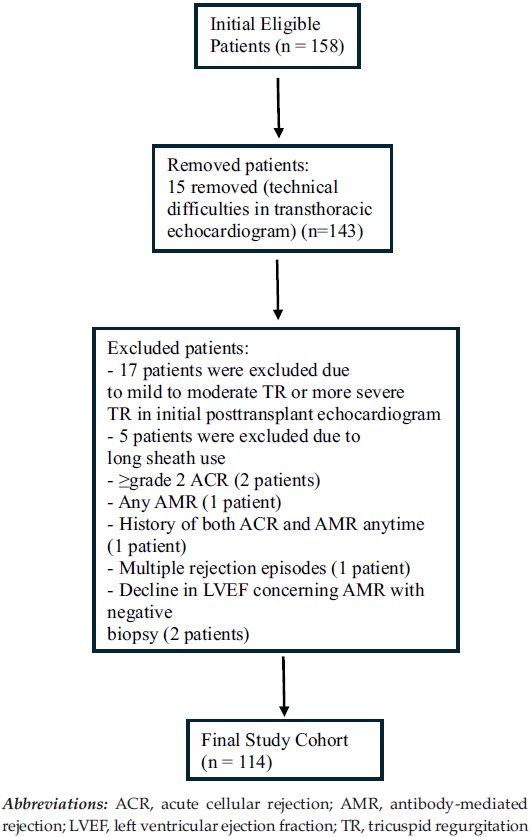
Figure 1. Flow Diagram
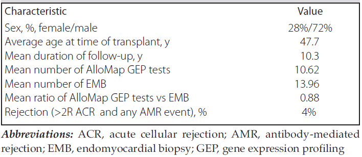
Table 1. Demographic Information of Patient Population
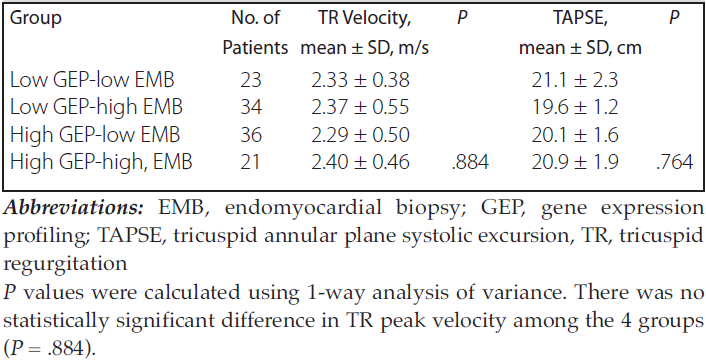
Table 2. Tricuspid Regurgitation Peak Velocities and Tricuspid Annular Plane Systolic Excursion Values at Last Follow-Up