Volume: 24 Issue: 5 May 2026
FULL TEXT
Fibrin Hydrogel Containing Microvascular Fragments for Improving Subcutaneous Islet Transplants in Diabetic Rats
Objectives: Subcutaneous tissue serves as a noninvasive and easily accessible site for pancreatic islets transplant; however, poor blood supply impedes survival of pancreatic islet cells and increases the number of islets necessary for normoglycemia. We evaluated the potential of fibrin hydrogel containing microvascular fragments to improve subcutaneous islet engraftment in diabetic rats.
Materials and Methods: Male Wistar rats were prevascularized subcutaneously for 7 days with fibrin hydrogel, 5000 microvascular fragments, or fibrin hydrogel + 5000 microvascular fragments. After streptozotocin injection to induce type 1 diabetes mellitus, 1500 IEQ were transplanted into prevascularized groups. A cotransplant group received 1500 IEQ together with fibrin hydrogel + 5000 microvascular fragments, and a control group received 3000 IEQ alone. Graft function was assessed through blood glucose monitoring, glucose tolerance tests, immunostaining, and plasma insulin concentration over 28 days.
Results: Immunohistochemical analysis revealed that prevascularization with the combination of fibrin hydrogel and microvascular fragments significantly increased CD31 expression compared with fibrin hydrogel alone and microvascular fragments alone (P < .05). Following transplantation of 1500 IEQ to diabetic rats, both the prevascularization and cotransplant groups, which received the combination of fibrin hydrogel and microvascular fragments, achieved normoglycemia within 28 days and both demonstrated superior glucose tolerance, higher plasma insulin levels, and increased CD31 and insulin expression compared with the other groups (P < .05).
Conclusions: We demonstrated that fibrin hydrogel containing microvascular fragments could significantly increase survival and function of subcutaneously transplanted islets, allowing for effective glycemic control with reduced islet mass.
Key words : Islet transplantation, Subcutaneous tissue, Type 1 diabetes mellitus, Vascularization
Introduction
Type 1 diabetes mellitus is an autoimmune disorder in which the immune system damages insulin-producing beta cells, necessitating lifelong insulin therapy.1 The standard treatment, intrahepatic islet transplant, involves substantial challenges, such as an instant blood-mediated inflammatory response (IBMIR), limited blood supply, and ischemic injury to the islets, leading to poor engraftment and function.2 To overcome these issues, researchers have been exploring extrahepatic transplant sites, such as subcutaneous tissue, which offer benefits such as easier access, noninvasive procedures, and reduced IBMIR risk.3 However, the hypoxic nature of the subcutaneous space negatively affects islet survival and function, making enhanced vascularization a crucial step for improvement of outcomes.4
Revascularization is crucial for the survival of transplanted beta cells; poor vascular integration results in hypoxia, impaired function, or cell death.5 Strategies to improve vascularization, including cotransplant with vascular endothelial cells (ECs) and the use of proangiogenic factors, are being investigated to increase therapeutic efficacy.6,7 Experimental investigations have shown that vascularization occurs with a delay between the graft and the host, initiating within 1 to 3 days and peaking approximately 14 days after transplant, with complete vascularization typically achieved by posttransplant day 28.8 To increase the success of subcutaneous islet transplant, prevascularization techniques have been developed.9,10 These methods aim to establish a suitable vascular bed prior to transplant, thereby improving the integration and functionality of the transplanted islets. A notable approach is device-free prevascularization, which minimizes the risks associated with surgical tools, such as infection or mechanical damage.11,12 In this study, we used fibrin hydrogel containing microvascular fragments (MVF) and compared the prevascularization and cotransplant methods for subcutaneous islet transplant in rats with type 1 diabetes mellitus.
Microvascular fragments are preformed vascular segments that are easily isolated from adipose tissue and maintain a well-defined vascular structure with ECs and pericytes.13 They serve as suitable vascularization units and are rich in proangiogenic factors.14 Compared with individual ECs, MVF can rapidly reassemble into functional microvascular networks, which are crucial for the survival of islets during the critical initial posttransplant phase.15 Studies have shown that MVF demonstrate improved engraftment efficiency and vasculogenic activity in vivo, making them valuable in regenerative medicine and tissue engineering.16
Fibrin is approved by the US Food and Drug Administration and is highly biodegradable, biocompatible, and biologically safe.17 Its structure facilitates interactions between cells and the extracellular matrix (ECM).18 Fibrin-based scaffolds can effectively integrate grafted islets into host tissue, enhancing diabetes management outcomes.19 When used as a carrier for pancreatic islets in subcutaneous grafts, fibrin stimulates local vascularization and prevents islet dispersion, making it an effective biomaterial for vascularizing pancreatic islets.20
We aimed to evaluate the potential of fibrin hydrogel containing MVF for subcutaneous prevascularization and cotransplant. We further hypothesized that the fibrin-MVF composite would reduce the marginal islet mass and improve glycemic control versus naked islets transplanted into unmodified subcutaneous tissue.
Materials and Methods
Chemical reagents and antibodies
Most of the chemical reagents used in this study were obtained from Sigma-Aldrich unless otherwise noted. The cell culture materials were sourced from Gibco. The antibodies we used included CD31 (orb10314; 1:100) and α-smooth muscle actin (α-SMA: orb195993; 1:100) from Biorbyt. The anti-insulin antibody (GTX34797; 1:100) was purchased from GeneTex. Secondary antibodies, including those for rabbits (orb688925) and mice (orb688924), were also obtained from Biorbyt.
Animals
Sixty adult male Wistar rats weighing 250 to 300 g were purchased from Pasteur Institute (Tehran, Iran). The animals were acclimated to the laboratory environment for 1 week. All rats were housed under constant temperature (23 ± 2 °C) and humidity (55% ± 5%), with a 12-h/12-h dark/light cycle and free access to food and water. The care and use of all animals were conducted in adherence to the Guide for the Care and Use of Laboratory Animals published by the National Institutes of Health (publication No. 86-23, revised 1985). All research procedures were approved by the ethics committee of Tehran University of Medical Sciences (ethical code No. IR.TUMS.AEC.1402.066).
Study design
This study consisted of 2 phases. The first phase was defined as the subcutaneous prevascularization phase (phase 1) from day -7 to day 0. Diabetes induction was initiated by streptozotocin (STZ) on day -3. The second phase was the subcutaneous islet transplant phase (phase 2) from day 0 to day 28 (Figure 1). Sixty rats were randomly allocated to 7 groups;13 6 animals from each prevascularized group were randomly selected on day 0 for immunohistology analysis of prevascularization efficacy. Therefore, experimental groups were as follows: (1) healthy control group (n = 6); (2) diabetic control group (n = 6); (3) fibrin hydrogel (day -7) + STZ (day -3) + 1500 islets (day 0), considered as the H1500 group (n = 12); (4) MVF (day -7) + STZ (day -3) + 1500 islets (day 0), considered as the MVF1500 group (n = 12); (5) fibrin hydrogel containing MVF (day -7) + STZ (day -3) + 1500 islets (day 0), considered as the HMVF1500 group (n = 12); (6) fibrin hydrogel containing MVF (day 0) + STZ (day 0) + 1500 islets (day 0), considered as the Co-HMVF1500 group (n = 6); and (7) islet-only group, which received STZ (day -3) and 3000 islets (day 0), considered as the Islet-only3000 group (n = 6).
Isolation and characterization of microvascular fragments
The MVF were freshly isolated from the epididymal fat tissue of donor rats via established protocols.21 Briefly, the rats were anesthetized via intraperitoneal injection of xylazine (10 mg/kg) and ketamine (100 mg/kg). Under aseptic conditions, epididymal fat pads were excised, minced, and subjected to enzymatic digestion using 2 mg/mL of type I collagenase with agitation for 7 minutes. The digestion process was stopped by the addition of Dulbecco's modified Eagle's medium (DMEM) supplemented with 20% fetal bovine serum (FBS). The suspension was then filtered sequentially through 500-μm and 30-μm nylon meshes and then washed twice by centrifugation at 600 g for 5 minutes at room temperature. The final MVF pellet was resuspended in DMEM supplemented with 10% FBS, 100 U/mL penicillin, and 0.1 mg/mL streptomycin and incubated at 37 °C with 5% CO2 for subsequent experiments.
Hydrogel preparation
The prevascularization involved the preparation of 100 μL of a mixture of 0.5% fibrinogen and 1 U/mL thrombin combined with 5000 MVF.20,21 Following a 2-cm skin incision, this mixture was injected into the flanks of rats and allowed to polymerize for 10 minutes before the incision was closed via nonabsorbable sutures.
Isolation and characterization of islets
Pancreatic islets were extracted from donor rat pancreas organs via a collagenase digestion method described in previous studies.22 After digestion, the islets were separated from the digested tissue through centrifugation via a stepwise Ficoll density gradient (Pan-Biotech). The isolated islets were subsequently cultured overnight in RPMI-1640 medium supplemented with 10% FBS, 100 U/mL penicillin, and 100 μg/mL streptomycin to facilitate recovery from the digestion procedure. The identity of the isolated islets was verified via dithizone staining, which specifically marks insulin-containing beta cells within the islets. Islet viability was evaluated via acridine orange/ethidium bromide staining, which distinguishes live cells (green in color) from dead cells (red in color).
Diabetes induction and islet transplant
Three days prior to islet transplant, the recipient rats received a single intraperitoneal injection of 50 mg/kg STZ dissolved in 0.1 mol/L sodium citrate buffer (pH 4.5). Diabetic rats were selected for transplant if blood glucose levels consistently ranged from 250 to 350 mg/dL for a period of 2 consecutive days following STZ injection. Diabetic animals were anesthetized with an intraperitoneal injection of ketamine at a dosage of 100 mg/kg and xylazine at 10 mg/kg. In the prevascularized groups and the Co-HMVF group, 1500 islets were transplanted, whereas the positive control group received 3000 free islets subcutaneously.
Recipient monitoring
After transplant, nonfasting blood glucose was monitored twice a week during the first week and then weekly. Blood samples were collected from the tail vein, and glucose levels were measured via a glucometer (Roche Diagnostics). Body weight was recorded weekly. Normoglycemia was defined as a blood glucose level <200 mg/dL. In addition, blood samples were collected from different groups via heart puncture at the end of phase 2. Plasma insulin levels were measured via a rat-specific enzyme-linked immunosorbent assay kit (model No. RK09278; Abclonal). To validate graft-dependent normoglycemia, the HMVF1500 and Co-HMVF1500 groups underwent glycemic control for 2 days after graft removal.
Assessment of graft function
To assess the capacity of the graft to respond to a glucose bolus, the animals were fasted for 4 hours on day 21 before being administered a glucose bolus of 20% glucose solution (2 g/kg) via intraperitoneal injection. Blood glucose levels were measured at each time point (before the test and after 30, 60, 90, and 120 minutes).
Immunohistochemical analysis
The subcutaneous implants were removed on day 28 for histological analysis. The subcutaneous tissue blocks were fixed in 4% paraformaldehyde at 4 °C for 24 hours. Following dehydration, the samples were embedded in paraffin, and 5-μm-thick sections were prepared. The sections were permeabilized with 2% Triton X-100 for 10 minutes and blocked with goat serum. Primary antibodies, including anti-insulin (1:100), anti-CD31 (1:100), and anti-α-SMA (1:100), were applied, and the sections were incubated overnight at 4 °C. Secondary antibodies were then used for detection. Nuclei were counterstained with 4′,6-diamidino-2-phenylindole dihydrochloride. Analysis was conducted via a fluorescence microscope (model BX60; Olympus, Japan). Every tenth section of the tissue sample was subjected to immunofluorescence staining. Positively stained cells relative to the total graft area in each section were quantified at 40× magnification via ImageJ software. The percentage of vascular density was acquired from 10 micrographs per rat.
Statistical analyses
Statistical analyses were performed via Prism software (version 8; GraphPad). For comparisons among multiple groups, 1-way analysis of variance was employed, followed by the Tukey post hoc test. The results are presented as means ± SD, and P < .05 was considered statistically significant.
Results
Characterization of isolated microvascular fragments and islets
The isolated MVF were characterized by CD31 and α-SMA surface markers, revealing a well-defined luminal structure (Figure 2A and 2B). Additionally, islets stained with dithizone were red in color. Islet viability assessment via acridine orange/ethidium staining indicated high viability (93%) of the isolated islets (Figure 2C and 2D ).
Immunofluorescence analysis of graft-bearing subcutaneous tissue revealed that the expression of CD31 in the HMVF group (37.94 ± 2.32%) was significantly greater than that in the H group (16.07 ± 1.08%; P < .0001) and MVF group (27.01 ± 2.39%; P < .01). Additionally, the MVF group had significantly greater numbers of CD31-positive cells versus the H group (P < .01) (Figure 3).
Immunofluorescence staining of graft-bearing subcutaneous tissue revealed a significant difference in CD31 expression between the HMVF1500 (34.68 ± 2.27%) and MVF1500 groups (26.82 ± 3.17%; P < .05), but this was not significant versus the H1500 group (31.32 ± 0.81%; P > .05) or the Co-HMVF1500 group (31.12 ± 1.98%; P > .05). As expected, the Islet-only3000 group had lower CD31 expression versus the other groups (20.99 ± 2.437%) (Figure 4).
Fibrin hydrogel containing microvascular fragments supports subcutaneous islet engraftment and function
Insulin expression in the HMVF1500 group (56.41 ± 0.79%) was significantly greater than insulin expression in the H1500 (41.32 ± 1.24%; P < .001) and MVF1500 groups (43.99 ± 2.66%; P < .001). No significant difference in insulin expression was shown between the HMVF1500 and Co-HMVF1500 groups (52.08 ± 1.36%; P > .05). Insulin expression in the Co-HMVF1500 group was significantly different from the insulin expression in the H1500 and MVF1500 groups (P < .001 and P < .001, respectively) (Figure 5).
In the second phase, the Islet-only3000 group did not exhibit a successful reversal of diabetes (Figure 6A). We observed that the HMVF1500 group showed superior results versus the H1500 and MVF1500 groups with regard to reduction of blood glucose levels (Figure 6A). The efficacy of the MVF1500 approach alone to lower blood glucose levels was not comparable to the efficacy of the cotransplant strategy (Figure 6A). Codelivery of the hydrogel and MVF in both the prevascularization and cotransplant methods led to euglycemia during the 28 days after islet transplant. Following graft removal, blood glucose in the HMVF1500 and Co-HMVF1500 groups increased within 2 days and showed graft-dependent insulin dependency (Figure 6A). Among the different transplant groups, the body weights of the MVF1500 and Islet-only3000 groups tended to decrease (Figure 6B). Moreover, no significant differences were observed in the body weights of the rats in either of the transplant groups over the 28-day study period.
The results of the intraperitoneal glucose tolerance tests revealed that both the HMVF1500 and Co-HMVF1500 groups were able to respond to the glucose bolus and reached normoglycemia within 120 minutes (Figure 6C, purple and green lines). The related area under the curve (AUC) results for the HMVF1500 and Co-HMVF1500 groups were smaller versus the Islet-only3000 group (Figure 6D). There was no significant difference in the AUC results between the HMVF1500 or Co-HMVF1500 group (P > .05) (Figure 6D). In the H1500 and MVF1500 groups, the blood glucose levels remained above 200 mg/dL (Figure 6C, blue and red lines). In addition, animals in the Islet-only3000 group were unable to respond to the glucose challenge and remained hyperglycemic (Figure 6C, gray line), as confirmed by the increased AUC of blood glucose (Figure 6D).
The same pattern was observed for the plasma insulin concentration, which was significantly greater in the HMVF1500 and Co-HMVF1500 groups (9.435 ± 0.24 and 9.258 ± 0.55 μU, respectively) versus the Islet-only3000 group (6.686 ± 0.30 μU; P < .0001) (Figure 6E). However, the insulin concentration in these groups was not significantly different from the insulin concentration in the healthy control group (10.27 ± 0.30 μU; P > .5) (Figure 6E). In addition, the HMVF1500 and Co-HMVF1500 groups presented superior results with regard to insulin concentration versus the H1500 and MVF1500 groups (Figure 6E). Overall, this codelivery approach led to a 50% reduction in the marginal islet mass required to reverse diabetes in rats.
Discussion
Subcutaneous tissue is gaining attention as an alternative site for extrahepatic islet transplant because of its accessibility and reduced risk of IBMIR.23 However, subcutaneous tissue requires a substantial number of islets to maintain normal glucose levels because of its low oxygen pressure and inadequate blood supply.12 In addition, the process of isolating islets, particularly through collagenase digestion, can adversely affect the intra-islet microvasculature.24 Therefore, it is essential to employ techniques for regeneration of the ECM and enhancement of islet vascularization.
To address these challenges, we investigated improvements in subcutaneous islet transplant via the vasculogenic potential of fibrin hydrogels containing MVF in diabetic rat models. To the best of our knowledge, this study is the first to demonstrate that the combination of fibrin hydrogel and MVF synergistically enhances vascularization for subcutaneous islet transplant.
Fibrin hydrogels not only provide structural support by delivering arginine-glycine-aspartic acid sequences that bind to integrins (αvβ1), maintaining the spatial organization of islets, but also degrade within 28 days without eliciting foreign body reactions.20,25 Studies conducted in porcine models indicate that fibrin alone can induce angiogenesis,26 although the effects of fibrin are enhanced when additional vasculogenic factors are included. Nalbach and colleagues demonstrated that, compared with the use of single fat-derived cells, prevascularized islets with MVF before transplant into diabetic mice improved vessel fusion, blood perfusion, islet viability, and function.27 We hypothesized that MVF could further enhance vasculogenic stimulation within the fibrin hydrogel.
Embedding islets with MVF in collagen hydrogels promotes in vitro islet vascularization. Within 8 days, MVF-derived ECs form capillary sprouts connecting to the islets, which maintain viability, function, and glucose-stimulated insulin secretion.28 Previous work has shown that MVF-derived ECs can establish functional capillaries within just 7 days, resulting in a 2.8-fold increase in capillary density, a 42% increase in blood flow velocity during the early posttransplant phase, and a 65% increase in the revascularized area, which reduces the marginal islet mass requirement from 600 to 250 islets in mouse models.27 Our results indicated that use of MVF combined with fibrin results in more favorable engraftment, which decreases the marginal islet mass by 50% in diabetic rats. The vascular density also significantly improved, with 34% and 31% in the HMVF groups versus 26% in the MVF group.
Interestingly, we observed no significant difference in islet function between the HMVF1500 (prevascularized) and Co-HMVF1500 (cotransplant) groups. Both groups benefited from the combined use of fibrin hydrogel and MVF, with fibrin providing a scaffold for vascular network formation while also supporting islet viability by mimicking the ECM. The MVF secrete proangiogenic factors such as hepatocyte growth factor and vascular endothelial growth factor, enhancing both vascularization and islet survival.29,30 This synergy likely created a sufficiently supportive microenvironment regardless of the timing of application. Moreover, the initial period of days after transplant represents a critical and vulnerable period for islet survival because of factors such as hypoxia and inflammation. Perhaps both our prevascularization and cotransplant strategies provided sufficient microvascular support to meet the metabolic demands of the islets, leading to similar outcomes at the day 28 endpoint through MVF-derived endothelial cell sprouting, reduced apoptosis via hepatocyte growth factor-mediated survival signals, and enhanced oxygen and nutrient diffusion through the porous structure of fibrin. Finally, we acknowledge that the lack of a statistically significant difference could be due to limitations in our study design, such as the sample size and the sensitivity of our methods for assessment of microvascular architecture. Future research could benefit from extended observation periods of more than 3 months to assess durability and more sophisticated imaging techniques.
In summary, our study demonstrated that the inherent proangiogenic properties of fibrin, combined with the vascularization potential of MVF, create a synergistic environment that promotes vascularization, improved islet survival and function. This approach addresses the critical challenge of inadequate vascularization in subcutaneous islet transplant and shows potential for reducing the quantity of donor islets required for successful outcomes.
References:
- Quattrin T, Mastrandrea LD, Walker LSK. Type 1 diabetes. Lancet. 2023;401(10394):2149-2162. doi:10.1016/S0140-6736(23)00223-4
CrossRef - PubMed - Addison P, Fatakhova K, Rodriguez Rilo HL. Considerations for an alternative site of islet cell transplantation. J Diabetes Sci Technol. 2020;14(2):338-344. doi:10.1177/1932296819868495
CrossRef - PubMed - Pellegrini S. Chapter 65: Alternative transplantation sites for islet transplantation. In: Orlando G, Piemonti L, Ricordi C, Stratta RJ, Gruessner RW, eds. Transplantation, Bioengineering, and Regeneration of the Endocrine Pancreas. Academic Press; 2019:833-847. doi:10.1016/B978-0-12-814833-4.00065-4
CrossRef - PubMed:
- Cayabyab F, Nih LR, Yoshihara E. Advances in pancreatic islet transplantation sites for the treatment of diabetes. Front Endocrinol (Lausanne). 2021;12:732431. doi:10.3389/fendo.2021.732431
CrossRef - PubMed - Pepper AR, Gala-Lopez B, Pawlick R, Merani S, Kin T, Shapiro AMJ. A prevascularized subcutaneous device-less site for islet and cellular transplantation. Nature Biotechnol. 2015;33(5):518-523. doi:10.1038/nbt.3211
CrossRef - PubMed - Dubiel EA, Jonathan, Lamb MW, Vermette P. Culturing free-floating and fibrin-embedded islets with endothelial cells: effects on insulin secretion and apoptosis. Cell Mol Bioeng. 2014;7(2):243-253. doi:10.1007/s12195-014-0332-0
CrossRef - PubMed - Bowers DT, Song W, Wang LH, Ma M. Engineering the vasculature for islet transplantation. Acta Biomater. 2019;95:131-151. doi:10.1016/j.actbio.2019.05.051
CrossRef - PubMed - Luciana Mateus Gonçalves, Almaça J. Functional characterization of the human islet microvasculature using living pancreas slices. Front Endocrinol (Lausanne). 2021;11:602519. doi:10.3389/fendo.2020.602519
CrossRef - PubMed - Luan NM, Iwata H. Long-term allogeneic islet graft survival in prevascularized subcutaneous sites without immun
osuppressive treatment. Am J Transplant. 2014;14(7):1533-1542. doi:10.1111/ajt.12739
CrossRef - PubMed - Später T, Frueh FS, Nickels RM, Menger MD, Laschke MW. Prevascularization of collagen-glycosaminoglycan scaffolds: stromal vascular fraction versus adipose tissue-derived microvascular fragments. J Biol Eng. 2018;12(1):24. doi:10.1186/s13036-018-0118-3
CrossRef - PubMed - Vlahos AE, Talior-Volodarsky I, Kinney SM, Sefton MV. A scalable device-less biomaterial approach for subcutaneous islet transplantation. Biomaterials. 2021;269:120499. doi:10.1016/j.biomaterials.2020.120499
CrossRef - PubMed - Zhou X, Xu Z, You Y, et al. Subcutaneous device-free islet transplantation. Front Immunol. 2023;14:1287182. doi:10.3389/fimmu.2023.1287182
CrossRef - PubMed - Frueh FS, Später T, Scheuer C, Menger MD, Laschke MW. Isolation of murine adipose tissue-derived microvascular fragments as vascularization units for tissue engineering. J Vis Exp. 2017;(122):55721. doi:10.3791/55721
CrossRef - PubMed - Laschke MW, Menger MD. Adipose tissue-derived microvascular fragments: natural vascularization units for regenerative medicine. Trends Biotechnol. 2015;33(8):442-448. doi:10.1016/j.tibtech.2015.06.001
CrossRef - PubMed - Nalbach L, Müller D, Wrublewsky S, et al. Microvascular fragment spheroids: three-dimensional vascularization units for tissue engineering and regeneration. J Tissue Eng. 2021;12:20417314211035593. doi:10.1177/20417314211035593
CrossRef - PubMed - Später T, Marschall JE, Brücker LK, et al. Vascularization of microvascular fragment isolates from visceral and subcutaneous adipose tissue of mice. Tissue Eng Regen Med. 2022;19(1):161-175. doi:10.1007/s13770-021-00391-8
CrossRef - PubMed - Ahmed TAE, Dare EV, Hincke M. Fibrin: A versatile scaffold for tissue engineering applications. Tissue Eng Part B Rev. 2008;14(2):199-215. doi:10.1089/ten.teb.2007.0435
CrossRef - PubMed - Riopel M, Stuart W, Wang R. Fibrin improves beta (INS-1) cell function, proliferation and survival through integrin αvβ3. Acta Biomater. 2013;9(9):8140-8148. doi:10.1016/j.actbio.2013.05.035
CrossRef - PubMed - Najjar M, Manzoli V, Abreu M, et al. Fibrin gels engineered with pro-angiogenic growth factors promote engraftment of pancreatic islets in extrahepatic sites in mice. Biotechnol Bioeng. 2015;112(9):1916-1926. doi:10.1002/bit.25589
CrossRef - PubMed - Kim JS, Lim JH, Nam HY, et al. In situ application of hydrogel-type fibrin-islet composite optimized for rapid glycemic control by subcutaneous xenogeneic porcine islet transplantation. J Control Release. 2012;162(2):382-390. doi:10.1016/j.jconrel.2012.07.018
CrossRef - PubMed - Shepherd BR, Chen HYS, Smith CM, Gruionu G, Williams SK, Hoying JB. Rapid perfusion and network remodeling in a microvascular construct after implantation. Arterioscler Thromb Vasc Biol. 2004;24(5):898-904. doi:10.1161/01.ATV.0000124103.86943.1e
CrossRef - PubMed - Moazenchi M, Sadr Hashemi Nejad A, Izadi M, et al. Comparative study of the effects of confounding factors on improving rat pancreatic islet isolation yield and quality. Cell J. 2022;24(9):491-499. doi:10.22074/cellj.2022.8123
CrossRef - PubMed - Smink AM, Li S, Swart DH, et al. Stimulation of vascularization of a subcutaneous scaffold applicable for pancreatic islet‐transplantation enhances immediate post‐transplant islet graft function but not long‐term normoglycemia. J Biomed Mater Res A. 2017;105(9):2533-2542. doi:10.1002/jbm.a.36101
CrossRef - PubMed - Santini-González J, Simonovich JA, Castro-Gutiérrez R, et al. In vitro generation of peri-islet basement membrane-like structures. Biomaterials. 2021;273:120808. doi:10.1016/j.biomaterials.2021.120808
CrossRef - PubMed - Kuehn C, Lakey JR, Lamb MW, Vermette P. Young porcine endocrine pancreatic islets cultured in fibrin show improved resistance toward hydrogen peroxide. Islets. 2013;5(5):207-215. doi:10.4161/isl.26989
CrossRef - PubMed - Salama BF, Seeberger KL, Korbutt GS. Fibrin supports subcutaneous neonatal porcine islet transplantation without the need for pre‐vascularization. Xenotransplantation. 2020;27(4):e12575. doi:10.1111/xen.12575
CrossRef - PubMed - Nalbach L, Leticia Prates Roma, Schmitt BM, et al. Improvement of islet transplantation by the fusion of islet cells with functional blood vessels. EMBO Mol Med. 2021;13(1):e12616. doi:10.15252/emmm.202012616
CrossRef - PubMed - Salamone M, Rigogliuso S, Nicosia A, Campora S, Bruno CM, Ghersi G. 3D collagen hydrogel promotes in vitro Langerhans islets vascularization through ad-MVFs angiogenic activity. Biomedicines. 2021;9(7):739. doi:10.3390/biomedicines9070739
CrossRef - PubMed - Aghazadeh Y, Poon F, Sarangi F, et al. Microvessels support engraftment and functionality of human islets and hESC-derived pancreatic progenitors in diabetes models. Cell Stem Cell. 2021;28(11):1936-1949.e8. doi:10.1016/j.stem.2021.08.001
CrossRef - PubMed - Wrublewsky S, Weinzierl A, Hornung I, et al. Co-transplantation of pancreatic islets and microvascular fragments effectively restores normoglycemia in diabetic mice. NPJ Regen Med. 2022;7(1):67. doi:10.1038/s41536-022-00262-3
CrossRef - PubMed

Volume : 24
Issue : 5
Pages : 419 - 427
DOI : 10.6002/ect.2026.0024
From the 1Department of Anatomy, School of Medicine, Tehran University of Medical Sciences, Tehran, Iran; the 2Pharmaceutical Sciences Research Center, Tehran University of Medical Sciences (TUMS), Tehran, Iran; the 3Department of Pharmaceutical Biomaterials and Medical Biomaterials Research Center, Faculty of Pharmacy, Tehran University of Medical Sciences (TUMS), Tehran, Iran; the 4Department of Laboratory Sciences, School of Paramedicine, Dezful University of Medical Sciences, Dezful, Iran; the 5Department of Regenerative Medicine, Cell Science Research Center, Royan Institute for Stem Cell Biology and Technology, ACECR, Tehran, Iran; and the 6Department of Anatomy, School of Medicine, Lorestan University of Medical Sciences, Khorramabad, Iran
Acknowledgements: The current study was supported by a grant (No. 1402-3-410-68148) from the Tehran University of Medical Sciences and Health Services, Tehran, Iran. Other than described, the authors have not received any funding or grants in support of the presented research or for the preparation of this work and have no declarations of potential conflicts of interest.
Corresponding author: Bagher Minaei-Zangi, Department of Anatomy, School of Medicine, Tehran University of Medical Sciences, Tehran, Iran
E-mail: minaeibagher1@gmail.com
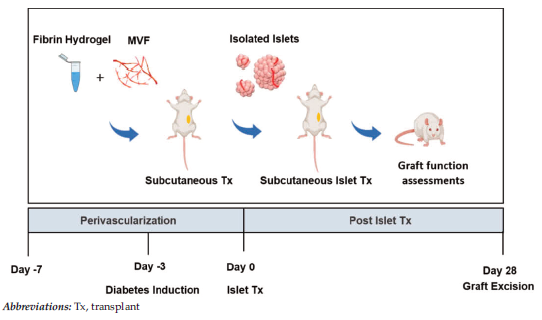
Figure 1. Study Design
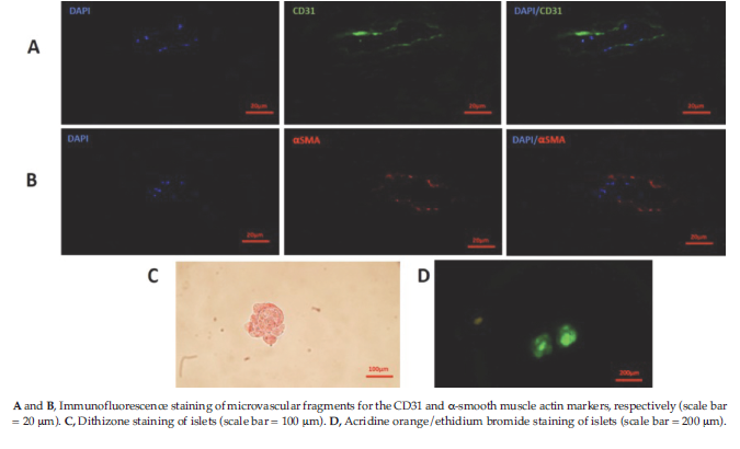
Figure 2. Characterization of Isolated Microvascular Fragments and Islets
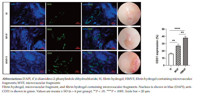
Figure 3. Immunofluorescence Staining of CD31 in the Prevascularized Groups at the End of the Phase 1
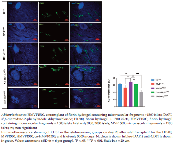
Figure 4. Immunohistochemical Analysis of Graft-Bearing Subcutaneous Tissue at the End of Phase 2
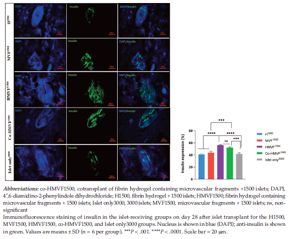
Figure 5. Immunohistochemical Analysis of Graft-Bearing Subcutaneous Tissue at the End of Phase 2
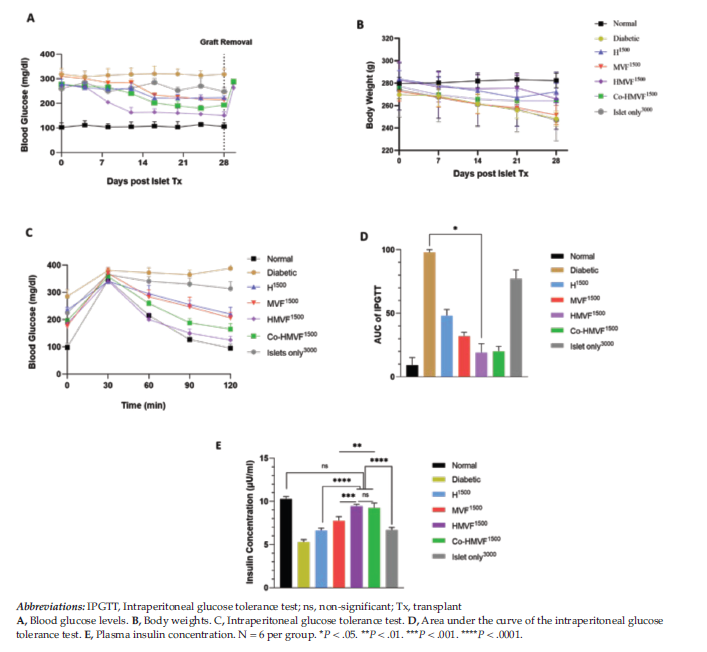
Figure 6. Graft Function Assessment