Volume: 22 Issue: 5 May 2024
FULL TEXT
Glycogen storage disease type 1 is a congenital abnormality of metabolism caused by the deficiency of the glucose-6-phosphatase enzyme, essential in glucose homeostasis. Patients with this disease are at high risk of developing hypoglycemia, hyperlipidemia, lactic acidemia, growth retardation, neutropenia, inflammatory bowel disease, and many other severe complications, such as hepatic adenomas converting into hepatocellular carcinomas. To prevent these complications, a liver transplant is the ultimate method of treatment. We present the successful anesthesia management for a 21-year-old man who had gross hepatomegaly, severe hypoglycemia, and hyperlactatemia and who received a liver transplant from his mother, which is a substantial challenge for anesthesiologists. Anesthesiologists should know the underlying pathophysiological condition and perform a comprehensive preoperative evaluation to determine the correct anesthesia plan in patients with glycogen storage disease type 1 who will undergo an orthotopic liver transplant due to multiple system disorders. Successful perioperative management of patients with glycogen storage disease type 1 relies on effective communication and collaboration between specialists through a multidisciplinary team approach.
Key words : Hyperlactatemia, Hypoglycemia, Perioperative treatment, von Gierke disease
Introduction
Glycogen storage disease type 1 (GSD-1), also known as von Gierke disease, is an autosomal recessive metabolic abnormality caused by the deficiency of the enzyme glucose-6-phosphatase, which is essen-tial for glucose homeostasis. The deficiency affects glycogenolysis and gluconeogenesis, causing fasting intolerance and severe hypoglycemia episodes. The abnormality affects many organs, such as the liver, kidneys, lungs, bowel, and immune system. As a result, these organs are at high risk of glycogen accumulation, and patients with GSD-1 experience hepatomegaly, nephromegaly, hypoglycemia, hyper-lipidemia, lactic acidemia, elevated serum triglycerides, growth retardation, trunk obesity, round doll-like face, neutropenia, inflammatory bowel, transformation of hepatic adenomas into hepatocellular carcinoma (HCC), and many other serious complications. To prevent these complica-tions, patients with GSD-1 must maintain a strict diet consisting of frequent feedings day and night, continuous gastric tube feeding, or administration of uncooked cornstarch; otherwise, hypoglycemia and seizures leading to death may occur.2,3 Despite the development of new medical treatment methods, liver transplant (LT) is the definitive treatment for patients with GSD-1. Most patients undergo LT between the ages of 4.3 and 50 years.1
Perioperative management of patients with GSD-1 can be highly challenging, even for minor elective surgeries. Major procedures, such as LT, which have a high risk of mortality and morbidity, present challenges for anesthesiologists and require mainte-nance of metabolic homeostasis, close glycemic control, management of bleeding due to platelet dysfunction, treatment of lactic acidosis, varying doses of anesthetic drugs due to liver impairment.4,5 Herein, we present our anesthetic management in an adult patient with von Gierke disease who underwent LT.
Case Report
A 21-year-old patient (weight 78 kg, height 155 cm, body mass index 32.5) diagnosed with GSD type 1a (GSD-1a) was referred to our hospital for LT surgery. The patient experienced many episodes of hypoglycemic seizure, and he was under a particular diet therapy that contained cornstarch. He was also receiving oral sodium bicarbonate, vitamin D, amlodipine, and testosterone because of hypogonadism. He had hypertension, hypothyroidism adenoma, and renal unilateral nephrolithiasis. Unlike other cases in the literature with GSD-1a, our patient was diagnosed with the disease at 14 years of age. Because his hypoglycemic episodes were becoming more frequent and because liver biopsy, as well as differentiated liver neoplasia, concluded a risk of converting to HCC, the patient was scheduled for LT at 5 years after primary diagnosis.
On physical examination, the patient had short stature, a rounded doll-like face, a fatty and distended abdomen due to gross hepatomegaly, and ascites. The patient's renal function tests were within reference ranges. The preoperative laboratory values were as follows: hemoglobin 8.5 g/dL, hematocrit 25.5%, platelets 213 × 103 cells/mL, alanine amino-transferase 46 IU/L, and aspartate transaminase 95 IU/L, with hyperlipidemia (triglyceride 932 mg/dL, cholesterol 299 mg/dL) and hyperuricemia (uric acid 9.3 mg/dL). Serum alkaline phosphatase, prothrom-bin time, total and direct bilirubin, albumin, sodium, potassium, blood urea nitrogen, and creatinine levels were all within normal limits. Preoperative electrocardiography, echocardiography, and thorax computed tomography were normal. His abdomen computed tomography showed hepatomegaly and nephromegaly (Figure 1). His Child-Pugh score was 5A, and his model for end-stage liver disease sodium score was 11. American Society of Anesthesiologists physical classification was class III, and Mallampati score was II. Written consent was obtained from the patient and his relatives to publish this case report.
The patient was prescribed a preoperative fasting time of 6 hours. A 10% dextrose solution infusion was started to prevent hypoglycemia, and close blood glucose monitoring with a glucometer every 2 hours was performed. His fasting blood glucose level at the beginning of the operation was 52 mg/dL, and he had lactic acidosis (lactic acid 9.2 mmol/L, pH 7.32). Standard monitoring was applied during the induction phase, including 5-lead electrocardiography, heart rate, peripheral oxygen saturation, noninvasive blood pressure (mean arterial pressure), end-tidal carbon dioxide, and nasopharyngeal temperature. After preoxygenation with 80% oxygen for 3 minutes, general anesthesia was induced with propofol of 1 to 2 mg/kg, fentanyl 1 ?g/kg, and rocuronium 0.6 mg/kg intravenously (IV).
After general anesthesia was administered, we performed rapid sequence induction and intubation with cricoid pressure to minimize the chance of pulmonary aspiration, and the patient was intubated with a size 7.5 cuffed endotracheal tube without difficulty. We administered a right and a left subclavian central venous catheter, left femoral artery catheterization for pulse index continuous cardiac output monitoring, and a nasogastric tube. Anesthesia was maintained by infusion of remifentanil 0.02 to 0.2 ?g/kg/min with 2% sevoflurane and 50% oxygen-50% air mixture and rocuronium infusion at 0.3 mg/kg/h. Surgical prophylaxis was done with 4.5 g IV piperacillin-tazobactam every once at 8 hours.
At the beginning of the operation, severe hypoglycemia (48 mg/dL) and lactic acidosis were observed; thus, we provided an aggressive IV infusion of 10% dextrose at 50 mL/h and sodium bicarbonate. Arterial blood gas samples were received every 90 minutes. Intraoperatively, lactate levels peaked at 16 mmol/L. The patient had episodes of hypotension requiring significant circulatory support with norepinephrine infusion (0.01-0.2 ?g/kg/min) to maintain a mean arterial pressure >60 mm Hg. The patient was given 10 mg/kg of prednisolone, 100 mg/kg of N-acetyl-cysteine, 500 mg of vitamin C, and 25 mg of IV heparin. The patient had a high central venous pressure of 23 mm Hg and oliguria with a urine output of 50 mL every 4 hours; therefore, we infused a furosemide stress test dose of 40 mg IV mannitol 20%.
Liver transplant surgery was mechanically complex because of gross hepatomegaly, and the dissection phase lasted 80 minutes (Figure 2). Because of platelet dysfunction and the nature of major surgery, the patient had a total of 1300 mL of bleeding and required a transfusion of 3 units of red blood cells (RBCs) and 3 units of fresh frozen plasma (FFP). We intraoperatively provided autologous blood salvage (ABS) and administered 300 mL of blood. Because the patient's blood glucose levels improved in the neohepatic phase, dextrose infusion was stopped.
Total fluid therapy, during 10 hours of surgery, included infusion of 5000 mL of crystalloids, 1000 mL of 10% dextrose, 2400 mL of 5% albumin-containing colloid, 3 units of RBCs, 3 units of FFP, and 300 mL of ABS. We observed 1000 mL of urine output. The explanted liver weighed 7165 g, and the new graft from the left lobe of the patient's mother weighed 665 g. At the end of the operation, the patient was weaned from inotropes and fulfilled standard clinical criteria for safe extubation. Sugammadex (4 mg/kg IV) was administered.
The patient was extubated in the operating room without complications and then discharged to the intensive care unit (ICU). His lactate level was 12 mmol/L in the ICU but normalized over the next 24 hours. After 6 days of follow-up, he was discharged from the ICU; on day 8, post-operative, he was discharged from the hospital as an outpatient.
Discussion
Patients with GSD-1a face various anesthesia and perioperative challenges.6 Patients with GSD-1 have various complications, such as hyperlipidemia, hyperuricemia, hypoglycemia, growth failure, and HCC, which is the most fatal complication.3 The incidence of GSD-1 is estimated at 1 in 100?000 births.4 The glucose-6-phosphatase enzyme is lacking in these patients, and glucose-6 phosphate cannot be converted to glucose, causing hypoglycemia, hepatomegaly, hepatic adenomas, and HCC.2,3
In the perioperative management of patients, particular attention is needed for blood glucose monitoring because of the risk of severe hypoglycemia episodes. Hypoglycemia causes increased lactate production by activating the glycolytic and gluconeogenesis pathways and can lead to severe lactic acidosis. Despite careful monitoring and treatment, physicians must be aware of the risk of metabolic coma episodes in patients with GSD-1.2,4,5
Current practice guidelines from the American College of Medical Genetics and Genomics recom-mend a continuous infusion of 10% dextrose in 0.45% saline, stimulating age-appropriate liver glucose output and adjusting the intravenous fluid rate to maintain blood glucose above 70 mg/dL. Frequent monitoring of blood sugar and lactate levels is also recommended.2 By the guidelines, our patient was hospitalized 2 days before the surgery, and blood glucose balance was achieved with the endocrinologist.
Wilke and colleagues5 recommended total exogenous carbohydrate intake before LT exceeding estimated glucose requirements corresponding to 1.5 mL/kg/h of 10% dextrose. We continued our patient's diet until midnight and then started the infusion of dextrose 10 containing 0.45% saline with a glucose infusion rate of 2.3 mg/kg/min. Our patient had high central venous pressure, hyponatremia, and low blood osmolarity because of dilution and hypervolemia; thus, we infused 10% dextrose preoperatively in volumes low enough to maintain normoglycemia.
Patients with GSD-1 have increased abdominal pressure due to ascites and hepatomegaly, and a short fasting period could cause an increased risk of pulmonary aspiration.2,6,7 We performed a rapid sequence induction and intubation to minimize this risk.
Surgical stress during the intraoperative period can cause profound lactic acidosis. In general, the literature recommends that infusion containing dextrose, which is started in the preoperative period, should be continued throughout the intraoperative period to prevent worsening of lactic acidosis.2,4,7 In our patient, at the beginning of the operation, severe hypoglycemia (48 mg/dL) and lactic acidosis were observed; thus, the patient was given an aggressive IV infusion of 10% dextrose at 50 mL/h and sodium bicarbonate. Gurrieri and colleagues7 used Ringer lactate solution in their GSD-1 patients. Our patient had high serum lactate levels, so we used a plas-malyte A and a 5% albumin infusion solution.
Although neutropenia does not often occur in patients with GSD-1a, anemia and prolonged bleeding times are expected. Because of decreased concentrations of von Willebrand factor and bleeding during surgery in many patients, blood preparations should include RBCs, FFP, and thrombocyte sus-pension preoperatively.6-8
The use of ABS may lead to an increased risk of mortality. Klein and colleagues6 stated that active malignancy is a relative contraindication to autologous transfusion because cells in ABS may contain tumor cells and metastasize after reinfusion. In our case, the patient had no malignancy detected on liver biopsy and computed abdominal tomography. We used 300 mL of ABS to inhibit complications related to blood transfusion.
Conclusions
Anesthesia management in patients with GSD-1a requires special perioperative planning due to multisystemic involvement. A liver transplant is the definitive treatment option in patients with GSD-1a, and anesthesia management in these patients is highly challenging as a result of both resistant hypoglycemia episodes and severe lactic acidosis triggered by existing enzyme deficiency and surgical stress. Anesthesia management of patients with GSD-1a who undergo LT requires aggressive blood glucose and lactic acidosis monitoring. Therefore, a meticulous preoperative evaluation and a multidisciplinary team approach should be performed.
References:
-
Boers SJ, Visser G, Smit PG, Fuchs SA. Liver transplantation in glycogen storage disease type I. Orphanet J Rare Dis. 2014;9:47. doi:10.1186/1750-1172-9-47
CrossRef - PubMed -
Kishnani PS, Austin SL, Abdenur JE, et al. Diagnosis and management of glycogen storage disease type I: a practice guideline of the American College of Medical Genetics and Genomics. Genet Med. 2014;16(11):e1. doi:10.1038/gim.2014.128
CrossRef - PubMed -
Jang HJ, Yang HR, Ko JS, Moon JS, Chang JY, Seo JK. Development of hepatocellular carcinoma in patients with glycogen storage disease: a single center retrospective study. J Korean Med Sci. 2020;35(1):e5. doi:10.3346/jkms.2020.35.e5
CrossRef - PubMed -
Beyzaei Z, Shamsaeefar A, Kazemi K, et al. Liver transplantation in glycogen storage disease: a single-center experience. Orphanet J Rare Dis. 2022;17(1):127. doi:10.1186/s13023-022-02284-y
CrossRef - PubMed -
Wilke MV, de Kleine RH, Wietasch JKG, et al. Orthotopic liver transplantation in glycogen storage disease type 1a: perioperative glucose and lactate homeostasis. J Inborn Errors Metabol Screen. 2016;4. doi:10.1177/2326409816649599
CrossRef - PubMed -
Klein AA, Bailey CR, Charlton AJ, et al. Association of Anaesthetists guidelines: cell salvage for perioperative blood conservation 2018. Anaesthesia. 2018;73(9):1141-1150. doi:10.1111/anae.14331
CrossRef - PubMed -
Gurrieri C, Sprung J, Weingarten TN, Warner ME. Patients with glycogen storage diseases undergoing anesthesia: a case series. BMC Anesthesiol. 2017;17(1):134. doi:10.1186/s12871-017-0428-x
CrossRef - PubMed -
Wang DQ, Carreras CT, Fiske LM, et al. Characterization and pathogenesis of anemia in glycogen storage disease type Ia and Ib. Genet Med. 2012;14(9):795-799. doi:10.1038/gim.2012.41
CrossRef - PubMed

Volume : 22
Issue : 5
Pages : 402 - 405
DOI : 10.6002/ect.2024.0052
From the 1Department of Anesthesiology and Intensive Care Unit and the 2Department of General Surgery and Transplantation Unit, Baskent University, Faculty of Medicine, Ankara, Turkey
Acknowledgements: The authors have not received any funding or grants supporting the presented research or preparing this work and have no declarations of potential conflicts of interest.
Corresponding author: Denada Haka, Baskent University, Faculty of Medicine, Department of Anesthesiology and Intensive Care Unit, Fevzi Cakmak Caddesi 10, Sokak No:45 Bahcelievler, 06490 Ankara, Turkey
E-mail: denadahaka97@gmail.com
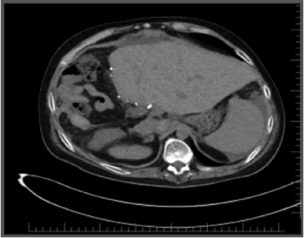
Figure 1.Computed Tomography Showing Hepatomegaly and Nephromegaly
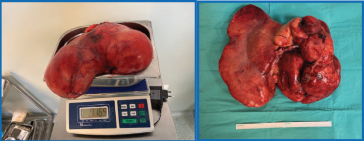
Figure 2.Massive Hepatomegaly of Size 15 cm and Weight 7.165 g