Volume: 22 Issue: 5 May 2024
FULL TEXT
Objectives: Heart transplant is the most effective treatment in patients with advanced heart failure who are refractory to medical treatment. The brain death interval and type of inotrope We assessed the effects of these parameters on heart transplant outcomes.
Materials and Methods: In this follow-up study, we followed heart transplant recipients for 1 year to study patient survival, ejection fraction, adverse events, and organ rejection. We evaluated follow-up results on time from brainstem death test to the cross-clamp placement, as well as the type of inotrope used.
Results: Our study enrolled 54 heart transplant candi-dates. The inotrope dose was 3.66 ± 0.99 μg/kg/min, and the most used inotrope, with 28 cases (51.9%), was related to dopamine. Six cases (11.1%) of death and 1 case of infection after transplant were observed in recipients. The average ejection fraction of transplanted hearts before transplant, instantly at time of transplant, and 1 month, 6 months, and 1 year after transplant was 54.9 ± 0.68, 52.9 ± 10.4, 51.9 ± 10.7, 50.1 ± 10.9, and 46.8 ± 17, respectively; this decreasing trend over time was significant (P =.001). Furthermore, ejection fraction changes following transplant did not differ significantly in transplanted hearts regarding brain death interval and type of inotrope used.
Conclusions: Our study revealed that cardiac output of a transplanted heart may decrease over time and the time elapsed from brain death, and both dopamine and norepinephrine could have negligible effects on cardiac function.
Key words : Dopamine, Ejection fraction, Heart transplantation, Norepinephrine
Introduction
Each year, more than 5000 heart transplants occur worldwide.1 Heart transplants are the most efficacious long-lasting treatment in terms of mortality and quality of life in patients with advanced heart failure who are refractory to medical treatment.2 Most transplanted organs are from donors after neurological deter-mination of death or, simply put, brain death (BD).3 Brain death is a pathophysiological event with catastrophic consequences, and, even if optimal care is provided, asystole will usually occur as a result of deterioration in cardiorespiratory function.4 The period between the certification of BD and subsequent asystole is usually short, in the order of, at most, days.5 The brain death interval (BDI) is frequently overlooked as a potential variable influencing graft function, despite the fact that the process of BD, especially during the initial stages, involving increased sympathetic activity induces deterioration of a donor’s myocardium6 and, as a result, predicts a poor prognosis for the recipient.7 More studies are required to investigate the relationship between the BDI and transplant outcomes.
Vasoactive agents are used in organ donors to treat the pathophysiological processes that occur during the progression of BD.8 To better understand the vitality of this intervention, a better understanding of hemodynamic decompensation after BD is important. In the initial stages of BD, a catecholamine surge and dysfunction of the parasympathetic centers of the brainstem lead to unopposed sympathetic activity, often referred to as “the sympathetic storm”.9 The sympathetic storm in turn may cause several unwanted and deleterious effects in the body: decreased tissue perfusion, increased inotropy and chronotropy, cardiac cell death, and impaired cardiac function.10,11 Closer to the completion of BD, however, a loss of sympathetic activity and a concurrent shift from aerobic to anaerobic metabolism take place, which lead to hormonal failure and metabolic acidosis, and, as a result, vasoconstriction and hypertension start to fade out and become replaced by vasodilation and hypotension. Hence, relative hypovolemia occurs, which in turn decreases the cardiac output.12 Therefore, from a pathophysiological standpoint and to preserve cardiac output, vasoconstrictor inotropic catecho-lamines, such as dopamine and norepinephrine, play key roles in optimal donor management.8 However, a lack of consensus remains in the guidelines on use of either a specific agent or combination therapy.
A review of the current literature has revealed a paucity of studies on the relationship between the BDI and the outcomes of heart transplants, and the reports that do assess it have often arrived at conflicting conclusions.13-16 Furthermore, studies comparing the use of different vasoactive agents are also sparse and contradictory.17,18 In this study, our goal is to evaluate the relationship between BDI scores and the type of inotropic agent used, and our primary outcomes were the effects of these factors on both short-term and long-term outcomes after heart transplant We also aimed to identify any additional predictors of outcome and assess the correlation with rejection and mortality rates among recipients, which served as secondary outcomes.
Materials and Methods
Study design
This follow-up study adheres to the principles outlined in the Declaration of Helsinki for medical research and was approved by the Iran University of Medical Sciences Ethics Committee (IR.IUMS.FMD. REC.1400.376). Informed consent was obtained from either the participants themselves or their legal representatives prior to any interventions.
Inclusion and exclusion criteria
Participants were selected based on the following inclusion criteria: (1) adults of any sex aged 18 years and older and (2) individuals undergoing heart transplant. Exclusion criteria included (1) presence of significant cognitive impairment affecting participation, (2) history of psychiatric disorders that may confound psychological assessments, and (3) patients undergoing retransplant.
Interventions
Participants were monitored over a period of 1 year after heart transplant. Assessments were conducted immediately after surgery and at 1 month, 6 months, and 12 months after transplant. The key parameters included patient survival, ejection fraction (EF), occurrence of adverse events, and incidence of organ rejection. The time interval between brainstem death and the first cross-clamp placement during transp-lant, as well as the type of inotropic agent utilized, were also analyzed.
Primary outcomes
The primary outcomes were patient survival rates at various posttransplant time point; changes in ejection fraction over the 1-year follow-up period; incidence of adverse events and organ rejection after transplant; correlation between the time elapsed from BD to the first cross-clamp placement and posttransplant outcomes; and association between the type and dosage of inotropic agents used during transplant and transplant outcomes.
Secondary outcomes
As secondary outcomes, we identified factors influencing transplant rejection by using multivariate regression analysis.
Standard definitions of parameters
Patient survival was defined as the proportion of transplant recipients who remained alive at specified posttransplant time points.
The EF was a measure of the percentage of blood pumped out of the heart’s left ventricle with each heartbeat, assessed with echocardiography or other imaging techniques.
We monitored any adverse events or untoward medical occurrence in patients after surgery, including complications such as infections, bleeding, or other perioperative complications.
Organ rejection was defined as the immune-mediated response leading to the destruction or damage of transplanted organs, typically diagnosed through biopsy or clinical assessment.
Statistical analyses
We used the mean (with SD) and frequency (percentage) to describe quantitative and qualitative variables, respectively. We applied the t-test to compare quantitative variables and the chi-square test or the Fisher exact test to compare qualitative variables. We also evaluated the study results based on time elapsed from BD to first cross-clamp placement (BDI) with 2 subgroups: (1) less than 3 days and (2) more than 3 days. We also identified the type of inotrope used, including dopamine nd norepinephrine subgroups. We chose a 1-way repeated analysis of variance test to investigate EF changes during the follow-up period, and we used simple multiple logistic regression to investigate the factors affecting transplant rejection. A significance level of 5% was considered. All analyses were performed with SPSS software (version 22).
Results
Our study included 54 cases of heart transplant. The average age of transplant donors was 27.7 ± 10.9 years. Thirty-two donors (59.3%) were male, and the rest were female. The O positive blood group with 20 cases (37%) was the most frequent among organ donors. Unintentional/unexpected trauma was the most common cause of BD among organ donors, with 30 cases (55.6%). No significant difference was seen among the variables of age, sex, and blood groups based on BDI in the donors. The average age of transplant recipients was 30.7 ± 17.7 years. Thirty-four recipients (63%) were male patients, and the rest were female patients. No significant difference was seen in comparisons of age and sex based on the BDI in transplant recipients (Table 1).
The average EF of transplanted hearts before, immediately after, and 1 month, 6 months, and 1 year after transplant was 54.9 ± 0.68, 52.9 ± 10.4, 51.9 ± 10.7, 50.1 ± 10.9, and 46.8 ± 17, respectively (Table 2). Furthermore, EF changes after transplant did not differ significantly in transplanted hearts with regard to BDI in donors. On the other hand, the EF value had a decreasing trend based on the elapsed time, which was significant (P = .001) (Table 2).
The dose of inotrope used in the patients was equal to 3.66 ± 0.99 μg/kg/min, and the most used inotrope, with 28 cases (51.9%), was related to dopamine. Six cases (11.1%) of death and 1 case of infection after transplant were observed in recipients. Inotrope dose, type of inotrope, the number of deaths among the patient groups, and transplant rejection based on BDI showed no significant difference among the recipient cases (Table 2).
According to the results of the Bonferroni post hoc test, this significant trend was caused by the difference in the EF value of the patient after the operation, 6 months, and 1 year after transplant (Table 3).
For the comparisons of the EF index at different times after transplant, inotrope dose, complications, and rejection of transplant according to the type of ino-trope, no significant differences were seen between the 2 groups of norepinephrine and dopamine (Table 4).
Based on the results of multivariate regression, donor age (OR, 1.01; 95% CI, 0.93-1.10), donor sex (OR, 0.94; 95% CI, 0.10-8.37), time elapsed until donation (OR, 1.20; 95% CI, 0.87-1.66), type of inotrope (OR, 0.19; 95% CI, 0.02-1.93), dose of inotrope (OR, 1.91; 95% CI, 0.69-5.26), age of recipient (OR, 0.99; 95% CI 0.93-1.05), and sex of recipient (OR, 0.88; 95% CI, 0.09-8.57) had no significant effect on transplant rejection (Table 5).
Discussion
We discovered that the groups showed similarities in both primary factors, such as the correlation between BDI scores and the type of inotrope used, and secondary outcomes, such as the correlation between predictors and rejection and mortality rates among recipients, with no significant differences noted between the 2 groups identified as early and late recipients of heart transplants. We also found that, although rejection and complication rates were higher among late recipients, these results were also not significant. In our examination of the literature, we found conflicting results regarding the influence of donor BDI on outcomes in heart transplants. In a 10-year-long retrospective study by Ramjug and colleagues involving 157 patients, the interval between the second brainstem death test and the cross-clamp placement time at organ retrieval was compared with survival time. The investigators found that the time between brainstem death test and organ retrieval cross-clamp placement time predicted greater recipient mortality (hazard ratio = 1.15; P = .001).13 It is important to note that the given hazard ratio for the BDI is small and may be due to the relatively small sample size or could merely suggest a weak correlation. In addition, in a study by Cantin and colleagues among 475 transplant cases, a management time longer than 72 hours was associated with poorer outcome, but no differences were observed in survival based on the time from BD declaration to cross-clamp placement.14 Furthermore, in a study of 215 heart transplants, Marasco and colleagues also reported that BDI was not associated with rejection or mortality.15 Another report analyzed BDI and outcomes of pediatric cardiac transplants from national registry data, and among 2565 patients, the authors concluded that there was no clear relationship between BDI and recipient or graft survival after pediatric cardiac transplant.19 Also, a compelling analysis of data from the United Network for Organ Sharing registry for 22 960 patients was performed by Jawitz and colleagues, who concluded that a longer duration of BD was not associated with worse survival after heart transplant.16 It is important to note that BD donors whose organs were not successfully transplanted were excluded in this analysis, introducing a marked potential for selection bias.
These observations of either a weak association or no association at all can be justified by considering the pathophysiological steps of BD. Granted, the sympathetic storm as a step of BD is highly detrimental to the donor myocardium,20 but an increased BDI may allow hemodynamic recovery from the catastrophic events of this step of BD, thereby mitigating the damage to the donor heart.16 Moreover, a longer time period could enable the clinicians to take on a more active role in the treatment of the donor and correct the possible injurious effects of BD.21 Our results and the conclusions of these studies convey that donor organs undergoing a longer BDI should not be hastily dismissed as less suitable or second rate, and that BDI should not serve as the sole basis for rejection of a donor heart.16,21 This is especially important when there is a pronounced discrepancy between the number of patients in need of transplants and the number of donors.22
In the comparison of the groups that received either norepinephrine or dopamine, no statistically significant difference was observed. These results are similar to those of a study by von Ziegler and colleagues, who reported the long-term outcomes of norepinephrine versus dopamine pretreatment among 936 potential heart donors. Neither norepi-nephrine nor dopamine pretreatment of potential heart donors demonstrated superior overall survival. However, in a subpopulation of long-term survivors, pretreatment with norepinephrine was correlated with better survival.18 On the other hand, a meta-analysis based on the registry of the Eurotransplant International Foundation involving 720 heart transplants found that the use of norepinephrine, versus other vasoactive agents, was predictive of initial nonfunction after heart transplants.17 Investigations in this aspect of heart transplant are scarce, and this signals a need for both more descriptive studies and more clinical trials regarding the choice of vasoactive agents.
Our study had some limitations. First, the sample size of this report was small, and the study was conducted in a single center; these associations or lack thereof should be investigated in larger and more statistically sound samples. Second, the 3-day cutoff that divided the patients into 2 groups is rather limiting, and more studies can be undertaken using earlier or later cutoffs. Third, for the comparison of the effects of vasoactive agents, retrospective study designs are prone to an array of biases, and randomized clinical trials would be useful to discover whether the type of the agent affects patients in a meaningful way. The same logic could also be applied to comparisons of the effects of BDI in the recipients, as there is little consistent evidence that organs procured with shorter or longer BDIs will significantly harm the patients.
Conclusions
Our study revealed that the cardiac output of a transplanted heart might be decreased over time, which implies the need for complete cardiology follow-up and supportive care for the transplanted heart. Moreover, the time elapsed from BD and each dopamine and norepinephrine treatment may have negligible effects on cardiac function. Thus, surgeons could have a more flexible window to prescribe norepinephrine or dopamine according to the patients situation and patient comorbidities.
References:
- Taylor DO, Edwards LB, Boucek MM, et al. Registry of the International Society for Heart and Lung Transplantation: twenty-fourth official adult heart transplant report—2007. J Heart Lung Transplant. 2007;26(8):769-781. Doi:10.1016/j.healun.2007.06.004
CrossRef - PubMed - Wever-Pinzon O, Edwards LB, Taylor DO, et al. Association of recipient age and causes of heart transplant mortality: Implications for personalization of post-transplant management-an analysis of the International Society for Heart and Lung Transplantation Registry. J Heart Lung Transplant. 2017;36(4):407-417. Doi:10.1016/j.healun.2016.08.008
CrossRef - PubMed - Girlanda R. Deceased organ donation for transplantation: Challenges and opportunities. World J Transplant. 2016;6(3):451-459. Doi:10.5500/wjt.v6.i3.451
CrossRef - PubMed - Maciel CB, Greer DM. ICU Management of the potential organ donor: State of the art. Curr Neurol Neurosci Rep. 2016;16(9):86. Doi:10.1007/s11910-016-0682-1
CrossRef - PubMed - Power BM, Van Heerden PV. The physiological changes associated with brain death: current concepts and implications for treatment of the brain dead organ donor. Anaesth Intensive Care. 1995;23(1):26-36. Doi:10.1177/0310057X9502300107
CrossRef - PubMed - Szabo G, Hackert T, Sebening C, Melnitchuk S, Vahl CF, Hagl S. Role of neural and humoral factors in hyperdynamic reaction and cardiac dysfunction following brain death. J Heart Lung Transplant. 2000;19(7):683-693. Doi:10.1016/s1053-2498(00)00129-7
CrossRef - PubMed - Mehra MR, Uber PA, Ventura HO, Scott RL, Park MH. The impact of mode of donor brain death on cardiac allograft vasculopathy: an intravascular ultrasound study. J Am Coll Cardiol. 2004;43(5):806-810. Doi:10.1016/j.jacc.2003.08.059
CrossRef - PubMed - Lazzeri C, Bonizzoli M, Guetti C, Fulceri GE, Peris A. Hemodynamic management in brain dead donors. World J Transplant. 2021;11(10):410-420. Doi:10.5500/wjt.v11.i10.410
CrossRef - PubMed - Novitzky D. Selection and management of cardiac allograft donors. Curr Opin Cardiol. 1996;11(2):174-182. Doi:10.1097/00001573-199603000-00011
CrossRef - PubMed - Watts RP, Thom O, Fraser JF. Inflammatory signalling associated with brain dead organ donation: from brain injury to brain stem death and posttransplant ischaemia reperfusion injury. J Transplant. 2013;2013:521369. Doi:10.1155/2013/521369
CrossRef - PubMed - Schrader H, Hall C, Zwetnow NN. Effects of prolonged supratentorial mass expansion on regional blood flow and cardiovascular parameters during the Cushing response. Acta Neurol Scand. 1985;72(3):283-294. Doi:10.1111/j.1600-0404.1985.tb00872.x
CrossRef - PubMed - Grissom TE, Richards JE, Herr DL. Critical care management of the potential organ donor. Int Anesthesiol Clin. 2017;55(2):18-41. Doi:10.1097/AIA.0000000000000137
CrossRef - PubMed - Ramjug S, Hussain N, Yonan N. Prolonged time between donor brain death and organ retrieval results in an increased risk of mortality in cardiac transplant recipients. Interact Cardiovasc Thorac Surg. 2011;12(6):938-942. Doi:10.1510/icvts.2010.252809
CrossRef - PubMed - Cantin B, Kwok BW, Chan MC, et al. The impact of brain death on survival after heart transplantation: time is of the essence. Transplantation. 2003;76(9):1275-1279. Doi:10.1097/01.TP.0000093445.50624.5A
CrossRef - PubMed - Marasco S, Kras A, Schulberg E, et al. Donor brain death time and impact on outcomes in heart transplantation. Transplant Proc. 2013;45(1):33-37. Doi:10.1016/j.transproceed.2012.08.008
CrossRef - PubMed - Jawitz OK, Raman V, Barac YD, et al. Influence of donor brain death duration on outcomes following heart transplantation: a United Network for Organ Sharing Registry analysis. J Thorac Cardiovasc Surg. 2020;159(4):1345-1353 e2. Doi:10.1016/j.jtcvs.2019.04.060
CrossRef - PubMed - Schnuelle P, Berger S, de Boer J, Persijn G, van der Woude FJ. Effects of catecholamine application to brain-dead donors on graft survival in solid organ transplantation. Transplantation. 2001;72(3):455-463. Doi:10.1097/00007890-200108150-00017
CrossRef - PubMed - von Ziegler F, Helbig S, Kreissl N, Meiser B, Becker A, Kaczmarek I. Norepinephrine versus dopamine pretreatment of potential heart donors: impact on long-term outcome. Ann Transplant. 2013;18:320-326. Doi:10.12659/AOT.883960
CrossRef - PubMed - Grosshuesch C, Johnson WK, DeVogel N, et al. Brain death interval and relationship to outcomes of pediatric cardiac transplantation. Pediatr Transplant. 2019;23(4):e13426. Doi:10.1111/petr.13426
CrossRef - PubMed - Wilhelm MJ, Pratschke J, Beato F, et al. Activation of the heart by donor brain death accelerates acute rejection after transplantation. Circulation. 2000;102(19):2426-2433. Doi:10.1161/01.cir.102.19.2426
CrossRef - PubMed - Kilic A. Commentary: Duration of brain death and heart transplant outcomes: the devil is in the detail. J Thorac Cardiovasc Surg. 2020;159(4):1356. Doi:10.1016/j.jtcvs.2019.05.023
CrossRef - PubMed - Evans RW, Orians CE, Ascher NL. The potential supply of organ donors. An assessment of the efficacy of organ procurement efforts in the United States. JAMA. 1992;267(2):239-246.
CrossRef - PubMed

Volume : 22
Issue : 5
Pages : 386 - 391
DOI : 10.6002/ect.2024.0008
From the 1Department of Anesthesiology and Pain Medicine, School of Medicine, Rasool-E Akram Hospital, Iran University of Medical Sciences, Tehran; the 2Cardiovascular Surgery Research and Development Committee, Iran University of Medical Sciences, Tehran; the 3Rockingham Medical Research Centre, Rockingham, Western Australia, Australia; the 4Department of Surgery, Surgery Research Center, School of Medicine, Rasool-E Akram Hospital, Iran University of Medical Sciences, Tehran; the 5Loghman-e Hakim Hospital, Department of Intensive Care Unit and Critical Care, Faculty of Medicine, Shahid Beheshti University of Medical Science, Tehran; and the 6Pain Research Center, Department of Anesthesiology and Pain Medicine, School of Medicine, Iran University of Medical Sciences, Tehran, Iran
Acknowledgements: The authors have not received any funding or grants in support of the presented research or for the preparation of this work and have no declarations of potential conflicts of interest.
†Mehrdad Mesbah Kiaei and Mohammadhosein Akhlaghpasand contributed equally to this work.
Corresponding authors: Mohammadhosein Akhlaghpasand, Cardiothoracic research associate at Rockingham Medical Research Centre, Rockingham, Australia; and Dr. Hanieh Hatefifard, Department of Anesthesiology and Pain Medicine, School of Medicine, Rasool-E Akram Hospital, Iran University of Medical Sciences, Tehran
E-mail: Akhlaghpasandm@yahoo.com, Hani.hatef@gmail.com
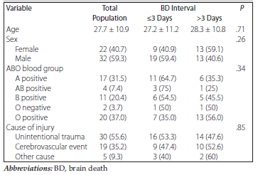
Table 1. Comparison of Basic Variables Based on the Elapsed Time From Hospitalization to Organ Transplant
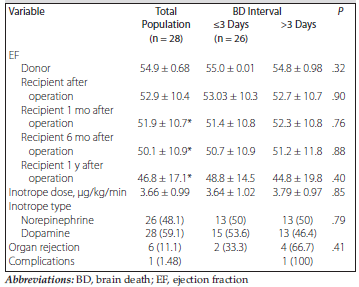
Table 2. Comparison of Cardiac Output, Type of Inotrope, and Outcomes Based on Time Elapsed From Hospitalization to Organ Transplant
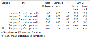
Table 3. Results of Bonferroni Post Hoc Test Comparing Two Different Times in Terms of Cardiac Output in Transplant Recipients
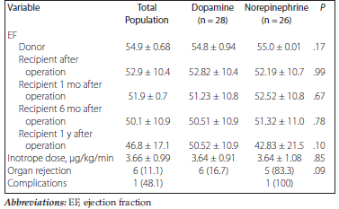
Table 4. Comparison of Cardiac Output, Type of Inotrope, and Outcomes Based on Type of Inotrope
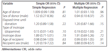
Table 5. Factors Affecting Transplant Rejection and Death of Transplant Patients