Volume: 22 Issue: 2 February 2024
FULL TEXT
Objectives: Vascularized composite allotransplantation is a reconstructive option after severe injury but is fraught with complications, including transplant rejection due to major histocompatibility complex mismatch in the context of allogeneic transplant, which in turn is due to altered immuno-inflammation secondary to transplant. The immunosuppressant tacrolimus can prevent rejection. Because tacrolimus
is metabolized predominantly by the gut, this immunosuppressant alters the gut microbiome in multiple ways, thereby possibly affecting immuno-inflammation.
Materials and Methods: We performed either allogeneic or syngeneic transplant with or without tacrolimus in rats. We quantified protein-level inflammatory mediators in the skin, muscle, and plasma and assessed the diversity of the gut microbiome through 16S RNA analysis at several timepoints over 31 days posttransplant.
Results: Statistical analysis highlighted a complex interaction between major histocompatibility complex and tacrolimus therapy on the relative diversity of the microbiome. Time-interval principal component analysis indicated numerous significant differences in the tissue characteristics of inflammation and gut microbiome that varied over time and across experimental conditions. Classification and regression tree analysis suggested that both inflammatory mediators in specific tissues and changes in
the gut microbiome are useful in characterizing the temporal dynamics of posttransplant inflammation.
Dynamic network analysis highlighted unique changes in Methanosphaera that were correlated with Peptococcus in allogeneic transplants with and without tacrolimus versus Prevotella in syngeneic transplant with tacrolimus, suggesting that alterations in Methanosphaera might be a biomarker of vascularized composite allotransplant rejection.
Conclusions: Our results suggest a complex interaction among major histocompatibility complex, local and systemic immuno-inflammation, and tacrolimus therapy and highlight the potential for novel insights into vascularized composite allotransplant from computational approaches.
Key words : Allogeneic transplant, Dynamic network analysis, Syngeneic transplant, Systems biology, Tacrolimus
Introduction
Vascularized composite allotransplantation (VCA) offers the potential to restore the appearance, anatomy, and function of debilitating injuries that are not conducive to conventional reconstruction. Although more than 200 VCAs have been performed successfully in the past decade, local and systemic immuno-inflammation leading to acute and chronic transplant rejection remains a critical limitation of the success of VCA.1 Tacrolimus (FK506) is the mainstay drug used to suppress the innate and adaptive immune response to the major histocompatibility complex (MHC)-mismatched graft and mitigate transplant rejection.1,2 However, tacrolimus is associated with complications including, but not limited to, the development of opportunistic infections, metabolic disorders, and gut dysbiosis (ie, the disruption of gut flora homeostasis).3-5 In addition to these side effects, tacrolimus administration in the setting of VCA is associated with altered cross-tissue inflammation, likely given the complexity of the multiple transplanted tissues inherent to VCA.6 Given the complications associated with VCA and tacrolimus, there remains a need to identify opportunities to increase the success of VCA, in part through under-standing the effects of tacrolimus on downstream homeostatic regulators.
In transplantation, tacrolimus has a suppressive effect on both the systemic (host) and local (graft) inflammatory responses, mitigating the host-versus-graft (rejection) and graft-versus-host immune reactions. Also, when administered orally in transplant patients, tacrolimus is known to alter the gut microbiome, a phenomenon that has been linked to worse overall transplant outcomes.5,7
One mechanism through which tacrolimus may affect the gut microbiome is through induction of gut dysbiosis, which is thought to perpetuate systemic inflammation.8,9 In addition to dysbiosis, where tacrolimus disrupts the balance of the gut microbiome, this drug also reduces overall diversity of flora; both phenomena have been linked to systemic immune dysregulation. Specifically, tacrolimus can increase the presence of certain bacterial taxa, such as Allobaculum, and decrease the presence of other bacterial taxa such as Clostridium and Bifidobacterium.10-12 Tacrolimus also disrupts lipid, glucose, and short fatty acid metabolism, which in turn alters the availability of nutrients for the various microbiota and can lead to a change in microbiota levels and ultimately, an altered gut microbiome.10-12 Such an altered microbiome (which is imbalanced and less diverse) can then change the metabolism of tacrolimus in the gut, adversely influencing its therapeutic efficacy or toxicity. Furthermore, in skin-containing VCA, such as face or hand transplants, the gut-skin axis of the microbiome may have a complex impact in influen-cing local immune activity and systemic immune responses.13,14
In the context of transplantation, VCA is unique because it involves multiple tissues and can be either syngeneic or allogeneic.15 We hypothesized that tacrolimus had nuanced effects on the gut microbiome that were dependent on whether a syngeneic or allogeneic transplant was performed. Specifically, we hypothesized that differences in the gut microbiome of rats subjected to syngeneic compared with allogeneic transplant were exacerbated by tacrolimus administration. To better understand the effects of VCA, transplant type, and immunosuppressive therapy on the gut microbiome, we subjected rats to either allogeneic or syngeneic hindlimb transplant with or without tacrolimus therapy and quantified changes in tissue-level inflammatory mediators and the gut microbiota over the course of transplant rejection.
We also leveraged advancements in dynamic systems modeling to model the temporal and tissue-specific effects that alterations in the gut microbiome in response to tacrolimus administration had on local inflammatory mediators. Computational modeling, including time-interval principal component analysis (TI-PCA) and dynamic network analysis (DyNA), were used to model the temporal and spatial sensitivity of protein-level inflammatory mediators in VCA with and without tacrolimus.6 Here, we sought to use a similar systems biology approach to understand how MHC mismatch, the transplant of multiple tissue units, and/or immunosuppression with tacrolimus affected the gut microbiome. The goal of this body of work is to begin to probe the complex interactions between the gut microbiome, immunosuppressive therapy, and tissue-specific inflammation inherent to syngeneic and allogeneic transplant.
Materials and Methods
Animals
Major histocompatibility complex-mismatched male Brown-Norway and Lewis rats were used in this study. Rats received a syngeneic (Lewis-Lewis) or allogeneic (Brown-Lewis) hindlimb transplant and were given either 1 mg/kg of tacrolimus immuno-suppressive therapy or no immunosuppressive treatment. We performed transplant procedures according to previously published methods6,16 and as approved by the University of Pittsburgh Institutional Animal Care and Use Committee. Experimental groups included the following: syngeneic transplant without tacrolimus (Syn – Tac; n = 7), syngeneic transplant with tacrolimus (Syn + Tac; n = 8), allogeneic transplant without tacrolimus (Allo – Tac; n = 6), and allogeneic transplant with tacrolimus (Allo + Tac; n = 7). Skin, muscle, and peripheral blood samples were collected on the following days posttransplant: 0, 3, 7, 11, 20, 25, and 31. In accordance with the Institutional Animal Care and Use Committee, rats subjected to allotransplant without tacrolimus therapy were euthanized on day 11.
Analysis of inflammatory mediators
Transplanted tissue and peripheral blood samples were assayed using Luminex (MagPix) for the following mediators: granulocyte colony-stimulating factor, eotaxin, granulocyte macrophage colony-stimulating factor, macrophage inflammatory protein-1α (MIP-1α/CCL3), monocyte chemoat-tractant protein 1(MCP-1/CCL2), leptin, interleukin (IL)-1α, IL-1β, IL-2, IL-4, IL-5, IL-6, IL-10, IL-12p70, IL-13, IL-17A, IL-18, interferon (IFN)-γ, IFN-γ-inducible protein 10 (IP-10CXCL10), epidermal growth factor, GRO-KC, tumor necrosis factor α, fractalkine, LIX, MIP-2, and regulated on activation normal T cell expressed and secreted (RANTES/CCL5). Concentrations were quantified (expressed as either pg/mg total protein [for tissue] or as pg/mL [for plasma]).
Processing stool samples for 16S rRNA gene sequencing
Genomic DNA was extracted from rat stool samples from which the V4 region of the 16S rRNA gene was amplified and processed for sequencing on an Illumina MiSeq platform as previously described.17 Sequences were processed through the University of Pittsburgh Center for Medicine and the Microbiome in?house sequence quality control pipeline, which includes low complexity filtering, quality value (<30) trimming, trimming of primers used for 16S rRNA gene amplification, and minimum read-length filtering as previously described.14,15 The additive log ratio transformation to relative abundances was applied to break the spurious correlation among taxa in compositional data, allowing each taxon to be analyzed as independent and normally distributed.18
Statistical and computational analyses
Factorial analysis of variance
Time-dependent changes in the taxa of the gut microbiome based on transplant type (allogeneic vs syngeneic), adminis-tration of tacrolimus, and pretransplant and posttransplant days were assessed using a factorial analysis of variance (ANOVA) with correction for multiple comparisons. The ANOVA utilized the following parameters: a = 0.05, type 2 sum of squares, and the Tukey honestly significant difference procedure for multiple comparisons. Analysis of the same timepoints between experi-mental groups was conducted to identify any taxa that differed significantly (P < .05) between experi-mental groups.
Time-interval principal component analysis
We used TI-PCA, a modified PCA that highlights variables that contribute the most to variance in treatment response across consecutive timepoints in a dataset with temporal resolution.19 We calculated the principal components for subsets of data consisting of 2 consecutive timepoints as previously described.19-21 In our study, the data analyzed by TI-PCA included additive log ratio quantifications of taxa in the gut microbiome and Luminex quantification of inflammatory mediators in the plasma, muscle, and skin across 7 timepoints. We calculated the sum of all principal components for each variable and ranked the variables from greatest to least. We selected variables whose sum of principal components was in the upper quartile of the sum of all variables’ principal components for further analysis.
For each experimental condition with rats, a leave-one-out approach to TI-PCA was used such that for any TI-PCA analysis, “n – 1” rats were analyzed according to the above procedure. Mediators in the upper quartile of the TI-PCA were then categorized by their location (microbiome, plasma, skin, or muscle). We calculated the mean and SD of the number of mediators in each location across the number of TI-PCA iterations and conducted t tests between experimental conditions and locations to identify significant differences in the number of mediators contributing to variance.
Classification and regression tree analysis with bagging
A decision tree was created from an independently drawn bootstrap replica of the input data using a classification and regression tree analysis approach to predict the time at which the data were sampled (MATLAB). The data that were withheld were “out-of-bag,” and the misclassification rate of each trained model was known as the “out-of-bag” error (OOBE). To find the optimal decision tree, we used 50 weak learners. The optimal decision tree was the decision tree with the lowest OOBEs. If multiple trees had the same OOBE, the first decision tree of this set was used for further analysis.
Dynamic network analysis
We used DyNA, a network analysis performed by creating a correlation matrix between the entire dataset and itself over defined time intervals.22 The correlation between any 2 mediators over 2 consecutive timepoints, whether they be bacterial taxa and/or inflammatory mediators, was calculated. Mediators with an absolute correlation coefficient >0.99 were indicated by an edge connecting the 2 nodes, indicating that the mediators exhibited correlated temporal changes over the 2 consecutive timepoint intervals studied. We conducted DyNA between the top 40 taxa in the gut microbiome and separately between the mediators identified by TI-PCA in the corresponding time interval.
Results
Rats subjected to allogeneic or syngeneic hindlimb transplant with peripheral nerve repair, with or without tacrolimus, were biopsied at serial timepoints. Fecal samples were collected to assess inflammation and gut microbiome composition posttransplant. We hypothesized that following transplant, as inflam-matory mediators rose in the plasma and throughout tissues,6,23 the gut microbiome would be altered in a manner that reflected the temporal changes in systemic inflammation.
Allogeneic composite tissue transplant and tacro-limus administration impact the gut microbiome
We initially conducted a factorial ANOVA with correction for multiple comparisons to analyze significant differences in relative abundance of bacterial taxa between experimental conditions and to determine the effect, if any, of VCA with or without tacrolimus on the gut microbiome. Most of the variance in relative abundance of taxa occurred between day 5 and day 9, regardless of whether rats received a syngeneic or allogeneic transplant. No notable differences in relative abundance of taxa were shown between the Syn – Tac and the Allo – Tac groups, suggesting that MHC mismatch alone had no effect on the gut microbiota (Table 1). Notably, the relative abundance of Clostridium sensu stricto 1 differed at all timepoints between the Syn + Tac versus Syn – Tac groups and the Syn + Tac versus Allo + Tac groups (Table 1). Thus, the variance in Clostridium sensu stricto 1 density appeared to be a consequence of both tacrolimus therapy and MHC mismatch.
A closer analysis of the effect of MHC mismatch and tacrolimus therapy highlighted a complex inte-raction among tacrolimus, MHC mismatch, and the gut microbiome. The taxa that varied in density bet-ween the Syn – Tac and Syn + Tac groups were entirely different from the set of taxa that varied in density between the Allo – Tac and Allo + Tac groups. That is, although tacrolimus was associated with differences in the gut microbiome, the interaction bet-ween MHC compatibility and tacrolimus was dif-ferentially related to the specific taxa that differed in samples from animals treated with tacrolimus (Table 1).
Time-interval principal component analysis highlights temporally sensitive differences in micro-biome composition and inflammation dependent on major histocompatibility complex mismatch
We have previously defined important differences in dynamic networks and trajectories of tissue-level inflammatory mediators in this rat model of VCA, utilizing computational methods, including TI-PCA and DyNA.6,23 Therefore, we next assessed the salient, dynamic characteristics of tissue-specific inflammatory mediators and the gut microbiome using TI-PCA. Across all timepoints and experimental conditions, allogeneic transplant without tacrolimus was associated with the greatest number of total mediators in the upper quartile of TI-PCA. The number of total mediators in the upper quartile of TI-PCA significantly different between the Allo + Tac condition and the Allo – Tac condition on days 0 to 3 and days 3 to 7 (Figure 1). Between these two allogeneic conditions, there was a significant difference in the gut microbiome and inflammatory mediators in both plasma and muscle, but not in skin, across all 3 time intervals (days 0-3, days 3-7, and days 7-11). Interestingly, more mediators contributed to variance in the gut microbiome and plasma in the Allo + Tac versus the Allo – Tac condition across all time intervals (Figure 1 and Figure 2). However, in the muscle, we observed more mediators in the Allo – Tac condition contributing to variance (Figure 1).
We next further assessed the effect of tacrolimus on the gut microbiome by analyzing the data up to day 11 versus after day 11 (when tacrolimus was withdrawn). In the syngeneic conditions with and without tacrolimus, the total number of taxa in the upper quartile of TI-PCA significantly different between conditions after removal of tacrolimus (Figure 2A). More taxa were shown in the upper quartile of TI-PCA of the gut microbiome contributing to variance in the Syn – Tac condition than the Syn + Tac condition at all time intervals, except from day 7 to day 11, during which there was also no significant difference between the 2 conditions (Figure 2B). During all intervals, there were more taxa in the upper quartile of TI-PCA in the muscle in the Syn + Tac compared with the Syn – Tac condition, and the difference in the number of inflammatory mediators was always significant (Figure 1). Through day 25, there was a significant difference in the number of inflammatory mediators in the plasma contributing to variance in the syngeneic conditions. However, there were more inflammatory mediators in the Syn – Tac condition contributing to variance up until day 11, after which there were more inflammatory mediators in the Syn + Tac condition contributing to variance (Figure 2E).
From day 0 through day 11, a significantly different total number of mediators contributed to variance in the Allo + Tac condition compared with the Syn + Tac condition, as well as the Allo – Tac and Syn – Tac conditions. This suggested that the variance captured by TI-PCA was driven by MHC mismatch more than sensitivity to tacrolimus. In both groups with and without tacrolimus, there was a significant difference in the number of mediators contributing to variance in the muscle and plasma on day 0 through day 11 (Figure 2, C and E). However, there was only a significant difference in the number of mediators contributing to variance in skin and the microbiome on day 0 through day 7 (Figure 1).
Decision tree analysis captures the variability in effect of major histocompatibility complex mismatch and tacrolimus
We next conducted decision tree analyses to identify which mediators might be key differentiating factors in predicting the posttransplant day. In 3 of 4 experimental conditions, the root of the decision tree was an inflammatory mediator located in the plasma (Figure 3). In the Syn + Tac and Syn – Tac conditions, terminal branches of the decision tree were a mix of inflammatory mediators and microbiota. In the Allo + Tac condition, nearly all terminal branches were inflammatory mediators in the muscle, skin, or plasma (Figure 3). In the Allo – Tac condition, the decision tree was remarkably simple, as there were only 4 nodes, only 1 of which was a bacterium (Figure 3). The OOBE for syngeneic transplant, with and without tacrolimus, was 0.60. The minimum OOBE for Allo – Tac was 0.41 and the OOBE for Allo + Tac was 0.42. The minimum OOBE was found approximately halfway through the 50 trees (27 for Syn – Tac, 47 for Syn + Tac, 28 for Allo – Tac, 29 for Allo + Tac).
Dynamic network analysis highlights lack of coordination between the inflammatory response and the microbiome and a potential microbiome-regulatory role of Methanosphaera after composite tissue transplant
Given the known association between the gut microbiome and the inflammatory response in various contexts,8,15,23-25 we next sought to determine whether cross-tissue, dynamic inflammation net-works induced by composite tissue transplant were intertwined with dynamic changes in the gut microbiome. Across all 4 experimental conditions, DyNA of the mediators in the upper quartile from TI-PCA showed no connections or correlations between mediators in the plasma, skin, or muscle and taxa from the microbiome, suggesting the absence of a direct link between the inflammatory pathways assessed and the gut microbiome. However, DyNA of the top 40 taxa in the gut microbiome alone demonstrated that changes in Methanosphaera were correlated with either changes in Peptococcus in allogeneic transplant with or without tacrolimus or changes in Prevotella in syngeneic transplant without tacrolimus (Table 2). Further analysis of the additive log ratio data quantifying the taxa in the gut microbiome at each timepoint showed a weak decrease in Methanosphaera across a subset of datapoints from each condition.
Discussion
The effect that an altered gut microbiome has on inflammation is poorly understood but may be linked to the fact that an altered gut microbiome affects the innate immune response.26 Prior studies have suggested prominent changes in the immune landscape following VCA and TAC,6 as well as significant differences in the responsiveness of TLR4 and receptors to pathogen-associated molecular patterns and microbial-associated molecular patterns,26 which differentially affects the host’s systemic inflammatory state.
Murine skin transplant models have highlighted the possible role for manipulating the gut microbiome to favorably skew host-versus-graft immune responses. In 1 model, supplementation with specific gut microbiota induced tolerogenic responses toward fully mismatched skin grafts, with attendant prolongation of survival.11 In contrast, altering the growth of microbiota, such as Faecalibacterium prausnitzii, reduced the gut metabolism and clearance of tacrolimus, thereby allowing for a reduction of the dose and frequency of treatment and associated toxicity.27,28 Studies with germ-free or antibiotic-pretreated skin transplants in mice have reported different survival patterns. For example, mice that received a germ-free transplant had accelerated rates of graft rejection after fecal transplants from pathogen-free mice but not antibiotic-pretreated mice. This study highlights that the constituents of the gut microbiota affect the rate of graft rejection.23 Another study found that, regardless of treatment duration, mice treated with antibiotics following aortic grafting had increased systemic inflammation and faster rejection than controls.24 Overall, increasing evidence has suggested complex roles of the gut microbiome vis-à-vis transplant outcomes.
In the present study, we used a systems biology approach to model the effects of MHC mismatch and tacrolimus on the gut microbiome. A multifaceted computational analysis identified nuanced dif-ferences in the diversity of the gut microbiota of rats that received allogeneic versus syngeneic transplant in response to tacrolimus. Furthermore, this methodology highlighted a potential biomarker, Methanosphaera, whose temporally sensitive upregulation could indicate transplant rejection. Our study analyzed the composite effects of allogeneic versus syngeneic transplant as well as tacrolimus therapy on the gut microbiome. Nuanced differences in gut metabolites have been shown following allogeneic compared with syngeneic bone marrow transplant in the absence of tacrolimus therapy,29 as well as the effect of the gut microbiome on tacrolimus-induced immunosup-pression.11 To the best of our knowledge, however, this is the first body of work to analyze differences in the gut microbiota associated with limb transplants and tacrolimus.
Statistical analysis (ANOVA) highlighted that the set of taxa that were differentially expressed in response to tacrolimus was different between allogeneic and syngeneic transplants. The set of taxa that were upregulated differentially in response to syngeneic transplant with versus without tacrolimus was entirely different from the set of taxa that were differentially upregulated in response to allogeneic transplant with versus without tacrolimus. Thus, we conclude that the changes in the gut microbiome in response to tacrolimus were dependent on the type of transplant performed in a complex manner that also involved the impact of tacrolimus.
Thus, tacrolimus, which modulates the immune response, may affect systemic inflammation and this effect may be intertwined in a complex manner with the microbiome and ultimately transplant rejection. To better understand whether there was a difference in the variance in inflammatory mediators or microbiota driving rejection in allogeneic and syngeneic transplant in response to tacrolimus, we utilized TI-PCA. During the day 0 to 3, day 3 to 5, and day 5 to 11 intervals, there was a difference in the number of inflammatory mediators in the upper quartile of TI-PCA in the plasma and muscle in rats that did not receive tacrolimus. Interestingly, the Syn – Tac condition was characterized by more drivers of inflammation in the plasma than the Allo – Tac condition; however, the Allo – Tac condition was characterized by more drivers of inflammation in the muscle than the Syn – Tac condition. A greater number of taxa that varied in expression were shown over days 0 to 3 and days 3 to 5 in the Syn – Tac condition than in the Allo – Tac condition. Notably, we inferred a greater number of drivers of inflammation in the plasma of the Allo + Tac group than the Syn + Tac group and a greater number of drivers of inflammation in the muscle of the Syn + Tac than the Allo + Tac group. Based on the combined ANOVA and TI-PCA, we infer that tacrolimus affects the diversity of the gut microbiome and the number of drivers of inflammation in the muscle and plasma differently, depending on whether an allogeneic or syngeneic transplant was performed.
Decision tree analysis, another dynamic compu-tational tool, had poor overall performance in predicting the days posttransplant (as a surrogate for degree of rejection) based on data on inflammatory mediators and microbiome composition; nonetheless, the overall structure of the decision trees was informative. In both the syngeneic and allogeneic transplants with and without tacrolimus, both the taxa from the microbiome and the inflammatory mediators served as terminal nodes. This finding suggested that, in the context of syngeneic transplant, there were certain timepoints that were defined by the state of the gut microbiome and others that were charac-terized by inflammation within a tissue.
Decision tree analysis strongly suggested that both changes in the gut microbiota and inflammation are temporally sensitive hallmarks of transplant rejection. For example, in the setting of Allo + Tac, the expression of IL-17A was useful in differentiating the inflam-matory profile of day 3 from day 11. The decision tree also highlighted that, in the Syn + Tac condition, the density of Methanosphaera was useful in differentiating the inflammatory profile of day 0 from day 25.
Although there were numerous differences in the makeup of the gut microbiome of rats receiving allogeneic or syngeneic VCA with or without tacrolimus, most experimental groups shared 1 common feature: the coordinated upregulation of Methanosphaera with 1 other taxon near the end of the experimental time course. Methanosphaera had coordinated upregulation with Peptococcus over the day 7 to day 9 interval in the context of allogeneic transplant with or without tacrolimus. On day 20 to day 25 Methanosphaera exhibited coordinated upregulation with Prevotella in the Syn − Tac condition. Notably, in the ostensibly least proinflam-matory condition (Syn + Tac), Methanosphaera did not exhibit coordinated upregulation with any other taxon.Of the 40 taxa analyzed, only Methanosphaera appeared as a key biomarker of transplant rejection. Methanosphaera, a methane-producing archaea, has been found to be associated with inflammatory diseases such as irritable bowel disease.30 Other methane-producing microbiota, such as Methanobre-vibacter smithii, have been linked to inflam-matory bowel disease and altered systemic inflammatory states. Compared with healthy controls, renal transplant recipients had lower concentrations of Methanobrevibacter, another methane-producing archaea, in their feces and exhibited significantly reduced exhaled methane.31-33 This preliminary work suggests that future studies should be aimed at carefully quantifying changes in Methanosphaera compared with other taxa associated with VCA, tacrolimus, and transplant rejection. Future directions may include quantification of temporal changes in exhaled methane in transplant patients as a proxy for transplant rejection status.
Additional limitations should be addressed in the future. Although an opportunity remains to study the behavior of a larger set of inflammatory mediators, perhaps the largest limitation is the lack of immune profiling of the gut itself. The gut’s local lymphoid tissue, Peyer’s patches, are sensitive to changes in the gut microbiome.34 Future studies should investigate the effects of VCA and tacrolimus on the gut microbiome and the changes in inflammation in the gut itself. Furthermore, repeating the our present study in humans is pertinent as there exists nonnegligible differences in the composition of the gut microbiome and inflammatory profiles in rats versus humans.35 Our current study examined only the composition of the gut microbiome as inferred from 16s RNA sequencing; no data were obtained on protein- or metabolite-level changes that might yield nuanced and important insights as to how the functional state of the microbiome might impact VCA-induced inflammation and vice versa.
Through computational approaches that were leveraged to model dynamic changes in inflammation and gut dysbiosis, a complex but discernible interac-tion between MHC mismatch and tacrolimus therapy was identified as a significant trigger of deranged homeostasis of the host microbiome and systemic immunity. Furthermore, the frequency, intensity, or grade of transplant rejection, as investigated through a decision tree analysis, highlighted that both changes in the gut microbiome and systemic and tissue-specific inflammatory mediators were important in charac-terizing the time course of transplant rejection. Interestingly, Methanosphaera emerged as a sug-gested biomarker of immune inflammation and thereby possibly also of transplant rejection. We hypothesize that changes in methane production by Methanosphaera could be considered as a surrogate biomarker of either early or ensuing transplant rejection and merit further investigation in carefully designed small and large animals of VCA and solid-organ transplant. Future applications of dynamic systems biology, ‘omic analysis of the gut microbiome, and quantification of protein-level inflammatory mediators in the context of VCA and tacrolimus therapy have the potential to identify the spatiotemporal patterns of biomarkers of transplant rejection and pinpoint specific targets for directed immunosuppression. The computational approach outlined here should be leveraged in large-scale human studies to better identify microbiome-based biomarkers of transplant rejection and facilitate understanding of the complex interplay between systemic inflammation and gut dysbiosis following tacrolimus administration.
References:
- Schneeberger S, Gorantla VS, van Riet RP, et al. Atypical acute rejection after hand transplantation. Am J Transplant. 2008;8(3):688-696. doi:10.1111/j.1600-6143.2007.02105.x
CrossRef - PubMed - Lee WPA, Shores JT, Brandacher G. From Auto- to Allotransplantation: immunomodulatory protocol for hand and arm transplantation. J Reconstr Microsurg. 2018;34(9):683-684. doi:10.1055/s-0038-1651524
CrossRef - PubMed - Ong PW, Kee T, Ho QY. Impact of tacrolimus versus cyclosporine on one-year renal transplant outcomes: A single-centre retrospective cohort study. Proceedings of Singapore Healthcare. 2020;29(4):217-222. doi:10.1177/2010105820957370
CrossRef - PubMed - Ling Q, Huang H, Han Y, et al. The tacrolimus-induced glucose homeostasis imbalance in terms of the liver: from bench to bedside. Am J Transplant. 2020;20(3):701-713. doi:10.1111/ajt.15665
CrossRef - PubMed - Gabarre P, Loens C, Tamzali Y, Barrou B, Jaisser F, Tourret J. Immunosuppressive therapy after solid organ transplantation and the gut microbiota: bidirectional interactions with clinical consequences. Am J Transplant. 2022;22(4):1014-1030. doi:10.1111/ajt.16836
CrossRef - PubMed - Aral AM, Zamora R, Barclay D, et al. The effects of tacrolimus on tissue-specific, protein-level inflammatory networks in vascularized composite allotransplantation. Front Immunol. 2021;12:591154. doi:10.3389/fimmu.2021.591154
CrossRef - PubMed - Noce A, Marchetti M, Marrone G, et al. Link between gut microbiota dysbiosis and chronic kidney disease. Eur Rev Med Pharmacol Sci. 2022;26(6):2057-2074. doi:10.26355/eurrev_202203_28354
CrossRef - PubMed - Kim D, Zeng MY, Nunez G. The interplay between host immune cells and gut microbiota in chronic inflammatory diseases. Exp Mol Med. 2017;49(5):e339. doi:10.1038/emm.2017.24
CrossRef - PubMed - Yamashiro K, Tanaka R, Urabe T, et al. Gut dysbiosis is associated with metabolism and systemic inflammation in patients with ischemic stroke. PLoS One. 2017;12(2):e0171521. doi:10.1371/journal.pone.0171521
CrossRef - PubMed - Toral M, Romero M, Rodriguez-Nogales A, et al. Lactobacillus fermentum improves tacrolimus-induced hypertension by restoring vascular redox state and improving eNOS coupling. Mol Nutr Food Res. 2018;62(14):e1800033. doi:10.1002/mnfr.201800033
CrossRef - PubMed - Zhang Z, Liu L, Tang H, et al. Immunosuppressive effect of the gut microbiome altered by high-dose tacrolimus in mice. Am J Transplant. 2018;18(7):1646-1656. doi:10.1111/ajt.14661
CrossRef - PubMed - Tourret J, Willing BP, Dion S, MacPherson J, Denamur E, Finlay BB. Immunosuppressive treatment alters secretion of ileal antimicrobial peptides and gut microbiota, and favors subsequent colonization by uropathogenic Escherichia coli. Transplantation. 2017;101(1):74-82. doi:10.1097/TP.0000000000001492
CrossRef - PubMed - De Pessemier B, Grine L, Debaere M, Maes A, Paetzold B, Callewaert C. Gut-Skin axis: current knowledge of the interrelationship between microbial dysbiosis and skin conditions. Microorganisms. 2021;9(2):353. doi:10.3390/microorganisms9020353
CrossRef - PubMed - Byrd AL, Belkaid Y, Segre JA. The human skin microbiome. Nat Rev Microbiol. 2018;16(3):143-155. doi:10.1038/nrmicro.2017.157
CrossRef - PubMed - Friedman O, Carmel N, Sela M, et al. Immunological and inflammatory mapping of vascularized composite allograft rejection processes in a rat model. PLoS One. 2017;12(7):e0181507. doi:10.1371/journal.pone.0181507
CrossRef - PubMed - Shah AM, Aral AM, Zamora R, et al. Peripheral nerve repair is associated with augmented cross-tissue inflammation following vascularized composite allotransplantation. Front Immunol. 2023;14:1151824. doi:10.3389/fimmu.2023.1151824
CrossRef - PubMed - Li K, Epperly MW, Barreto GA, Greenberger JS, Methe BA. Longitudinal fecal microbiome study of total body irradiated mice treated with radiation mitigators identifies bacterial associations with survival. Front Cell Infect Microbiol. 2021;11:715396. doi:10.3389/fcimb.2021.715396
CrossRef - PubMed - Tarabichi Y, Li K, Hu S, et al. The administration of intranasal live attenuated influenza vaccine induces changes in the nasal microbiota and nasal epithelium gene expression profiles. Microbiome. 2015;3:74. doi:10.1186/s40168-015-0133-2
CrossRef - PubMed - Zamora R, Korff S, Mi Q, et al. A computational analysis of dynamic, multi-organ inflammatory crosstalk induced by endotoxin in mice. PLoS Comput Biol. 2018;14(11):e1006582. doi:10.1371/journal.pcbi.1006582
CrossRef - PubMed - Abboud A, Namas RA, Ramadan M, et al. Computational analysis supports an early, type 17 cell-associated divergence of blunt trauma survival and mortality. Crit Care Med. 2016;44(11):e1074-e1081. doi:10.1097/CCM.0000000000001951
CrossRef - PubMed - Tohme S, Yazdani HO, Sud V, et al. Computational analysis supports IL-17A as a central driver of neutrophil extracellular trap-mediated injury in liver ischemia reperfusion. J Immunol. 2019;202(1):268-277. doi:10.4049/jimmunol.1800454
CrossRef - PubMed - Mi Q, Constantine G, Ziraldo C, et al. A dynamic view of trauma/hemorrhage-induced inflammation in mice: principal drivers and networks. PLoS One. 2011;6(5):e19424. doi:10.1371/journal.pone.0019424
CrossRef - PubMed - Lei YM, Chen L, Wang Y, et al. The composition of the microbiota modulates allograft rejection. J Clin Invest. 2016;126(7):2736-2744. doi:10.1172/JCI85295
CrossRef - PubMed - Rey K, Manku S, Enns W, et al. Disruption of the gut microbiota with antibiotics exacerbates acute vascular rejection. Transplantation. 2018;102(7):1085-1095. doi:10.1097/TP.0000000000002169
CrossRef - PubMed - Sepulveda M, Pirozzolo I, Alegre ML. Impact of the microbiota on solid organ transplant rejection. Curr Opin Organ Transplant. 2019;24(6):679-686. doi:10.1097/MOT.0000000000000702
CrossRef - PubMed - Nellore A, Fishman JA. The microbiome, systemic immune function, and allotransplantation. Clin Microbiol Rev. 2016;29(1):191-199. doi:10.1128/CMR.00063-15
CrossRef - PubMed - Lee JR, Muthukumar T, Dadhania D, et al. Gut microbiota and tacrolimus dosing in kidney transplantation. PLoS One. 2015;10(3):e0122399. doi:10.1371/journal.pone.0122399
CrossRef - PubMed - Guo Y, Crnkovic CM, Won KJ, et al. Commensal gut bacteria convert the immunosuppressant tacrolimus to less potent metabolites. Drug Metab Dispos. 2019;47(3):194-202. doi:10.1124/dmd.118.084772
CrossRef - PubMed - Mathewson ND, Jenq R, Mathew AV, et al. Gut microbiome-derived metabolites modulate intestinal epithelial cell damage and mitigate graft-versus-host disease. Nat Immunol. 2016;17(5):505-513. doi:10.1038/ni.3400
CrossRef - PubMed - Blais Lecours P, Marsolais D, Cormier Y, et al. Increased prevalence of Methanosphaera stadtmanae in inflammatory bowel diseases. PLoS One. 2014;9(2):e87734. doi:10.1371/journal.pone.0087734
CrossRef - PubMed - Dridi B, Henry M, El Khechine A, Raoult D, Drancourt M. High prevalence of Methanobrevibacter smithii and Methanosphaera stadtmanae detected in the human gut using an improved DNA detection protocol. PLoS One. 2009;4(9):e7063. doi:10.1371/journal.pone.0007063
CrossRef - PubMed - Eckburg PB, Bik EM, Bernstein CN, et al. Diversity of the human intestinal microbial flora. Science. 2005;308(5728):1635-1638. doi:10.1126/science.1110591
CrossRef - PubMed - Gandhi A, Shah A, Jones MP, et al. Methane positive small intestinal bacterial overgrowth in inflammatory bowel disease and irritable bowel syndrome: a systematic review and meta-analysis. Gut Microbes. 2021;13(1):1933313. doi:10.1080/19490976.2021.1933313
CrossRef - PubMed - Lin S, Mukherjee S, Li J, Hou W, Pan C, Liu J. Mucosal immunity-mediated modulation of the gut microbiome by oral delivery of probiotics into Peyer's patches. Sci Adv. 2021;7(20). doi:10.1126/sciadv.abf0677
CrossRef - PubMed - Nagpal R, Wang S, Solberg Woods LC, et al. Comparative microbiome signatures and short-chain fatty acids in mouse, rat, non-human primate, and human feces. Front Microbiol. 2018;9:2897. doi:10.3389/fmicb.2018.02897
CrossRef - PubMed

Volume : 22
Issue : 2
Pages : 137 - 147
DOI : 10.6002/ect.2023.0312
From the 1Department of Surgery, University of Pittsburgh, Pittsburgh, Pennsylvania, USA; the 2Department of Surgery, Wake Forest Institute for Regenerative Medicine, Wake Forest Baptist Medical Center, Winston Salem, North Carolina, USA; and the 3Center for Inflammation and Regeneration Modeling, McGowan Institute for Regenerative Medicine, University of Pittsburgh, Pittsburgh, Pennsylvania, USA
Acknowledgements: The authors thank Kelvin Li and Barbara Methé (University of Pittsburgh Center for Medicine and the Microbiome) for carrying out 16S RNA assays and associated bioinformatics analyses of gut microbiome composition. This work was funded by Department of Defense grant W81 XWH-15-1-0336 (YV and VG). Y Vodovotz is a cofounder of, and stakeholder in, Immunetrics, Inc.
*Ashti M. Shah and Ali M. Aral are first coauthors.
#Vijay Gorantla and Yoram Vodovotz are senior coauthors.
Author contributions: A. M. Shah and R. Zamora conducted computational and statistical analyses, data interpretation, and writing. A. M. Aral conducted animal experiments and data collection. D. A. Barklay and J. Yin analyzed inflammatory mediators. V. Gorantla and Y. Vodovotz performed study design, data interpretation, and writing.
Corresponding author: Yoram Vodovotz, Department of Surgery, University of Pittsburgh, W944 Biomedical Science Tower, 200 Lothrop Pittsburgh, PA 15213, USA
E-mail: vodovotzy@upmc.edu
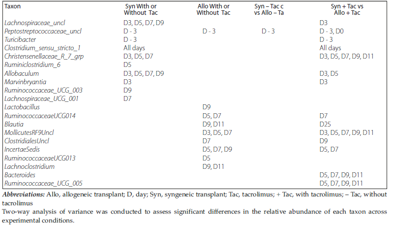
Table 1. Factorial Analysis of Variance to Assess Differences in Taxon Abundance Among Different Experimental Conditions
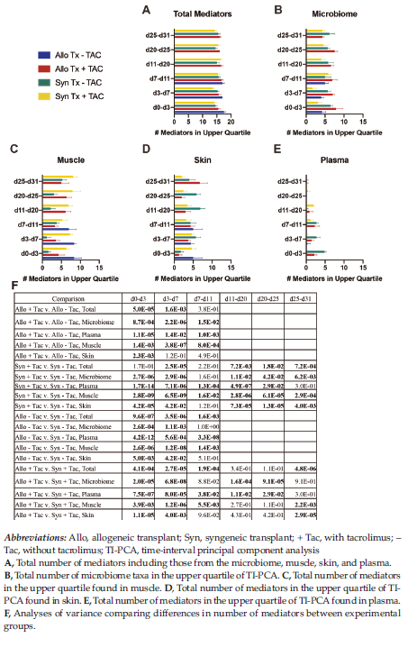
Figure 1. Analysis of Mediators in the Upper Quartile of Time-Interval Principal Component Analysis Before and After the Cessation of Tacrolimus Therapy
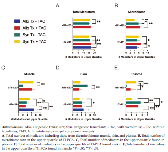
Figure 2. Focused Analysis of Mediators in the Upper Quartile of Time-Interval Principal Component Analysis Before and After the Cessation of Tacrolimus Therapy on Day 11
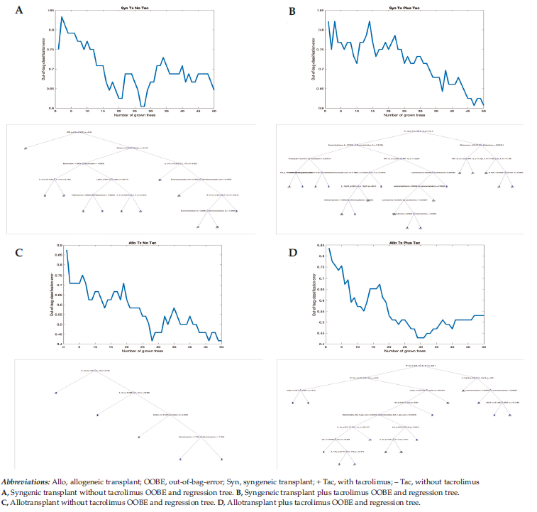
Figure 3. Classification and Regression Tree Analysis With Bagging

Table 2. Dynamic Network Analysis Summary of Correlated Microbiota in the Gut Microbiome for Different Experimental Conditions