Volume: 21 Issue: 5 May 2023
FULL TEXT
Genetically engineered pigs with multiple gene deletions and insertions are predicted to extend porcine to human xenograft survival. Several genes have been successfully knocked out and inserted, yet more have failed to produce viable animals for unexplained reasons. The effects of gene editing on cellular homeostasis may be the cause of reduced embryo fitness, failed pregnancies, or poor piglet viability. The elements of cellular dysfunction such as endoplasmic reticulum stress and oxidative stress induced by gene editing may additively affect the quality of genetically engineered cells to be used for cloning. Evaluating the impact of each gene edit on cellular fitness for cloning will allow researchers to maintain the cellular homeostasis of engineered cells that were validated as candidates for cloning and the production of porcine organ donors.
Key words : Endoplasmic reticulum stress, Genetic engineering, Nonsense-mediated RNA decay, Organ Donation, Transplantation
Genetic Engineering in Xenotransplantation
The availability of genetically engineered porcine donors may solve the global shortage of human donor organs and reduce the ever-increasing wait list of recipients.1 The use of pigs for xenotransplantation is well established as they share similarities with humans in organ size and function.2 This can be inferred from the recent success of porcine to rhesus macaque kidney transplantation and orthotopic porcine heart xenografts.3-5
Comparative genomic analysis revealed that porcine DNA sequences are more similar to humans than most other species.6 Yet, there are some differences that contribute to xenograft immune rejection, underscoring the important role of genetic engineering (GE) in xenotransplantation.7-9 Genetically engineered porcine cells play a pivotal role in mutating genes naturally deleted in humans, while inserting human genes missing in pigs to potentially extend long-term graft survival following xenotransplant. This explains the need for GE multimodified porcine organ donors. The impor-tance of combining GE approaches, including knock out (KO) of genes encoding predominant antigens and knock in of genes, providing normal receptor-ligand interactions (those that are active at the cell surface). Multiple gene edits have been suggested for producing donors that can contribute to the success of the initial clinical pig kidney or heart transplant models, which included 9 genetic modifications.10,11
Cloning Porcine Donors as a Source of Organs for Transplant
Advancements in cloning techniques have increased the possibility of producing these hypothesized multimodified genetically engineered piglets. Unfortunately, attempts at producing genetically engineered piglets resulted in early embryonic fetal loss, stillbirth, and early death after birth from abnormalities,12-14 as well as loss of transgenic gene expression and function.14,15
Importantly, living genetically engineered piglets generated from cloning must exhibit a normal cellular and microenvironmental homeostasis to provide healthy organs that are candidates for transplant. The graft stress response in donor organs plays a role in graft survival and function16-19 and must be considered in clinical xenotransplant, which underscores the critical role of providing healthy genetically engineered porcine cells for cloning.
Impact of Genetic Engineering on Cellular Health
An important consideration when genetically modifying porcine cells is the maintenance of cellular homeostasis and microenvironment that will enable multimodified gene-edited piglets to be generated more efficiently.
Nuclease-based genome editing has simplified the manipulation of mammalian genomes, enabling the disruption of porcine genes and the insertion of human genes in porcine genome.20-22 Gene editing with CRISPR/Cas9 is a fast and efficient tool to delete genes that encode for antigens and insertion of transgenes.23 Nevertheless, this could have other molecular effects that need to be addressed and investigated.
Knocking out genes
In porcine cells, ?-1,3-galactosyltransferase (GGTA1) KO has been generated with high efficiency in our laboratory and in other laboratories to produce pigs.24-26 Scarce information is available on the microenvironmental health of GE cells. Although the results of xenotransplant with GGTA1 KO organs are encouraging,13,26,27 determining the long-term health and function of donor organs is critical. The diseases that necessitate organ transplant often have 1 or more elements of dysfunction, such as endoplasmic reticulum (ER) stress and oxidative stress.28-32 Thereafter, transplant of a GE donor organ would contribute to additional stress responses that may affect organ function.33
Investigators created GGTA1 KO cells or piglets by single-guided RNA (sgRNAs) designed to target exon 1 of the GGTA1 gene24 or exon 3.26 Strategies of gene KO designed to target exon 1 should be recommended to mutate genes as early as possible, resulting in a premature termination codon (PTC) that can leave a short, truncated mRNA on transcription that could be degraded quickly by nonsense-mediated decay (NMD), possibly before or at the start of protein translation. Nevertheless, not all sgRNA designs result in gene deletion at exon 1, because additional factors, namely, the proximity of the protospacer adjacent motif site to the promotor, guide efficiency and off-target mutations. Targeting deletions downstream of exon 1 in the genomic sequence will lead to the transcription of longer mRNAs with subsequent protein translation that may persist in the cytoplasm, leading to possible effects on protein trafficking, misfolding, unfolding or any other unaddressed effects of the truncated mRNAs and proteins.
With the advent of the CRISPR/Cas9 tools and the insights generated from preclinical trials of xenotransplantation, more KOs have been added (CMAH and ß4galNT2 KOs)26 to the GGTA1 KO background. The accumulation of truncated mRNAs and proteins resulting from those KOs may end up as a burden on the NMD pathway and/or cellular homeostasis, thereby adversely impacting the gene-tically engineered cells, cloning, and donor organs.
Nonsense-mediated decay
The NMD pathway is an mRNA surveillance system and part of a general cellular quality control system that typically degrades transcripts containing PTCs introduced through DNA mutations to prevent translation of unnecessary or aberrant transcripts. Failure to eliminate these mRNAs with PTCs may result in the accumulation of such transcripts, promoting synthesis of potentially abnormal proteins that are toxic to cells.34 The typical target for the NMD has a termination codon positioned more than 50 to 55 nucleotides upstream of the last exon-exon junction (Figure 1) or has a long 3? untranslated region (UTR).35 Thus, mRNAs with nonsense codons present in other positions can escape the NMD pathway. Such an “escape from NMD surveillance” may cause expression of large amounts of aberrant truncated proteins with potential dominant-negative or gain-of-function effects in cells.34 The gain-of-function mechanism occurs particularly if the targeted cut site is present further on the genome and hence on the transcribed mRNA. Designing sgRNAs to mutate exon 1 should be a priority followed by extensive investigations to ensure maintainance of cellular homeostasis.
Human gene insertion
A component of the strategy to develop multimodified piglets is inserting human gene “transgenes” to normalize cellular functions or reduce inflammation. Transgene design is usually achieved by inserting the coding sequence of the desired gene with minimal regulatory domains that are required for proper transgene expression, that is, trying to include the most effective domain sequence with the shortest possible vector length to attain a high transfection efficiency.36 Previous transgenic studies in porcine cells have used a minimal gene coding sequence preceded by a strong promoter, followed by a polyA sequence for inducing human protein expression in the porcine model.12,37,38 These strategies did not take into consideration the need for additional regulatory elements present in mammalian or more specifically the porcine genome.
Controlling factors
Several regulatory domains have been proven to be critical for proper protein surface expression, namely, signal peptides, the C-terminal domain, and UTRs.39-41 Proper protein expression requires correct protein folding to form a 3-dimensional strucure, post-translational modifications, trafficking, localization, and processing active functional motifs, all of which should be considered in transgene design.
Xenogeneic protein expression requires codon optimization of the construct to rebalance codon usage needed from one species to another. Additioinally more than one isoform could be expressed either by only including specific introns or including all of the genomic sequences (coding and noncoding) at the expense of transfection efficiency. Investigating whether the expression construct creates a single or multiple functional proteins is an additional factor to be considered during transgene construct design.
Unfolded protein response and endoplasmic reticulum stress
Although limited data are available on the effects of gene insertion and its subsequent protein expression and trafficking on the cellular microenvironment and ER homeostasis,42 it is well-documented that imbalance or dysfunction in any of the above factors leads to protein accumulation in unfolded, misfolded, aberrant structure, or malfunctional proteins, triggering a defense mechanism known as the unfolded protein response (UPR) and ER stress. Unfolded protein response is important to restore ER homeostasis and to reestablish normal ER function. Unfolded protein response allows cells to overcome ER stress; however, overwhelming protein aggre-gation and excessive ER stress cannot always be countered by intrinsic cellular mechanisms, resulting in cell death.43
The mammalian cell senses UPR that these insults generate through the action of 3 canonical ER-resident transmembrane proteins: protein kinase R-like ER kinase (PERK), inositol requiring enzyme 1 (IRE1), and activating transcription factor 6 (ATF6).44 Unfolded protein response activation leads to the dissociation of BiP from 3 transducers (PERK, IRE1, and ATF6).45 The protein PERK homodimerizes and phosphorylates eukaryotic initiation factor 2? (eIF2?) to inhibit general protein translation. It also regulates several transcription factors, including NRF2 to upregulate the antioxidant response and ATF4, which can lead to both protective and apoptotic signaling via stimulating CHOP expression, also called growth arrest, and DNA damage-inducible gene 153, a key transcription factor for initiating the apoptotic program in extreme ER stress conditions.46,47
Activation of IRE1 results in the unconventional splicing of Xbp1, with unspliced Xbp1 (Xbp1u) mRNA removing a 26-base nucleotide intron resulting in a subsequent frame shift and then converting to the Xbp1s,48 which induces the transcription of several molecular chaperones, such as BiP, and stimulates protein degradation via ER-associated degradation.
Activated ATF6 is moved to the Golgi apparatus for translocation.45 Translocated ATF6 regulates the expression of several genes, including XBP1 and CHOP49 (Figure 2). Additional players recently discovered in ER stress include CRELD2 and SEL1L, with CRELD2 implicated in the processing and trafficking of proteins through the ER-Golgi apparatus and upregulated by ATF650 and SEL1L involved in ER-associated degradation51 and targeting of misfolded secretory and membrane proteins in the ER for proteasomal degradation.52
Human CD47+ (With and Without the 3?-Untranslated Region) Transgene on Porcine Cells
Recently, Hosny and colleagues, in a study of the effects of the 3?-UTR on human CD47 (hCD47) protein cell surface expression on porcine cells, reported the importance of including 3?-UTR for cell surface protein expression and the impact of the vector without 3?-UTR on the cellular microenvironment and homeostasis of the transfected cells in vitro.36 Berkovits and Mayr demonstrated the genetic modulation of the hCD47 construct to increase surface expression in human cells and observed that human cell lines with alternative 3?-UTRs regulated the localization and function of membrane proteins differently.39
Hosny and colleagues genetically enginereed porcine fetal fibroblasts using 2 vectors expressing hCD47, with and without the proposed uridine-rich element functional domain. The role of transgenic hCD47 encoded by mRNA containing the 3?-UTR to the hCD47 was compared with mRNA lacking the 3?-UTR domain and had greater cell surface expression, reduced impact on the cellular microenvironment, and improved immune function. Both vectors were inserted at the GGTA1 gene to create an ?Gal null background (GTKO), and the phenotypes of the genetically engineered cells generated from both vectors were compared along with the vectors’ effects on cellular homeostasis by measuring levels of ER stress markers. The ability of the GE cells to inhibit human macrophage phagocytosis and inhibit immune responses in human or nonhuman primates in coculture with peripheral blood mononuclear cells was determined.36 The investigators reported differential growth rates and viability between transfected cells of both vectors (with and without 3?-UTR) in culture.
Upon further characterization and cell surface and intracellular expression from both vectors, the cells transfected with hCD47+ with the 3?-UTR-containing construct exhibited more cell surface protein expression, whereas cells transfected with hCD47+ without the 3?-UTR vector had hCD47 protein expression that was mostly confined to the ER. The investigators found that the effects of the ER retained expression hCD47 on porcine cells and the cause of the observed differential cellular growth rates between cells derived from both vectors on the cellular microenvironment manifested in the form of UPR, ER stress, or through intertwined pathways (mitochondrial dysfunction, autophagy, and oxidative stress).53-55 These factors are considered detrimental for cellular homeostasis and animal development and may eventually affect donor organ production or function.56-58
Endoplasmic reticulum stress
Hosny and colleagues characterized the central regulators of the 3 ER stress pathways.59 Cells with hCD47+ and without 3?-UTR exhibited upregulated expression of ER stress markers such as XPBs, BiP, CHOP, ATF4, and SEL1L. No significant differences were observed for ATF6, IRE-1, and CRELD2 levels, indicating a differential activation of the 3 axes of the ER stress response. These results demonstrated the effects of ER protein retention in triggeringUPRs. Moreover, the upregulated expression of a proapop-totic CHOP, indicated a cell stress from severe ER stress response that may result in cell death.
Mitochondrial biogenesis
Peroxisome proliferator-activated receptor ? coac-tivator 1? (PGC1?) demonstrates substantially downregulated expression in cells that express hCD47 encoded by mRNA without the 3?-UTR compared with cells with hCD47 encoded by mRNA with the 3?-UTR. Levels of mitochondrial TFAM and NRF1, both regulators of mitochondrial DNA replication and transcription, demonstrated a decreased level of expression, albeit both were not statistically significant. The overall effect remains to be seen as it was reported that ER stress is associated with increased PGC1a to induce mitochondrial biogenesis as a compensatory mechanism in cellular stress leading to mitochondrial proliferation.60 These findings demonstrate that sustained activation of the UPR might cause a decrease in mitochondrial metabolism, suggesting metabolic insufficiency as a potential eventual cause of ER stress-associated cell death.61,62
Autophagy
Activation of ER stress can trigger changes not only in mitochondrial function but also in autophagy.63 Autophagy is a major catabolic process that delivers proteins, cytoplasmic components and organelles to lysosomes for degradation and recycling. While autophagy is a critical cytoprotective mechanism, it has been suggested to also lead to cellular death depending on cell circumstances.64 Hosny and colleagues observed a significantly higher expression level of autophagy regulatory molecules MAP1LC3B and SQSTM1 in cells with hCD47 encoded by mRNA without the 3?-UTR.
Oxidative stress
Oxidative stress is closely related to ER stress induction and is also reported to be involved in ischemic injury and hyperacute humoral rejection in the cardiac xenotransplant model.19 An increased expression of p22phox and NOX4 subunits of NADPH oxidase in cells with hCD47 encoded by mRNA without the 3?-UTR has been reported.36 NFE2L2, an antioxidant mediator and a downstream player of ATF4, has also been reported to have higher expression levels than cells with hCD47 encoded by mRNA with the 3?-UTR. In contrast, gene expression of both SOD1 and GPX3 antioxidant enzymes did not show any significant changes.36 Endoplasmic reticulum stress is known to upregulate NADPH oxidase expression and activity and is a major source of reactive oxygen species.65
Collectively, cells with transgenic surface protein that is localized internally suffer from microenvi-ronmental insult in the form of UPR, ER stress, dysregulated mitochondrial biogenesis, autophagy, and oxidative stress.36 Under normal conditions, successful UPR signaling results in cell survival, whereas severe and prolonged ER stress results in cell death through CHOP, one of the best characterized proapoptotic signals.
Although no data are available describing the order of involvement among these pathways, when one pathway is dysregulated, others will follow, creating a self-perpetuating cellular stress loop (Figure 3).
Impact of Endoplasmic Reticulum Stress on Embryonic Development
Endoplasmic reticulum stress has deleterious effects on genetically engineered cells used in creation of organ donors, with 1 report showing that ER stress perturbs porcine embryonic development.66 In addition, inhibition of Xbp1 splicing was shown to positively affect porcine embryo development.67 Similar results were observed in mouse preimplan-tation embryos, wherein inhibition of Xbp1 splicing improved embryo development and decreased apoptosis.68,69 Further evidence suggests that activated BiP causes arrest of embryonic development.70
Mitochondrial-Associated Membranes in Endoplasmic Reticulum Stress
Organelle interconnection is critical in maintaining cellular function and homeostasis.71 One of the most studied interactions is the interaction between mitochondria and ER; both are physically connected through mitochondria-associated ER membrane (MAM). This membrane is involved in calcium signaling, lipid metabolism, autophagy, and apoptosis.72 Even more, the configuration and the distance between these 2 organelles changes during environmental stress.73 Inositol-requiring kinase 1 and PERK, which are known for their effector control in ER stress and UPR, are present at MAM, suggesting an involvement of those controllers in intertwined effects between these organelles.71
Inositol-requiring kinase 1? in endoplasmic reticulum stress
Inositol-requiring kinase 1? activity is critical for the normal functioning of cells, especially during stress adaptation.7 The function of the IRE1a is to activate cell death when the threshold of cellular stress is reached or the death signals are activated. However, their function is regulated by regulating partner molecules. Inositol-requiring kinase 1a triggers cell death by promoting the intrinsic apoptosis pathway through interaction with tumor necrosis factor (TNF) receptor-associated factor 2 (TRAF).75 The association between IRE1? and TRAF2 is commonly implicated in induction of the proapoptotic signaling pathway through caspase 12 activation.76,77 In addition, another association of IRE1? with XBP1 has been demonstrated to induce apoptosis of hepatocytes in an ER stress-dependent pathway by inhibiting Akt through Pleckstrin homology-like domain family A member 3 (PHLDA3) expression.78 The IRE1?-mediated regulated IRE1?-dependent decay (RIDD) process has also been implied in cell apoptosis through mitochondrial apoptotic pathway of caspase 2 and BH3- interacting domain activation.79,80
PERK a major signaling pathway for ER stress
The protein kinase PERK belongs to the ?-subunit of eIF2?, a PERK downstream effector, and is composed of cytoplasmic and kinase domains. The cytoplasmic domain detects the accumulation of unfolded/ misfolded protein in the ER lumen.81 This protein kinase is located on the ER membrane as a homodimer, and the cytoplasmic domain is bound to the ER chaperone GRP78 under ER stress-free conditions. After the dissociation of glucose regulated protein 78 (GRP78), an ER-resident protein, the activation of PERK signaling is initiated upon its dimerization and autophosphorylation.82 Next, eIF2? is phosphorylated to suppress the general gene translations by inhibiting ribosome transportation of initiator methionyl-tRNAiMet.83 CHOP (also called growth arrest and DNA damage 153) is activated by ATF6 and PERK.84,85 PERK activates the downstream proapoptotic molecules ATF4, CHOP, CD95/Fas, and RyR2, leading to apoptosis and autophagy.
To our knowledge, no study has addressed the impact of GE on MAMs. However, it is well-documented that, under stressful stimuli (internal or external), MAMs play a fundamental role in regulating mitochondrial quality control to ensure the functional integrity of mitochondria, mastering the effect of ER stress on mitochondrial quality control.86
For example, MAMs participate in adaptive responses to mild ER stress by controlling protein homeostasis and mitochondrial dynamics to improve mitochondrial function and ensure cell survival.87 In addition, MAMs can enhance stress signals that cause apoptosis and cell death when cells experience severe ER stress.86,88 Ultimately, further investi-gations are required, especially in the context of GE, to completely understand the mechanisms controlling MAMs and the precise signal transmission between ER and mitochondria.
Consideration for Transgene Design
Based on the above-mentioned results, we encourage optimizing the construct design of the transgene to be used in generating genetically engineered porcine cells that can maintain cellular homeostasis to qualify as candidates for somatic cellular nuclear transfer (SCNT) and the creation of donor organs.
Other trangenes, particularly surface proteins, should be tested with 3?-UTR regulatory element and other regulatory and controlling factors. Several questions need to be raised before applying the construct design for pig production. An example is whether to direct surface protein expression using the human CD47 signaling peptide sequence, as shown previously,12,36,37 or use the porcine CD47 signaling peptide, which will likely yield variable results regarding extent, level, or distribution of cell surface hCD47 protein expression on porcine cells.
Molecular studies of the impact of transgene expression need to be investgated in vitro prior to the creation of embryos or porcine pregnancies to avoid costly and often unexplainable failures. Taking the time to evalute cellular fitness will allow researchers to attain cells that have maintained microenviroment homeostatis and would qualify as candidates for cloning and the production of organ donors.
Considerations for Generating Multimodified Piglets
The generation of multimodified porcine organ donors necessitates the addition of >1 KO and insertion of multiple transgenes either together or sequentially. These gene edits might additively affect both NMD and UPR in a form that might further control the timing and magnitude of the UPR, ER, and cellular stresses. This can be demonstrated by the symbiotic regulation between the UPR and NMD.89 Endoplasmic reticulum stressors that trigger the UPR can themselves inhibit NMD (Figure 4),90-92 likely because PERK is able to phosphorylate eIF2?. This mutual regulation (NMD inhibits the UPR and the UPR inhibits NMD) normally creates several desirable characteristics for cellular homesostasis. First, it allows the cell to achieve a rapid, switch-like response where insults below a certain threshold will not trigger the UPR (because NMD suppresses it). Second, when appropriate stresses are encountered, the UPR is activated in full (because the UPR attenuates NMD allowing full production of its effectors). Third, when stresses cease, the UPR fully shuts down (because NMD presumably resumes). If the stresses encountered exceed the threshold that the cell can tolerate, even with all of its adaptive mechanisms in place, it must terminate itself through apoptosis, correlating with the role of NMD.
Of importance, these mutimodified genetically engineered cells will face strong stresses passing through transfection, recovery, sorting, the cloning process, and potentially transplantation of a developed organ, a complicated process with many factors affecting outcomes.17,93 The addition of an improperly designed gene deletion or insertion will only add to the challenge of creating porcine organ donors.
Conclusions
Although some of the gene edits in the porcine genome have been successfully engineered, many unpublished attempts by our group and others were likely the results of our unwitting induction of cellular stress pathways. Successful production of genetically cloned piglets have been hindered due to the extensive application of GE. Therefore, we highly recommend adequate assessments and quality control of every gene edit before adding another, to ensure cellular homeostasis.
References:
- Ekser B, Ezzelarab M, Hara H, et al. Clinical xenotransplantation: the next medical revolution? Lancet. 2012;379(9816):672-683. doi:10.1016/S0140-6736(11)61091-X
CrossRef - PubMed - Dyson MC, Alloosh M, Vuchetich JP, Mokelke EA, Sturek M. Components of metabolic syndrome and coronary artery disease in female Ossabaw swine fed excess atherogenic diet. Comp Med. 2006;56(1):35-45.
CrossRef - PubMed - Kim SC, Mathews DV, Breeden CP, et al. Long-term survival of pig-to-rhesus macaque renal xenografts is dependent on CD4 T cell depletion. Am J Transplant. 2019;19(8):2174-2185. doi:10.1111/ajt.15329
CrossRef - PubMed - Langin M, Mayr T, Reichart B, et al. Consistent success in life-supporting porcine cardiac xenotransplantation. Nature. 2018;564(7736):430-433. doi:10.1038/s41586-018-0765-z
CrossRef - PubMed - Adams AB, Lovasik BP, Faber DA, et al. Anti-C5 antibody tesidolumab reduces early antibody-mediated rejection and prolongs survival in renal xenotransplantation. Ann Surg. 2021;274(3):473-480. doi:10.1097/SLA.0000000000004996
CrossRef - PubMed - Wernersson R, Schierup MH, Jorgensen FG, et al. Pigs in sequence space: a 0.66X coverage pig genome survey based on shotgun sequencing. BMC Genomics. 2005;6:70. doi:10.1186/1471-2164-6-70
CrossRef - PubMed - Le Bas-Bernardet S, Anegon I, Blancho G. Progress and prospects: genetic engineering in xenotransplantation. Gene Ther. 2008;15(18):1247-1256. doi:10.1038/gt.2008.119
CrossRef - PubMed - Yang YG. CD47 in xenograft rejection and tolerance induction. Xenotransplantation. 2010;17(4):267-273. doi:10.1111/j.1399-3089.2010.00601.x
CrossRef - PubMed - Kemter E, Denner J, Wolf E. Will genetic engineering carry xenotransplantation of pig islets to the clinic? Curr Diab Rep. 2018;18(11):103. doi:10.1007/s11892-018-1074-5
CrossRef - PubMed - Cooper DKC, Hara H, Iwase H, et al. Justification of specific genetic modifications in pigs for clinical organ xenotransplantation. Xenotransplantation. 2019;26(4):e12516. doi:10.1111/xen.12516
CrossRef - PubMed - Yamamoto T, Hara H, Iwase H, et al. The final obstacle to successful pre-clinical xenotransplantation? Xenotransplantation. 2020;27(5):e12596. doi:10.1111/xen.12596
CrossRef - PubMed - Tena A, Kurtz J, Leonard DA, et al. Transgenic expression of human CD47 markedly increases engraftment in a murine model of pig-to-human hematopoietic cell transplantation. Am J Transplant. 2014;14(12):2713-2722. doi:10.1111/ajt.12918
CrossRef - PubMed - Lai L, Kolber-Simonds D, Park KW, et al. Production of alpha-1,3-galactosyltransferase knockout pigs by nuclear transfer cloning. Science. 2002;295(5557):1089-1092. doi:10.1126/science.1068228
CrossRef - PubMed - Park KW, Lai L, Cheong HT, et al. Mosaic gene expression in nuclear transfer-derived embryos and the production of cloned transgenic pigs from ear-derived fibroblasts. Biol Reprod. 2002;66(4):1001-1005. doi:10.1095/biolreprod66.4.1001
CrossRef - PubMed - Tena AA, Sachs DH, Mallard C, et al. Prolonged survival of pig skin on baboons after administration of pig cells expressing human CD47. Transplantation. 2017;101(2):316-321. doi:10.1097/TP.0000000000001267
CrossRef - PubMed - Negi S, Park SH, Jetha A, Aikin R, Tremblay M, Paraskevas S. Evidence of endoplasmic reticulum stress mediating cell death in transplanted human islets. Cell Transplant. 2012;21(5):889-900. doi:10.3727/096368911X603639
CrossRef - PubMed - Pallet N, Fougeray S, Beaune P, Legendre C, Thervet E, Anglicheau D. Endoplasmic reticulum stress: an unrecognized actor in solid organ transplantation. Transplantation. 2009;88(5):605-613. doi:10.1097/TP.0b013e3181b22cec
CrossRef - PubMed - Graham ML, Bellin MD, Papas KK, Hering BJ, Schuurman HJ. Species incompatibilities in the pig-to-macaque islet xenotransplant model affect transplant outcome: a comparison with allotransplantation. Xenotransplantation. 2011;18(6):328-342. doi:10.1111/j.1399-3089.2011.00676.x
CrossRef - PubMed - Charniot JC, Bonnefont-Rousselot D, Albertini JP, et al. Oxidative stress implication in a new ex-vivo cardiac concordant xenotransplantation model. Free Radic Res. 2007;41(8):911-918. doi:10.1080/10715760701429775
CrossRef - PubMed - Li P, Estrada JL, Burlak C, Tector AJ. Biallelic knockout of the alpha-1,3 galactosyltransferase gene in porcine liver-derived cells using zinc finger nucleases. J Surg Res. 2013;181(1):e39-e45. doi:10.1016/j.jss.2012.06.035
CrossRef - PubMed - Tan W, Carlson DF, Lancto CA, et al. Efficient nonmeiotic allele introgression in livestock using custom endonucleases. Proc Natl Acad Sci U S A. 2013;110(41):16526-16531. doi:10.1073/pnas.1310478110
CrossRef - PubMed - Reyes LM, Estrada JL, Wang ZY, et al. Creating class I MHC-null pigs using guide RNA and the Cas9 endonuclease. J Immunol. 2014;193(11):5751-5757. doi:10.4049/jimmunol.1402059
CrossRef - PubMed - Sandrin MS, Loveland BE, McKenzie IF. Genetic engineering for xenotransplantation. J Card Surg. 2001;16(6):448-457. doi:10.1111/j.1540-8191.2001.tb00549.x
CrossRef - PubMed - Kumbha R, Hosny N, Matson A, Steinhoff M, Hering BJ, Burlak C. Efficient production of GGTA1 knockout porcine embryos using a modified handmade cloning (HMC) method. Res Vet Sci. 2020;128:59-68. doi:10.1016/j.rvsc.2019.10.021
CrossRef - PubMed - Petersen B, Frenzel A, Lucas-Hahn A, et al. Efficient production of biallelic GGTA1 knockout pigs by cytoplasmic microinjection of CRISPR/Cas9 into zygotes. Xenotransplantation. 2016;23(5):338-346. doi:10.1111/xen.12258
CrossRef - PubMed - Estrada JL, Martens G, Li P, et al. Evaluation of human and non-human primate antibody binding to pig cells lacking GGTA1/CMAH/beta4GalNT2 genes. Xenotransplantation. 2015;22(3):194-202. doi:10.1111/xen.12161
CrossRef - PubMed - Zhang R, Wang Y, Chen L, et al. Reducing immunoreactivity of porcine bioprosthetic heart valves by genetically-deleting three major glycan antigens, GGTA1/beta4GalNT2/CMAH. Acta Biomater. 2018;72:196-205. doi:10.1016/j.actbio.2018.03.055
CrossRef - PubMed - Oyadomari S, Araki E, Mori M. Endoplasmic reticulum stress-mediated apoptosis in pancreatic beta-cells. Apoptosis. 2002;7(4):335-345. doi:10.1023/a:1016175429877
CrossRef - PubMed - Scheuner D, Kaufman RJ. The unfolded protein response: a pathway that links insulin demand with beta-cell failure and diabetes. Endocr Rev. 2008;29(3):317-333. doi:10.1210/er.2007-0039
CrossRef - PubMed - Abe M, Cheng J, Qi J, et al. Elimination of porcine hemopoietic cells by macrophages in mice. J Immunol. 2002;168(2):621-628. doi:10.4049/jimmunol.168.2.621
CrossRef - PubMed - Seddon M, Looi YH, Shah AM. Oxidative stress and redox signalling in cardiac hypertrophy and heart failure. Heart. 2007;93(8):903-907. doi:10.1136/hrt.2005.068270
CrossRef - PubMed - Endo M, Oyadomari S, Suga M, Mori M, Gotoh T. The ER stress pathway involving CHOP is activated in the lungs of LPS-treated mice. J Biochem. 2005;138(4):501-507. doi:10.1093/jb/mvi143
CrossRef - PubMed - Siedlecki A, Irish W, Brennan DC. Delayed graft function in the kidney transplant. Am J Transplant. 2011;11(11):2279-2296. doi:10.1111/j.1600-6143.2011.03754.x
CrossRef - PubMed - Khajavi M, Inoue K, Lupski JR. Nonsense-mediated mRNA decay modulates clinical outcome of genetic disease. Eur J Hum Genet. 2006;14(10):1074-1081. doi:10.1038/sj.ejhg.5201649
CrossRef - PubMed - Lykke-Andersen S, Jensen TH. Nonsense-mediated mRNA decay: an intricate machinery that shapes transcriptomes. Nat Rev Mol Cell Biol. 2015;16(11):665-677. doi:10.1038/nrm4063
CrossRef - PubMed - Hosny N, Matson AW, Kumbha R, et al. 3'UTR enhances hCD47 cell surface expression, self-signal function, and reduces ER stress in porcine fibroblasts. Xenotransplantation. 2021;28(1):e12641. doi:10.1111/xen.12641
CrossRef - PubMed - Ide K, Wang H, Tahara H, et al. Role for CD47-SIRPalpha signaling in xenograft rejection by macrophages. Proc Natl Acad Sci U S A. 2007;104(12):5062-5066. doi:10.1073/pnas.0609661104
CrossRef - PubMed - Iwase H, Ekser B, Satyananda V, et al. Pig-to-baboon heterotopic heart transplantation--exploratory preliminary experience with pigs transgenic for human thrombomodulin and comparison of three costimulation blockade-based regimens. Xenotransplantation. 2015;22(3):211-220. doi:10.1111/xen.12167
CrossRef - PubMed - Berkovits BD, Mayr C. Alternative 3' UTRs act as scaffolds to regulate membrane protein localization. Nature. 2015;522(7556):363-367. doi:10.1038/nature14321
CrossRef - PubMed - Smart ML, Gu B, Panchal RG, et al. P2X7 receptor cell surface expression and cytolytic pore formation are regulated by a distal C-terminal region. J Biol Chem. 2003;278(10):8853-8860. doi:10.1074/jbc.M211094200
CrossRef - PubMed - Shepard BD, Natarajan N, Protzko RJ, Acres OW, Pluznick JL. A cleavable N-terminal signal peptide promotes widespread olfactory receptor surface expression in HEK293T cells. PLoS One. 2013;8(7):e68758. doi:10.1371/journal.pone.0068758
CrossRef - PubMed - Bauer J, Bradl M, Klein M, et al. Endoplasmic reticulum stress in PLP-overexpressing transgenic rats: gray matter oligodendrocytes are more vulnerable than white matter oligodendrocytes. J Neuropathol Exp Neurol. 2002;61(1):12-22. doi:10.1093/jnen/61.1.12
CrossRef - PubMed - Maly DJ, Papa FR. Druggable sensors of the unfolded protein response. Nat Chem Biol. 2014;10(11):892-901. doi:10.1038/nchembio.1664
CrossRef - PubMed - Rutkowski DT, Kaufman RJ. A trip to the ER: coping with stress. Trends Cell Biol. 2004;14(1):20-28. doi:10.1016/j.tcb.2003.11.001
CrossRef - PubMed - Yoshida H. ER stress and diseases. FEBS J. 2007;274(3):630-658. doi:10.1111/j.1742-4658.2007.05639.x
CrossRef - PubMed - Fels DR, Koumenis C. The PERK/eIF2alpha/ATF4 module of the UPR in hypoxia resistance and tumor growth. Cancer Biol Ther. 2006;5(7):723-728. doi:10.4161/cbt.5.7.2967
CrossRef - PubMed - Nishitoh H. CHOP is a multifunctional transcription factor in the ER stress response. J Biochem. 2012;151(3):217-219. doi:10.1093/jb/mvr143
CrossRef - PubMed - Hetz C, Glimcher LH. Fine-tuning of the unfolded protein response: Assembling the IRE1alpha interactome. Mol Cell. 2009;35(5):551-561. doi:10.1016/j.molcel.2009.08.021
CrossRef - PubMed - Adachi Y, Yamamoto K, Okada T, Yoshida H, Harada A, Mori K. ATF6 is a transcription factor specializing in the regulation of quality control proteins in the endoplasmic reticulum. Cell Struct Funct. 2008;33(1):75-89. doi:10.1247/csf.07044
CrossRef - PubMed - Oh-hashi K, Koga H, Ikeda S, Shimada K, Hirata Y, Kiuchi K. CRELD2 is a novel endoplasmic reticulum stress-inducible gene. Biochem Biophys Res Commun. 2009;387(3):504-510. doi:10.1016/j.bbrc.2009.07.047
CrossRef - PubMed - Christianson JC, Shaler TA, Tyler RE, Kopito RR. OS-9 and GRP94 deliver mutant alpha1-antitrypsin to the Hrd1-SEL1L ubiquitin ligase complex for ERAD. Nat Cell Biol. 2008;10(3):272-282. doi:10.1038/ncb1689
CrossRef - PubMed - Horimoto S, Ninagawa S, Okada T, et al. The unfolded protein response transducer ATF6 represents a novel transmembrane-type endoplasmic reticulum-associated degradation substrate requiring both mannose trimming and SEL1L protein. J Biol Chem. 2013;288(44):31517-31527. doi:10.1074/jbc.M113.476010
CrossRef - PubMed - Zeeshan HM, Lee GH, Kim HR, Chae HJ. Endoplasmic reticulum stress and associated ROS. Int J Mol Sci. 2016;17(3):327. doi:10.3390/ijms17030327
CrossRef - PubMed - Marchi S, Patergnani S, Pinton P. The endoplasmic reticulum-mitochondria connection: one touch, multiple functions. Biochim Biophys Acta. 2014;1837(4):461-469. doi:10.1016/j.bbabio.2013.10.015
CrossRef - PubMed - Rashid HO, Yadav RK, Kim HR, Chae HJ. ER stress: Autophagy induction, inhibition and selection. Autophagy. 2015;11(11):1956-1977. doi:10.1080/15548627.2015.1091141
CrossRef - PubMed - Ron D, Walter P. Signal integration in the endoplasmic reticulum unfolded protein response. Nat Rev Mol Cell Biol. 2007;8(7):519-529. doi:10.1038/nrm2199
CrossRef - PubMed - Wu J, Kaufman RJ. From acute ER stress to physiological roles of the unfolded protein response. Cell Death Differ. 2006;13(3):374-384. doi:10.1038/sj.cdd.4401840
CrossRef - PubMed - Schroder M, Kaufman RJ. The mammalian unfolded protein response. Annu Rev Biochem. 2005;74:739-789. doi:10.1146/annurev.biochem.73.011303.074134
CrossRef - PubMed - Lee AS. The ER chaperone and signaling regulator GRP78/BiP as a monitor of endoplasmic reticulum stress. Methods. 2005;35(4):373-381. doi:10.1016/j.ymeth.2004.10.010
CrossRef - PubMed - Wenz T. Regulation of mitochondrial biogenesis and PGC-1alpha under cellular stress. Mitochondrion. 2013;13(2):134-142. doi:10.1016/j.mito.2013.01.006
CrossRef - PubMed - Bravo R, Gutierrez T, Paredes F, et al. Endoplasmic reticulum: ER stress regulates mitochondrial bioenergetics. Int J Biochem Cell Biol. 2012;44(1):16-20. doi:10.1016/j.biocel.2011.10.012
CrossRef - PubMed - Nakashima A, Cheng SB, Kusabiraki T, et al. Endoplasmic reticulum stress disrupts lysosomal homeostasis and induces blockade of autophagic flux in human trophoblasts. Sci Rep. 2019;9(1):11466. doi:10.1038/s41598-019-47607-5
CrossRef - PubMed - Senft D, Ronai ZA. UPR, autophagy, and mitochondria crosstalk underlies the ER stress response. Trends Biochem Sci. 2015;40(3):141-148. doi:10.1016/j.tibs.2015.01.002
CrossRef - PubMed - Deegan S, Saveljeva S, Gorman AM, Samali A. Stress-induced self-cannibalism: on the regulation of autophagy by endoplasmic reticulum stress. Cell Mol Life Sci. 2013;70(14):2425-2441. doi:10.1007/s00018-012-1173-4
CrossRef - PubMed - Turner MD, Nedjai B, Hurst T, Pennington DJ. Cytokines and chemokines: At the crossroads of cell signalling and inflammatory disease. Biochim Biophys Acta. 2014;1843(11):2563-2582. doi:10.1016/j.bbamcr.2014.05.014
CrossRef - PubMed - Guo J, Niu YJ, Shin KT, Kwon JW, Kim NH, Cui XS. Fatty acid synthase knockout impairs early embryonic development via induction of endoplasmic reticulum stress in pigs. J Cell Physiol. 2018;233(5):4225-4234. doi:10.1002/jcp.26241
CrossRef - PubMed - Zhang JY, Lee KS, Kim JS, et al. Functional characterization of the ER stress induced X-box-binding protein-1 (Xbp-1) in the porcine system. BMC Mol Biol. 2011;12:25. doi:10.1186/1471-2199-12-25
CrossRef - PubMed - Zhang JY, Diao YF, Kim HR, Jin DI. Inhibition of endoplasmic reticulum stress improves mouse embryo development. PLoS One. 2012;7(7):e40433. doi:10.1371/journal.pone.0040433
CrossRef - PubMed - Zhang JY, Diao YF, Oqani RK, Han RX, Jin DI. Effect of endoplasmic reticulum stress on porcine oocyte maturation and parthenogenetic embryonic development in vitro. Biol Reprod. 2012;86(4):121-128. doi:10.1095/biolreprod.111.095059
CrossRef - PubMed - Basar M, Bozkurt I, Guzeloglu-Kayisli O, et al. Unfolded protein response prevents blastocyst formation during preimplantation embryo development in vitro. Fertil Steril. 2014;102(6):1777-1784. doi:10.1016/j.fertnstert.2014.09.004
CrossRef - PubMed - Kumar V, Maity S. ER Stress-sensor proteins and er-mitochondrial crosstalk-signaling beyond (ER) stress response. Biomolecules. 2021;11(2). doi:10.3390/biom11020173
CrossRef - PubMed - Gordaliza-Alaguero I, Canto C, Zorzano A. Metabolic implications of organelle-mitochondria communication. EMBO Rep. 2019;20(9):e47928. doi:10.15252/embr.201947928
CrossRef - PubMed - Giacomello M, Pellegrini L. The coming of age of the mitochondria-ER contact: a matter of thickness. Cell Death Differ. 2016;23(9):1417-1427. doi:10.1038/cdd.2016.52
CrossRef - PubMed - Coelho DS, Domingos PM. Physiological roles of regulated Ire1 dependent decay. Front Genet. 2014;5:76. doi:10.3389/fgene.2014.00076
CrossRef - PubMed - Urano F, Wang X, Bertolotti A, et al. Coupling of stress in the ER to activation of JNK protein kinases by transmembrane protein kinase IRE1. Science. 2000;287(5453):664-666. doi:10.1126/science.287.5453.664
CrossRef - PubMed - Nakagawa T, Zhu H, Morishima N, et al. Caspase-12 mediates endoplasmic-reticulum-specific apoptosis and cytotoxicity by amyloid-beta. Nature. 2000;403(6765):98-103. doi:10.1038/47513
CrossRef - PubMed - Yoneda T, Imaizumi K, Oono K, et al. Activation of caspase-12, an endoplastic reticulum (ER) resident caspase, through tumor necrosis factor receptor-associated factor 2-dependent mechanism in response to the ER stress. J Biol Chem. 2001;276(17):13935-13940. doi:10.1074/jbc.M010677200
CrossRef - PubMed - Han CY, Lim SW, Koo JH, Kim W, Kim SG. PHLDA3 overexpression in hepatocytes by endoplasmic reticulum stress via IRE1-Xbp1s pathway expedites liver injury. Gut. 2016;65(8):1377-1388. doi:10.1136/gutjnl-2014-308506
CrossRef - PubMed - Upton JP, Austgen K, Nishino M, et al. Caspase-2 cleavage of BID is a critical apoptotic signal downstream of endoplasmic reticulum stress. Mol Cell Biol. 2008;28(12):3943-3951. doi:10.1128/MCB.00013-08
CrossRef - PubMed - Upton JP, Wang L, Han D, et al. IRE1alpha cleaves select microRNAs during ER stress to derepress translation of proapoptotic Caspase-2. Science. 2012;338(6108):818-822. doi:10.1126/science.1226191
CrossRef - PubMed - Saito A, Ochiai K, Kondo S, et al. Endoplasmic reticulum stress response mediated by the PERK-eIF2(alpha)-ATF4 pathway is involved in osteoblast differentiation induced by BMP2. J Biol Chem. 2011;286(6):4809-4818. doi:10.1074/jbc.M110.152900
CrossRef - PubMed - Marciniak SJ, Garcia-Bonilla L, Hu J, Harding HP, Ron D. Activation-dependent substrate recruitment by the eukaryotic translation initiation factor 2 kinase PERK. J Cell Biol. 2006;172(2):201-209. doi:10.1083/jcb.200508099
CrossRef - PubMed - Zhao Y, Guo Z, Lin X, et al. Apolipoprotein E-Deficient Lipoproteins Induce Foam Cell Formation by Activation of PERK-EIF-2alpha Signaling Cascade. J Bioanal Biomed. 2010;2:113-120. doi:10.4172/1948-593x.1000033
CrossRef - PubMed - Hirsch I, Weiwad M, Prell E, Ferrari DM. ERp29 deficiency affects sensitivity to apoptosis via impairment of the ATF6-CHOP pathway of stress response. Apoptosis. 2014;19(5):801-815. doi:10.1007/s10495-013-0961-0
CrossRef - PubMed - Fang F, Gong PS, Song XF, Gong SL, Wang ZC. [Low-dose radiation induces endoplasmic reticulum stress and activates PERK-CHOP signaling pathway in mouse testicular cells]. Zhonghua Nan Ke Xue. 2012;18(9):777-782.
CrossRef - PubMed - Lan B, He Y, Sun H, Zheng X, Gao Y, Li N. The roles of mitochondria-associated membranes in mitochondrial quality control under endoplasmic reticulum stress. Life Sci. 2019;231:116587. doi:10.1016/j.lfs.2019.116587
CrossRef - PubMed - Glancy B, Balaban RS. Role of mitochondrial Ca2+ in the regulation of cellular energetics. Biochemistry. 2012;51(14):2959-2973. doi:10.1021/bi2018909
CrossRef - PubMed - Higuchi-Sanabria R, Frankino PA, Paul JW, 3rd, Tronnes SU, Dillin A. A futile battle? Protein quality control and the stress of aging. Dev Cell. 2018;44(2):139-163. doi:10.1016/j.devcel.2017.12.020
CrossRef - PubMed - Popp MW, Maquat LE. Nonsense-mediated mRNA decay and cancer. Curr Opin Genet Dev. 2018;48:44-50. doi:10.1016/j.gde.2017.10.007
CrossRef - PubMed - Gardner LB. Hypoxic inhibition of nonsense-mediated RNA decay regulates gene expression and the integrated stress response. Mol Cell Biol. 2008;28(11):3729-3741. doi:10.1128/MCB.02284-07
CrossRef - PubMed - Karam R, Lou CH, Kroeger H, Huang L, Lin JH, Wilkinson MF. The unfolded protein response is shaped by the NMD pathway. EMBO Rep. 2015;16(5):599-609. doi:10.15252/embr.201439696
CrossRef - PubMed - Usuki F, Fujimura M, Yamashita A. Endoplasmic reticulum stress preconditioning attenuates methylmercury-induced cellular damage by inducing favorable stress responses. Sci Rep. 2013;3:2346. doi:10.1038/srep02346
CrossRef - PubMed - Lee HY, Bae HK, Jung BD, et al. Analysis of Endoplasmic Reticulum (ER) Stress Induced during Somatic Cell Nuclear Transfer (SCNT) Process in Porcine SCNT Embryos. Dev Reprod. 2018;22(1):73-83. doi:10.12717/DR.2018.22.1.073
CrossRef - PubMed

Volume : 21
Issue : 5
Pages : 387
DOI : 10.6002/ect.2022.0357
From the 1Schulze Diabetes Institute, Department of Surgery, University of Minnesota, Minneapolis, Minnesota, USA; the 2Medical Biochemistry and Molecular Biology Department, Faculty of Medicine, Suez Canal University, Egypt; the 3Division of Solid Organ Transplantation, Department of Surgery, University of Minnesota, Minneapolis, Minnesota, USA; and the 4Department of Surgery, University of Miami, Miami, Florida, USA
Acknowledgements: The authors have not received any funding or grants in support of the presented research or for the preparation of this work and have no declarations of potential conflicts of interest.
Corresponding author: Joseph Sushil Rao, MD, Department of Surgery, University of Minnesota. 420 Delaware St. SE, Minneapolis, MN 55455, USA
Phone: +1 651 329 8892
E-mail:jrao@umn.edu
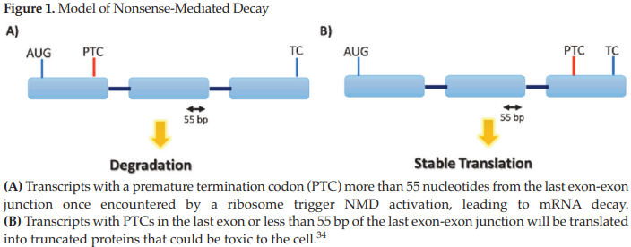
Figure 1. Model of Nonsense-Mediated Decay
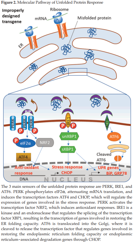
Figure 2. Molecular Pathway of Unfolded Protein Response
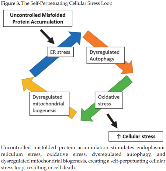
Figure 3. The Self-Perpetuating Cellular Stress Loop
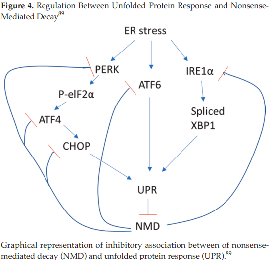
Figure 4. Regulation Between Unfolded Protein Response and Nonsense-Mediated Decay89