Volume: 21 Issue: 3 March 2023
FULL TEXT
Abstract
Objectives: In right lobe living donor liver transplant, proper reconstruction of the segment 5 vein and segment 8 vein is essential. Herein, we compared 2 different techniques for segment 5 vein reconstruction.
Materials and Methods: This prospective nonrando-mized study included all recipients of modified right lobe living donor liver transplant who had reconstruction of the segment 5 vein, with or without segment 8 veins, from October 2018 to October 2021. Patients were grouped into group A (classical technique) and group B (modified technique). For group A, the segment 5 (and segment 8, if present) vein was anastomosed in an end-to-side fashion to a polytetrafluoroethylene synthetic graft positioned parallel to the cut surface of the liver graft; then, during implant, its proximal end was anastomosed to recipient’s middle hepatic or middle-left hepatic veins unified orifice. In group B (modified technique), the stumps of segment 5 (and segment 8 if present) were anastomosed in an end-to-end fashion to 2 different polytetrafluoroethylene grafts; then during implant, the other ends of the segment 5 grafts were anastomosed directly to the inferior vena cava. Postoperative segment 5 vein patency and graft recovery were compared.
Results: Forty patients were included: 22 in group A and 18 group B. There were no significant differences in the demographic data or characteristics of donors, grafts, and recipients between the groups. There was better patency in segment 5 synthetic grafts in group A at all time points compared with group B, but this difference was statistically significant only at 1 month (18 [81.8%] vs 9 [50%, respectively; P = .046). There was no statistically significant difference in the markers of graft recovery in both groups.
Conclusions: Reconstruction of the segment 5 vein by polytetrafluoroethylene synthetic graft in a fashion to resemble the native middle hepatic vein in modified right lobe living donor liver transplant has better patency than anastomosis of the segment 5 vein in an end-to-end fashion to the synthetic graft and then to the inferior vena cava. Both techniques did not affect graft recovery.
Key words : Neo-middle hepatic vein, Polytetrafluoro-ethylene graft, Segment 5 vein
Introduction
Liver transplant is the standard of care for patients with end-stage liver failure. Due to the marked shortage of deceased donors and long wait lists for recipients, living donor liver transplant (LDLT) has become an important option for liver transplant programs around the world.1 Modified right lobe graft (right lobe graft without middle hepatic vein [MHV] inclusion) provides a sufficiently large graft to minimize the risk of the small-for-size syndrome in the recipient and also better ensure donor safety.2,3 To maximize the use of a modified right lobe graft, it should receive an optimized inflow and outflow.4,5 Proper reconstruction of the graft’s outflow is a crucial step for the success of the LDLT procedure. When modified right lobe grafts are used, it is essential to consider reconstruction of substantially large (more than 5 mm in diameter) segment 5 (S5) and segment 8 (S8) veins to the inferior vena cava (IVC) to avoid congestion of the graft’s anterior sector.6,7
Multiple reconstruction techniques and confi-gurations have been described in the literature for S5 and S8 reconstruction, and the most commonly described technique is the creation of neo-MHV by end-to-side anastomosis of both the S5 and S8 veins to a suitable homologous, autologous, or synthetic vascular graft. It has been reported that short-term and long-term patency of the S5 veins is less than that of the S8 veins with this classic technique.8,9
Herein, we introduce a simple modification that may improve S5 vein patency, and we compare this new technique to the classic technique.
Materials and Methods
This was a prospective nonrandomized study performed at the HPB Surgery and Liver Transplan-tation Unit, El-Rajhi Liver Hospital, Assiut University, Egypt. We obtained approval from the local ethical and scientific committee of our medical school (IRB No. 17200265) in accordance with the code of good practice and the guidelines of Declaration of Helsinki (7th revision, 2013, abide by the 2018 Declaration of Istanbul). All patients included in this research gave written informed consent to publish the data contained within this study. The study protocol has been registered at ClinicalTrials.gov (code NCT03666689). The study was conducted from October 2018 to October 2021. We included patients who had a modified right lobe LDLT during the study period and reconstruction of S5 veins, with or without S8 veins. All grafts were procured from living related donors (up to the fourth degree) who were over 18 years old.
All donors and recipients were evaluated according to hospital protocol. The surgical technique has been described previously.10,11 During the donor operation, in the step of parenchymal transection, the main trunk MHV was preserved on the donor side. We identified all S5 or S8 veins with diameters of 5 mm or larger, and these were preserved for subsequent reconstruction. During the back-bench preparation, the recipients were assigned to either group A (neo-MHV reconstruction, ie, the classic technique) or group B (the new technique) according to the method of right anterior sector outflow reconstruction. The methods of reconstruction were selected based on surgeon’s discretion.
In group A, S5 and S8 (or only S5, if S8 was too small) veins were anastomosed in an end-to-side fashion to a suitably sized (6 or 8 mm) ringed expanded polytetrafluoroethylene (PTFE) synthetic graft (JOTEC) positioned parallel to the cut surface of the liver graft, so that the synthetic graft would resemble a new MHV. The distal end of the PTFE graft was closed with a titanium nonabsorbable metal clip. During implant, reperfusion was performed after finishing and declamping both portal and right hepatic veins anastomoses, and then the proximal end of the PTFE graft was anastomosed to the recipient’s MHV or middle-left hepatic vein unified orifice (Figure 1).
In group B, the stumps of the S5 and S8 (if present) veins were anastomosed in an end-to-end fashion to 2 different suitably sized (6 or 8 mm) PTFE grafts. During implant, after reperfusion as in group A, the other end of the S8 synthetic graft was anastomosed to the recipient MHV or middle-left hepatic vein unified orifice. Finally, the other end of the S5 synthetic graft was anastomosed in an end-to-side fashion, directly to a venotomy created in the anterior wall of the IVC that was partially clamped. The length of the venotomy was 1.5 times larger than the diameter of the synthetic graft, and its site was selected with a goal to keep the length of the S5 synthetic graft as short as possible (Figure 2). All the anastomoses with the synthetic grafts were sutured with Prolene 6-0 (Ethicon) in continuous fashion.
Patency of the vascular graft(s), along with other vascular structures, was assessed intraoperatively and postoperatively by Doppler ultrasonography. For the purpose of the study, patency of the synthetic graft(s) was recorded at postoperative week 1 and then at month 3 and month 6. At the month 6 follow-up, patency of graft(s) was additionally assessed by computed tomography venography. In group A, any flow in the synthetic graft in the intervening segment between the orifice of the S5 vein and the orifice of the S8 vein was considered to be evidence of S5 vein patency, and any flow proximal to S8 vein orifice was considered to be evidence of S8 vein patency. In group B there were 2 separate synthetic grafts, so any flow was considered to be evidence of patency for the related vein.
Graft dysfunction was assessed by daily measu-rement of the international normalized ratio, aspartate aminotransferase, alanine aminotransferase, which are expressed as median values (with ranges) for week 1 and as mean values (with SD) for month 1 (excluding week 1) and for month 6 (excluding month 1). Small-for-size syndrome was diagnosed for any patient who developed coagulopathy, prolonged hyper-bilirubinemia, or ascites during the first 7 days after LDLT in the absence of liver ischemia.12
All patients in both groups received anticoagu-lation and antiplatelets in the first 3 months to maintain the international normalized ratio between 1.5 and 2 and the platelet count no higher than 75?000 platelets/mL, respectively.
We used SPSS software (version 22.0) for statistical analyses. Categorical data are expressed as number of patients (with percentage), with comparisons performed by the chi-square test or the Fisher exact test. Numerical data are expressed as mean values (with SD) or median values (with range), for which we used the Student t test or Mann-Whitney test as appropriate. P < .05 was considered significant.
Results
In the study period, a total of 59 patients underwent LDLT. Sixteen patients did not have S5 venous reconstruction and were therefore excluded from this study. The remaining 43 patients had S5 venous reconstruction, with or without the S8 vein. Three patients died in the early postoperative period: 1 from hepatic artery thrombosis at 20 days and 2 from multiorgan failure secondary to sepsis at 35 days and 3 months, respectively.
Forty patients were included in our study: 22 in group A and 18 in group B. There were no significant differences in the demographic data or the body mass index data (calculated as body weight in kilograms divided by the height in meters squared) between donors and recipients and between group A and group B (Table 1). There were also no significant differences in the indication for transplant or recipients’ Model for End-Stage Liver Disease (ie, MELD) scores between the groups. There was no significant difference in graft-to-recipient weight ratio between the 2 groups. In group A, 20 (90.9%) of 22 liver grafts had both S5 and S8 veins compared with 16 (88.9%) in group B, but this difference was not significant.
Patency of the S5 and S8 synthetic grafts is shown in Table 2. There was better patency in the S5 synthetic grafts in group A at all time points but was statistically significant only for the 1 month time point (Table 2). There was no difference in patency of the S8 synthetic grafts. There was no difference in the markers of graft recovery (Table 3). Small-for-size syndrome was detected in 2 (9.1%) patients in group A and 3 (16.7%) patients in group B.
Discussion
We designed this study to compare 2 reconstruction techniques of MHV tributaries in modified right lobe LDLT. The theory behind the classic reconstruction technique (group A) is to recreate an artificial conduit that has the shape and direction similar to native MHV, so that the blood that flows in S5 and S8 veins empties in a near-native manner. In this classic technique, both the S5 and S8 veins are connected to a single conduit, where the S5 vein orifice is set at a greater distance from the IVC than that of the S8 vein. This longer outflow pathway is associated with higher resistance and possibly explains the early occlusion of the S5 vein compared with the S8 vein.8 Hwang and colleagues, in a large study from Asan Medical Center, South Korea, reported their experience with the classic technique for MHV reconstruction with PTFE in LDLT; in their study, approximately two-thirds of S5 vein reconstructions and approximately one-fourth of S8 vein reconstruc-tions were occluded after 6 months.8
In contrast, the theory behind the modification that we propose here in the present study (group B) provides that the S5 and S8 veins are connected to 2 different conduits, with the synthetic graft set at a much shorter distance between the S5 vein and the IVC. Additionally, this new reconstruction technique abolishes the angle between the S5 vein and the synthetic graft. We theorized that the short and straight fashion of this new technique would enhance the flow in the vein, but we were not able to demonstrate such enhancement. We concluded that a plausible explanation is that the end-to-end anastomosis of the S5 vein to the synthetic graft developed an angular shape after the implant procedure, when the graft was anastomosed to the IVC, despite its straight (not angular) appearance during the back-bench preparation. Additionally, our endeavor to fashion the synthetic graft as short as possible led to accentuation of this angle. This new S5 vein/PTFE angle may have led to narrowing at the anastomosis site, particularly since the PTFE graft is ringed and less deformable compared with the delicate S5 vein. Our study shows that the better patency in the synthetic graft in group A (classic technique) is seen only at postoperative month 1. This finding was seen neither earlier (first week) nor later (3 and 6 months). Hwang and colleagues reported that synthetic graft occlusion occurs secondary to the inflammatory reaction at the anastomosis site and is due to endothelial cell hyperplasia.8 This endothelial hyperplasia requires a period of several days/weeks to develop.13 This may possibly explain our findings, where in the first week almost all the synthetic grafts in both groups were not yet occluded. At later follow-ups, at 3 and 6 months, this difference was not observed. The possible explanation is that most vascular grafts, irrespective of the technique, tend to become occluded over time, which has been reported in previous studies.9,14 The presence of heterogenicity between studies regarding patency rates of the synthetic grafts could be cause by differences in the following factors: sample size, patient criteria, reconstruction technique, surgical handling and expertise, type of vascular graft, use of heparin bonding, use of ringed PTFE grafts, postoperative anticoagulation and antiplatelet regimens, and follow-up duration.
There were no significant differences in the markers of recovery of the liver grafts between the 2 groups at different time points. Many factors can play a role in liver graft damage in the early postoperative period. Outflow reconstruction is perhaps the most essential element for liver graft recovery.1 The role of these reconstructed veins is probably greatest during the first few weeks postoperatively.13,14 It has been established that graft regeneration occurs rapidly within the first 2 weeks after LDLT,15,16 and we suggest that the patency of MHV tributaries could be more essential for regeneration without graft congestion in the first few weeks compared with later time points. Also, vascular graft occlusion occurs gradually, which may allow sufficient time for collaterals to develop between the MHV and the right hepatic vein and thereby relieve the consequent congestion.8 In the present study, at the early time point, there was no substantive difference in the patency with both techniques. Additionally, incidence of small-for-size syndrome was not different between the 2 groups.
In our study, we used synthetic grafts, which have been reported to be similar to autologous grafts regarding patency rates.8,9 We also preferred ringed PTFE grafts because these have higher patency than nonringed grafts.8 Park and colleagues used ringed PTFE grafts in MHV reconstruction in modified right lobe graft in LDLT, and they applied the same reconstruction method in their study that we applied in group A. They found that patency rates of the PTFE were 92.9%, 54.8%, 43.7%, and 36.5% at 3 months, 12 months, 24 months, and 36 months after transplant, respectively.17
We used 6-mm or 8-mm PTFE grafts. Hwang and colleagues hypothesized that, if the grafts were too large, then there would be a marked decrease in the velocity of blood flow, with a greater risk for thrombosis. Conversely, if the graft diameter is too small, then the resulting turbulence of blood flow may lead to thrombosis and occlusion.8
Our study has some limitations. The study is not randomized because we did not find sufficient data in the literature to support our proposed technique, so randomization was not possible. This study is from a single transplant center with a small sample of patients. The liver grafts sizes in our study are relatively large, whereas the role of MHV reconst-ruction would be more essential in smaller grafts. Volumetric imaging could provide insight about regeneration and volume changes, but this was not done in this study. Also, follow-up beyond 6 months is absent.
Conclusions
Reconstruction of the S5 vein by ringed PTFE synthetic graft in an end-to-side fashion to resemble native MHV in the modified right lobe LDLT has better patency than anastomosis of the S5 vein in an end-to-end fashion to the synthetic graft and then directly to the IVC. Both techniques did not affect graft recovery. Larger studies are required.
References:
- Lee SG. A complete treatment of adult living donor liver transplantation: a review of surgical technique and current challenges to expand indication of patients. Am J Transplant. 2015;15(1):17-38. doi:10.1111/ajt.12907
CrossRef - PubMed - Tan CHN, Hwang S, Bonney GK, Ganpathi IS, Madhavan K, Kow WCA. The influence of the middle hepatic vein and its impact on outcomes in right lobe living donor liver transplantation. HPB (Oxford). 2019;21(5):547-556. doi:10.1016/j.hpb.2018.09.003
CrossRef - PubMed - Zhang S, Dong Z, Zhang M, Xia Q, Liu D, Zhang JJ. Right lobe living-donor liver transplantation with or without middle hepatic vein: a meta-analysis. Transplant Proc. 2011;43(10):3773-3779. doi:10.1016/j.transproceed.2011.08.100
CrossRef - PubMed - Balci D, Kirimker EO. Hepatic vein in living donor liver transplantation. Hepatobiliary Pancreat Dis Int. 2020;19(4):318-323. doi:10.1016/j.hbpd.2020.07.002
CrossRef - PubMed - She WH, Chok KS, Fung JY, Chan AC, Lo CM. Outcomes of right-lobe and left-lobe living-donor liver transplantations using small-for-size grafts. World J Gastroenterol. 2017;23(23):4270-4277. doi:10.3748/wjg.v23.i23.4270
CrossRef - PubMed - Jeng LB, Thorat A, Yang HR, Li PC. Venous outflow reconstruction in living donor liver transplantation: dealing with venous anomalies. World J Transplant. 2015;5(4):145-153. doi:10.5500/wjt.v5.i4.145
CrossRef - PubMed - Thorat A, Jeng LB, Yang HR, et al. Outflow reconstruction for right liver allograft with multiple hepatic veins: “V-plasty” of hepatic veins to form a common outflow channel versus 2 or more hepatic vein-to-inferior vena cava anastomoses in limited retrohepatic space. Liver Transpl. 2016;22(2):192-200. doi:10.1002/lt.24342
CrossRef - PubMed - Hwang S, Jung DH, Ha TY, et al. Usability of ringed polytetrafluoroethylene grafts for middle hepatic vein reconstruction during living donor liver transplantation. Liver Transpl. 2012;18(8):955-965. doi:10.1002/lt.23456
CrossRef - PubMed - Singhal A, Makki K, Chorasiya V, et al. Venous outflow reconstruction using a polytetrafluoroethylene (PTFE) graft in right lobe living donor liver transplantation: a single center study. Surgery. 2021;169(6):1500-1509. doi:10.1016/j.surg.2021.01.011
CrossRef - PubMed - Taha AM, Hassan RA, Mahdy MM, Abbas MS, Makhlouf NA, Ibraheem TM. Arterial anastomosis without rotation in adult living donor liver transplantation: a modification to the back-wall-first technique. Egypt J Surg. 2021;40(3):1023-1027. doi:10.4103/ejs.ejs_178_21
CrossRef - PubMed - Taha AM, Hassan RA, Mahdy MM, Abbas MS, Makhlouf NA, Hassaneen SH, Ibraheem TM. Radio-opaque filaments guided bile duct division during living donor liver transplantation, a simple solution to an ongoing dilemma. Surg Gastroenterol Oncol. 2021;26(2):111-117. doi:10.21614/sgo-26-2-259
CrossRef - PubMed - Hernandez-Alejandro R, Sharma H. Small-for-size syndrome in liver transplantation: New horizons to cover with a good launchpad. Liver Transpl. 2016;22(S1):33-36. doi:10.1002/lt.24513
CrossRef - PubMed - Akamatsu N, Sugawara Y, Nagata R, et al. Adult right living-donor liver transplantation with special reference to reconstruction of the middle hepatic vein. Am J Transplant. 2014;14(12):2777-2787. doi:10.1111/ajt.12917
CrossRef - PubMed - Sakamoto K, Ogawa K, Matsui T, et al. Reconstruction of middle hepatic vein tributaries with artificial vascular grafts in living donor liver transplant using right lobe grafts: a case series. Transplant Proc. 2019;51(5):1506-1510. doi:10.1016/j.transproceed.2019.01.118
CrossRef - PubMed - Everson GT, Hoefs JC, Niemann CU, et al. Functional elements associated with hepatic regeneration in living donors after right hepatic lobectomy. Liver Transpl. 2013;19(3):292-304. doi:10.1002/lt.23592
CrossRef - PubMed - Emond JC, Fisher RA, Everson G, et al. Changes in liver and spleen volumes after living liver donation: a report from the Adult-to-Adult Living Donor Liver Transplantation Cohort Study (A2ALL). Liver Transpl. 2015;21(2):151-161. doi:10.1002/lt.24062
CrossRef - PubMed - Park GC, Hwang S, Ha TY, et al. Hemashield vascular graft is a preferable prosthetic graft for middle hepatic vein reconstruction in living donor liver transplantation. Ann Transplant. 2019;24:639-646. doi:10.12659/AOT.919780
CrossRef - PubMed

Volume : 21
Issue : 3
Pages : 245 - 250
DOI : 10.6002/ect.2022.0417
From the HPB Surgery and Liver Transplantation Unit, Surgery Department, El-Rajhi Liver Hospital, Assiut University, Egypt
Acknowledgements: The authors have not received any funding or grants in support of the presented research or for the preparation of this work and have no declarations of potential conflicts of interest.
Corresponding author: Ahmed M. Abdalla, Second floor, Surgery Department, Assiut University Hospital, Assiut, Egypt
Phone: +20 10 1894 2253
E-mail:aboabdalla_333@aun.edu.eg
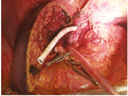
Figure 1. Anastomosis of Segment 5 and Segment 8 Veins With Polytetrafluoroethylene Graft in an End-to-Side Fashion: Group A
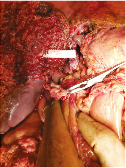
Figure 2. Anastomosis of Segment 5 Vein With Polytetrafluoroethylene Graft in an End-to-End Fashion: Group B
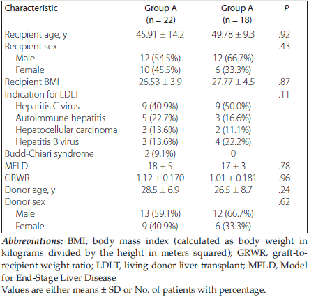
Table 1. Preoperative Characteristics of Donors, Grafts, and Recipients
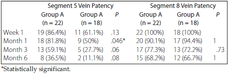
Table 2. Segment 5 Vein Patency Follow-Up of the Recipients
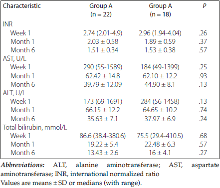
Table 3. Markers of Graft Recovery