Volume: 21 Issue: 3 March 2023
FULL TEXT
This study investigated the efficacy of stem cell transplantation for azoospermia, a major cause of male infertility. We conducted a systematic meta-analysis to assess the therapeutic effectiveness of stem cell transplant, using different transplant methods, injection sites, and stem cell types, and the reliability of this approach in different animal species. PubMed, the Cochrane Library, and Embase were searched for studies published from January 2006 to February 2022 that evaluated the use of stem cell transplant to treat azoospermia. We included 18 studies and conducted the analyses using Review Manager 5.2 software. Expression of the meiosis-related genes Vasa, Scp3, and Dazl and the average hematoxylin and eosin-positive staining area were improved after stem cell transplant. Subgroup analyses by mode of transplant showed higher expression of Scp3 and Dazl in the xenotransplant group. Although subgroup analyses by injection site showed that the seminiferous tubule group showed the most significant effect on Scp3 expression, spermatogenesis and repair of damaged testis were induced in the tunica albuginea group. The testicular torsion group also induced high levels of Scp3. Another subgroup analysis by stem cell type showed that umbilical cord mesenchymal stem cells promoted the highest expression of meiosis-related genes and successfully induced spermatogenesis and the repair of damaged testis. Urine-derived stem cells, spermatogonial stem cells, and amniotic fluid-derived stem cells showed significantly therapeutic effects; however, more studies are needed for definitive conclusions. Subgroup analyses by type of azoospermia animal model indicated that the use of stem cell transplant in rat or mouse models had an obvious therapeutic effect, but no significant therapeutic effect was seen in azoospermia hamsters. The meta-analysis confirmed that stem cell transplant can effectively treat azoospermia in animal models. Xenotransplant is shown to enhance the therapeutic effects of stem cell transplant on azoospermia.
Key words : Cellular therapy, Male infertility, Meiosis-related genes, Mesenchymal cells, Spermatogenesis
Introduction
Infertility, characterized by the failure to become pregnant after at least 12 months of regular and noncontraceptive sexual activity,1 is a common clinical condition affecting approximately 48.5 million human couples. The World Health Organization estimates that male-specific factors are the primary or contributing cause of approximately half of all infertility cases.2,3 Although there are many forms of male factor-related infertility, azoospermia is the most prevalent, accounting for almost 15% of cases.4 There are 2 types of azoospermia: nonobstructive azoospermia (NOA) and obstructive azoospermia (OA), which are based on pathology. In OA, the deferent duct is obstructed, but testicular spermatogenesis function is normal; obstruction can be treated with microsurgical anastomosis. In NOA, testicular abnormalities result in inadequate spermatozoa production, making it a challenge to treat. Moreover, NOA can be subdivided into idiopathic NOA and NOA with a history of cryptorchidism, that is, which can pass onto the next generation due to chromosomal gene deletion or induce testicular cancer.5 Sperm donation and assisted reproductive techniques are currently the most available types of treatment for male infertility. However, these methods are not suitable if no spermatozoa can be retrieved from the testicles, which is often the case for NOA patients. Thus, there is an urgent clinical demand for new therapies.
Stem cell transplant (SCT) is an advanced technology that is an effective treatment option for a variety of diseases.6 Stem cells are identified, isolated, purified from various tissues, and transfused into a patient, where they can repair damaged cells or rebuild tissues to reconstruct organ function. Stem cell transplant has recently been adapted to address male infertility caused by abnormalities in germ cells. Studies have indicated that different types of stem cells, including bone marrow mesenchymal stem cells (BM-MSCs),7 spermatogonial stem cells (SSCs),8 and adipose tissue-derived mesenchymal stromal cells (AT-MSCs),9 can have distinct therapeutic effects on azoospermia. Both xenotransplant and homotransplant methods are used to address this condition and are shown to impact SCT in different ways.
Spermatogenesis is a highly complex cell differentiation process. Multiple meiosis-related genes are involved, and the expression of these genes is often used to evaluate the therapeutic effect of spermatogenesis on azoospermia. Histological assessments have indicated that seminiferous tubules differ between healthy individuals and those with azoospermia.10-13 However, the results of these assessments are often presented in graphs and images and analyzed manually, reducing the accuracy and reliability of the results. Using computer image analysis systems to determine the average positive stained area may allow for a more advanced and intuitive observation of histological changes in seminiferous tubules.14
Although many studies have assessed the use of SCT to treat azoospermia, few have compared the efficacy of different SCT methods. We speculated that these differences may play a vital role in unders-tanding the therapeutic impact of this technique. To determine the most effective SCT-based therapeutic regimen for azoospermia, we performed a compre-hensive systematic review and meta-analysis that compared the efficacy of different SCT methods. Subgroup analyses were conducted by mode of transplant, injection site, induction of azoospermia, stem cell type, and the type of azoospermia animal model. The study findings could be of practical significance and serve as a reference for future stem cell research and experimental designs.
Materials and Methods
Search strategy
Three databases (PubMed, Embase, and the Cochrane Library) were systematically searched for original articles. The search terms consisted of free words and MeSH terms, including “stem cell transplantation” (MeSH unique identification [ID] D033581) or “stem cells” (MeSH unique ID D013234) and “azoospermia” (MeSH unique ID D009845). Studies published from January 2006 to February 2022 were included.
Exclusion and inclusion criteria
After removing duplicate articles using a reference management tool (Endnote X9.3.3), we screened the remaining full-text articles, which were further excluded if (1) only an abstract was presented; (2) the study was a review, editorial, reply letter, or note; (3) the study had no data or incomplete data; or (4) the study had no available full-text article. Studies were included if they (1) were carried out on azoospermia animal models, (2) the azoospermia models in the experimental group received any kind of stem cell used for stem cell therapy, (3) a control group was designed using the same animal, and (4) data on 1 or more of the following outcomes related to azoospermia recovery could be extracted: expression of the deleted in azoospermia-like (DAZL) gene, synaptonemal complex protein 3 (SCP3) gene, or DEAD-box helicase 4 (VASA) genes and average hematoxylin-eosin (HE)-positive staining area in the testes.
Data extraction
Two researchers independently extracted the following data from the retrieved studies: (1) authors, (2) publication year, (3) country in which the study was conducted, (4) azoospermia animal species, (5) cell sources, (6) injection site and injection dose, (7) induction of azoospermia, (8) number of animals in each group, and (9) outcomes. Outcomes included expression levels of the 3 meiosis-related genes and HE-positive staining area in the testes. Image-Pro Plus 6.0 and WebPlotDigitizer 4.5 were used to extract graphed data. Bar chart images were uploaded to WebPlotDigitizer 4.5 web-based software to calibrate the axes. After calibration, the researcher manually clicked each data point within the bar chart to acquire the extracted coordinates. The HE-stained histological images were imported into Image-Pro Plus 6.0 software. Seminiferous tubule images were manually depicted by clicking “Count and measure object → Manual → Select colors.” After applying the mask, the researcher selected “Automatic bright objects” and added “Per area (Obj./Total)” to the measurements filter. The “Area of object” range was edited beginning at 1, and “Count” was clicked to measure the segmentation results. “View → Statistics” was selected to show the measurements.
Quality appraisal
SYRCLE’s risk of bias tool for animal studies was used to assess the risk of bias in the included studies. This tool is an adapted version of the Cochrane risk of bias tool specifically used for animal intervention studies and contains 10 entries related to 6 types of bias: sequence generation (selection bias), baseline characteristics (selection bias), allocation concealment (selection bias), random housing (performance bias), blinding (performance bias), random outcome assessment (detection bias), blinding (detection bias), incomplete outcome data (attrition bias), selective outcome reporting (reporting bias), and other sources of bias (other biases). Each entry is judged as “low risk,” “high risk,” or “unclear risk.” Baseline characteristics between groups, including age and average weight of animals, feeding conditions, and randomized grouping design were examined by 2 independent authors to ensure baseline similarity and avoid selective bias. Studies were evaluated for having incomplete outcome data if (1) graphed or imaged data are poor and unclear that unable to extract accurate data and (2) standard deviations were not reported and could not be calculated from available data.
Data analyses and synthesis
The meta-analysis was performed using Review Manager version 5.2 software provided by the Cochrane Collaboration. For articles that presented only graphed or imaged data, 2 independent authors used tools to extract the mean and SD values from graphs and images. DAZL is a master translational regulator essential for spermatogenesis and is essential for embryonic germ cell development and differentiation. SCP3 is a structural component of the synaptonemal complex, which is essential
for meiosis. VASA, which encodes a DEAD-family protein of ATP-dependent RNA helicase, is specifically expressed in germ cells and has a central role in germ cell development. The upregulation of these genes can indicate the germ cell meiosis initiation and spermatogenesis activity. The average HE-positive staining area in the testes can intuitively and visually reveal the repairment of damaged testis. In our analysis, we determined the average HE-positive staining area as the primary endpoint and the expression of DAZL, SCP3, and VASA genes as the secondary endpoint. A random-effects model was used for the analysis, and the standard mean difference (SMD) with 95% CI was used as the pooled effect of the outcomes. The significance was set at P < .05, and heterogeneity values were calculated using I2 to quantify heterogeneity. If I2 > 50%, heterogeneity was considered substantial. Subgroup analyses were performed by the transplant method, the injection site, the induction of azoospermia, the azoospermia animal species, and the cell source to identify potential sources of heterogeneity. Heterogeneity for subgroup analysis was assessed using I2. A graphical funnel plot was used to investigate possible publication bias.
Results
Study selection and characteristics
We identified 471 possibly related studies, for which 78 duplicate records were excluded. Of these, 393 studies remained for further assessment after titles and abstracts were screened. Reviews, letters, and irrelevant studies were removed, and 177 studies were included for eligibility assessment by carefully screening the full-text articles. Studies that did not meet the inclusion criteria or did not have an available full-text article were excluded, and a total of 18 studies9,15-31 were selected for this meta-analysis. The screening process and results are shown in Figure 1, and the characteristics of the 18 selected studies are shown in Table 1. Of the studies, 10 used mouse models, 5 used rat models, 2 used hamster models, and 1 used a guinea pig model.
Assessment of bias
The risk of bias in the included studies was assessed using SYRCLE’s risk of bias tool (Figure 2, A and B). Four studies disclosed incomplete or poorly graphed data and thus were defined as “high risk” for incomplete outcome assessment. One study reported that hamster BM-MSCs resulted in reconstitution of the tubular microenvironment and proliferation of inactivated germinal cells in the host tubules without evidence of an identical spermatogenesis index in the seminiferous tubules of the control and treatment groups. Explanation with low confidence was shown and thus resulted in “high risk” for selective outcome reporting assessment. All included studies that lacked detailed descriptions or sufficient reporting and information were defined as “unclear risk” for blinding of the outcome, allocation concealment, and blinding assessment.
Publication bias was assessed and is summarized in a funnel plot (Figure 3). Funnel plots of Vasa, Scp3, and Dazl expression and average HE-positive staining area in the testes are shown in Figure 4, A-D, respectively. The possible presence of publication biases regarding these outcomes was indicated by obvious asymmetries in the funnel plots.
Analysis of Vasa expression
Five studies15,18,19,26,30 assessed the effects of SCT on azoospermia in animals by measuring Vasa gene expression (Figure 4A). As shown in the forest plot, all studies reported that higher Vasa expression was seen in the SCT group than in the control group (SMD = 16.94; 95% CI, 12.59-21.29; P < .001). Given that there was substantial heterogeneity in Vasa expression (I2 = 57%, P = .05), subgroup analyses were performed.
The 5 studies were divided into 2 groups based on their transplant method: 3 studies used xenotransplant (with human stem cells) and 2 used homotransplant. The 2 different transplant methods promoted Vasa expression (for xenotransplant: SMD = 21.43; 95%CI, 11.27-31.60; P < .001; for homotransplant: SMD = 18.71; 95%CI, 2.49-34.92; P = .02) (Figure 5A). There were no obvious differences between the subgroups (P = .78), and the heterogeneity remained substantial (for xenotransplant: I2 = 58%, P = .09; for homotransplant: I2 = 72%, P = .06).
The 5 studies were then divided into 3 groups based on the injection site used: 3 studies transplanted stem cells into seminiferous tubules, 1 study transplanted stem cells into the testicular artery and epididymis, and 1 study received stem cells by injection under the tunica albuginea. It is important to note that 2 of the injection site groups contained only 1 study; therefore, it was not possible to test for heterogeneity. Results showed that Vasa expression was elevated in all 3 groups (for seminiferous tubules: SMD = 21.78; 95% CI, 8.30-35.25; P = .002; for testicular artery and epididymis: SMD = 25.68; 95% CI, 14.41-36.95, P < .001; for tunica albuginea: SMD = 13.72; 95% CI, 5.90-21.55; P = .006) (Figure 5B). No obvious subgroup differences were detected among the groups (P = .20), and the seminiferous tubules group showed high levels of heterogeneity (I2 = 68%, P = .04).
The 5 studies were divided into 2 groups based on the method used to induce azoospermia: 4 studies used busulfan-induced azoospermia animal models and 1 study, which could not be tested for heterogeneity, performed a testicular torsion operation to induce azoospermia. The 2 different groups had no obvious subgroup differences (P = .06) (Figure 5C), and both the busulfan-induced azoospermia (SMD = 22.63; 95%CI, 13.98-31.29; P < .001) and testicular torsion-induced azoospermia (SMD = 11.94; 95%CI, 5.11-18.78; P < .001) animal models promoted Vasa gene expression. The busulfan-induced group had a lower level of heterogeneity (I2 = 49%, P = .12).
The 5 studies were then divided into 4 groups based on the type of stem cells used. The subgroup difference was significant (P = .02) (Figure 5D). Umbilical cord mesenchymal stem cells (UC-MSCs) had the most prominent effect on Vasa gene expression (SMD = 175.37; 95% CI, 76.63-274.10; P < .001), followed by umbilical cord blood-derived mesenchymal stem cells (UCB-MSCs) (SMD = 29.98; 95% CI, 13.05-46.90; P < .001), induced pluripotent stem cells (iPSCs) (SMD = 28.83; 95% CI, 12.55-45.11; P < .001), and adipose tissue-derived mesenchymal stem cells (AT-MSCs) (SMD = 17.42; 95% CI, 2.51-32.33; P = .02). The AT-MSC group had high heterogeneity (I2 = 80%, P = .03), and the remaining groups involving only 1 study could not be tested for heterogeneity. There were no differences in the therapeutic effects of iPSCs and the 3 different adult stem cell (ASC) types (P = .24) (Figure 5E).
Analysis of Scp3 expression
Six studies15-19,30 assessed the effects of SCT on azoospermia in animals by measuring Scp3 gene expression (Figure 4B). All 6 studies showed higher levels of Scp3 in the SCT group than in the control group (SMD = 6.64; 95% CI, 3.17-10.10; P < .001). Given that substantial heterogeneity was detected (I2 = 83%, P < .001), we performed subgroup analyses.
The 6 studies were divided into 2 groups based on the transplant method used: 4 studies used xenotransplant and 2 studies used homotransplant. All xenotransplant studies used human stem cells, and the results showed that xenotransplant promoted Scp3 gene expression (SMD = 7.65; 95% CI, 3.22-12.09; P < .001) (Figure 6A). Homotransplant, however, did not have a significant effect on Scp3 gene expression (SMD = 20.71; 95%CI, -18.66 to 60.08; P = .30). The subgroup differences (P = .52) were relatively indistinctive, and the heterogeneity remained substantial (for xenotransplant: I2 = 80%, P = .002; for homotransplant: I2 = 91%, P = .001).
The studies were then divided into 4 groups based on the injection site used: 2 studies transplanted stem cells into the seminiferous tubules, 1 study transplanted stem cells into the testis interstitium, 2 studies received stem cells by injection under the tunica albuginea, and 1 study transplanted stem cells into the testicular artery. The 2 injection site groups containing only 1 study could not tested for heterogeneity. Three groups showed induction of the Scp3 gene, of which the seminiferous tubules group had the most significant effect (SMD = 31.67; 95% CI, 16.53-46.81; P < .001) (Figure 6B), followed by the testicular artery and epididymis group (SMD = 4.80; 95% CI, 2.46-7.15; P < .001) and the testis interstitium group (SMD = 2.41; 95% CI, 0.92-3.89; P = .001). The tunica albuginea group did not have a significant effect on Scp3 gene expression (SMD = 8.81; 95% CI, -2.64 to 20.26; P = .13). Obvious subgroup differences were detected among the groups (P < .001); the seminiferous tubules group showed low hetero-geneity (I2 = 23%, P = .25), and the tunica albuginea group had high heterogeneity (I2 = 85%, P = .01).
The studies were then divided into 2 groups based on the methods used to induce azoospermia: 5 studies used busulfan-induced azoospermia animal models and 1 study used a testicular torsion operation to induce azoospermia. The latter could not be tested for heterogeneity. The 2 groups had obvious subgroup differences (P = .003) (Figure 6C); testicular torsion-induced azoospermia animal models (SMD = 42.75; 95% CI, 18.65-66.85; P < .001) were more effective at promoting Scp3 gene expression than busulfan-induced azoospermia animal models (SMD = 5.36; 95% CI, 2.45-8.26; P < .001). This was the opposite of the effect that these 2 methods had on Vasa gene expression. However, the busulfan-induced group still showed significant heterogeneity (I2 = 79%, P < .001).
The studies were divided into 5 groups based on the type of stem cells used, resulting in only 1 or 2 studies per group. The subgroup difference was significant (P < .001) (Figure 6D). We found that the UCB-MSC induced the highest level of Scp3 gene expression (SMD = 26.28; 95% CI, 11.43-41.13; P < .001), followed by UC-MSC (SMD = 15.53; 95% CI, 6.70-24.37; P < .001), urine-derived stem cells (SMD = 3.73; 95% CI, 1.95-5.52; P < .001), and BM-MSC (SMD = 2.41; 95% CI, 0.92-3.89; P = .001). With AT-MSC treatment, no prominent effect on Scp3 gene expression (SMD = 21.80; 95% CI, -15.19 to 58.79; P = .25) was shown, and high heterogeneity was still shown (I2 = 89%, P = .002). The remaining groups that only involved 1 study could not be tested for heterogeneity.
Analysis of Dazl expression
Three studies16,19,30 assessed the effects of SCT on azoospermia in animal models by measuring Dazl gene expression (Figure 4C). All 3 studies reported more or less positive effects on the expression of Dazl gene (SMD = 11.83; 95% CI, -1.61 to 25.28; P = .08) compared with the control group that was not treated with SCT. Substantial heterogeneity was detected (I2 = 92%, P < .001), and so subgroup analyses were performed.
Three studies were divided into 2 groups based on the transplant method used. Two studies used xenotransplant, both of which used human stem cells; the remaining study used homotransplant. Xenotransplant promoted a higher level of Dazl gene expression (SMD = 17.76; 95% CI, 11.50-24.02, P < .001) (Figure 7) than homotransplant (SMD = 1.30; 95% CI, -0.11 to 2.49; P = .03). Subgroup differences were significant (P < .001), and the heterogeneity was low in the xenotransplant group (I2 = 0%, P = .37).
Hematoxylin and eosin-positive staining in the testes
Twelve studies9,15,18,19,21,23,25,26,28-31 had available information after extracting graphed data (Figure 4D). According to the forest plot, all 12 studies reported more or less positive effects on the induction of spermatogenesis and repair of damaged testis (SMD = 64.40; 95% CI, 44.83-83.98; P < .001) compared with control groups that were not treated with SCT. Substantial heterogeneity was detected (I2 = 80%, P < .001), so subgroup analyses were performed.
All 12 studies were divided into 2 groups based on the transplant method used, of which 3 studies used xenotransplant and 9 used homotransplant. All xenotransplant studies used human stem cells. Both transplant methods were able to induce spermatogenesis and repair damaged testis (for xenotransplant: SMD = 101.13; 95% CI, 46.70-155.55; P = .001; for homotransplant: SMD = 55.89; 95% CI, 36.21-75.56; P < .001) (Figure 8A). Subgroup differences were not significant (P = .13). Hetero-geneity was high in both the xenotransplant and homotransplant groups (for xenotransplant: I2 = 64%, P = .06; for homotransplant: I2 = 79%, P < .001).
The 12 studies were divided into 4 groups based on the injection site used: the seminiferous tubules group involved 9 studies, with testicular artery and epididymis, the rete testis, and the tunica albuginea involved in 1 study each. Thus, tests for heterogeneity were not applicable. Subgroup differences were detected among the groups (P = .03) (Figure 8B), with the tunica albuginea group showing the highest level of spermatogenesis and repair of damaged testis (SMD = 187.87; 95% CI, 82.10-293.65; P < .001), followed by the testicular artery and epididymis groups (SMD = 88.80; 95% CI, 49.99-127.62; P < .001), rete testis group (SMD = 72.64; 95% CI, 40.88-104.39; P < .001), and seminiferous tubules group (SMD = 55.50; 95% CI, 35.76-75.23; P < .001). The heterogeneity of the seminiferous tubules group was substantial (I2 = 78%, P < .001).
The 12 studies were then divided into 2 groups based on the method used to induce azoospermia: 9 studies used busulfan-induced azoospermia animal models and 3 studies performed testicular torsion operations to induce azoospermia. The 2 groups did not have obvious subgroup differences (P = .68) (Figure 8C) but did show significant heterogeneity (for busulfan-induced group: I2 = 82%, P < .001; for testicular torsion-induced group: I2 = 63%, P = .07). Spermatogenesis was induced, and damaged testes were repaired in both groups (for busulfan-induced group: SMD = 62.09; 95% CI, 39.62-84.56; P < .001; for testicular torsion-induced group: SMD = 71.42; 95% CI, 32.83-110.01; P = .001).
The 12 studies were divided into 7 groups based on the type of stem cells used, with 5 groups that involved only 1 study and thus could not be tested for heterogeneity. Subgroup differences were significant (P = .002) (Figure 8D), with UC-MSC having the most prominent effect on the induction of spermatogenesis and repair of damaged testis (SMD = 249.29; 95% CI, 108.94-389.64; P = .001), followed by SSC (SMD = 117.78; 95% CI, 60.27-175.29, P < .001), amniotic fluid-derived stem cells (SMD = 86.82; 95% CI, 58.50-115.15; P < .001), BM-MSC (SMD = 76.59, 95% CI, 17.16-136.03; P = .01), UCB-MSC (SMD = 73.09; 95% CI, 31.92-114.25; P = .001), AT-MSC (SMD = 45.85, 95% CI, 22.27-69.43, P < .001), and iPSC (SMD = 37.24, 95% CI, 16.24-58.24, P < .001). Both the AT-MSC and the BM-MSC groups showed high heterogeneity (I2 = 63%, P = .05 and I2 = 89%, P = .001, respectively). The subgroup differences between the iPSC and the 6 ASC groups were significant (P = .04) (Figure 8E), with ASC having higher induction of spermatogenesis and repair of damaged testis than iPSC (for ASC: SMD = 69.17; 95% CI, 46.84-91.51; P < .001; for iPSC: SMD = 37.24; 95% CI, 16.24-58.24; P = .001).
The 12 studies were then divided into 3 groups based on the type of azoospermia animal model used. Both the mouse and rat groups had strong effects on the induction of spermatogenesis and repair of damaged testis (for mouse group: SMD = 75.80; 95% CI, 42.12-109.48; P < .001; for rat group: SMD = 55.17; 95% CI, 24.00-86.34; P = .001); the hamster group did not show significant improvement (SMD = 97.99; 95% CI, -50.25 to 246.24; P = .20). Subgroup differences were not significant (P = .62) (Figure 8F), and each group still showed high heterogeneity after the analysis (for mouse group: I2 = 74%, P = .002; for rat group: I2 = 84%, P < .001; for hamster group: I2 = 91%, P < .001).
Discussion
Azoospermia is characterized by the absence of normally formed and functioning mature sperm during ejaculation. Almost 60% of azoospermia patients have NOA, the most severe form of male infertility, which is associated with testicular failure.32 There are multiple causes of NOA. For example, cytotoxic drugs used in tumor therapy can cause NOA as a long-term side effect. Indeed, the increased use of chemotherapy and radiotherapy in tumor treatment that has raised cancer survival rates among male patients is associated with a higher prevalence of NOA.33 Chromosomal abnormalities, including Klinefelter syndrome and Y-chromosome microdeletions, can also promote this condition. Klinefelter syndrome is the most common chromosomal disorder among men, occurring in 1 of 650 newborn males,34 whereas Y-chromosome microdeletions, defined as deletions within the male-specific region of the Y-chromosome, are found in up to 24% of men.35,36 Several other genetic defects, including Kallmann syndrome, TEX11 mutation, and mild androgen insensitivity syndrome, are also associated with NOA. It is evident that a safe, effective, and efficient therapy for azoospermia is of great urgency and significance.
Stem cells are a self-replicating cell type that can show multidirectional differentiation, can reconstruct tissue function, and can induce differentiation into distinct germ layers under appropriate conditions. Because the clinical application of human embryonic stem cells is limited by ethical restrictions, tumori-genicity, and immune rejection, ASCs are most used for SCT. Studies have indicated that spermatogenesis is closely associated with microenvironments that involve multiple cytokines, including SCF, GDNF, FGF2, and BMP4,37-41 and transplanted ASCs can be the source of differentiated spermatogenic functional cells and cytokines that contribute to this environment.37,42 Mesenchymal stem cells derived from various tissues are considered the most appropriate stem cell type for SCT. These cells actively participate in tissue repair and have particularly significant multidirectional differen-tiation potential, including the ability to become functional germ-like cells.7,43 However, iPSCs also possess multidirectional differentiation without ethical constraints and can thus serve as an alternative stem cell for use in SCT.44
Xenotransplantation is an alternative and effective option for SCT when homotransplantation is not available for technical or ethical reasons.45 However, the differences between these transplant methods have not been well explored. Our meta-analysis compared the efficiency of different SCT methods by assessing 3 different outcomes. Subgroup analysis found that xenotransplant significantly increased the expression of all meiosis-related genes, whereas homotransplant had no significant effect on the expression of Scp3 and Dazl, suggesting that this method may have a lower therapeutic effect. Because the Scp3 gene encodes an essential structural component of the synaptonemal complex46,47 and the Dazl gene encodes a protein that is localized in the nucleus and cytoplasm of fetal germ cells and in the cytoplasm of developing oocytes, it was speculated that homotransplant may limit the formation of the synaptonemal complex. However, both transplant methods were associated with improved expression of the Vasa gene, spermatogenesis induction, and the repair of damaged testis.
The site of injection can influence the therapeutic effect of SCT for different diseases.48,49 To determine whether there is a similar association between the site of injection and SCT outcomes for azoospermia, we conducted a subgroup analysis by injection site type. Although stem cells injected into the seminiferous tubules induced the highest expression of Scp3 gene, those injected under the tunica albuginea showed improved repair of damaged testis and increased spermatogenesis. Scp3 is involved in synapsis, recombination, and segregation of meiotic chromosomes and meiosis occurs in seminiferous tubules; therefore, we assumed that injecting stem cells directly into seminiferous tubules can have the maximum possible effect to prevent stem cells from cellular inactivation or cell death. However, it is possible that, because few studies were included in other groups, a relatively objective comparison of results could not be obtained.
New techniques have been developed to induce an azoospermia animal model, including busulfan, radiation, and hyperthermia,50-52 but methods are associated with damaging side effects and prolonged treatment durations. Studies have shown that busulfan-treated animals are not suitable for SSC transplant because the SSC microenvironment in these animals prevents transplanted cells from differentiating and proliferating. Busulfan treatment can lead to severe bone marrow depression53,54 and cause death in some animals. We conducted a subgroup analysis to identify which induction model is more suitable for SCT. Testicular torsion-induced animals showed higher expression of Scp3 versus busulfan-induced animals. This may be because the seminiferous tubules, although severely damaged by busulfan treatment, are reversibly depleted by testicular torsion, allowing for improved outcomes. Additional studies are needed to further explore these differences.
As described above, many stem cell types have been used for SCT in the treatment of different diseases. Results of our meta-analysis showed that UC-MSC and UCB-MSC lead to significant impro-vements in azoospermia, either by promoting the expression of meiosis-related genes or inducing spermatogenesis and the repair of damaged testis. We found that use of BM-MSC was less effective than UC-MSC, followed by AT-MSC. There are also 3 newly discovered MSC types,17,55,56 urine-derived stem cells, SSC, and amniotic fluid-derived stem cells, of which SSC has the greatest potential for treating male infertility.57,58 Although our results indicated that all 3 MSC types had significant therapeutic effects on azoospermia, too few related studies were assessed and more data are needed to reach a definitive conclusion. As a newly discovered source of cells used for cellular therapy, the use of iPSCs has not been standardized in production conditions because it has been reported to have different differentiation propensities that were largely attributable to their donor differences, which means that different iPSC lines could have distinct differentiation properties.59 Even so, iPSCs indeed showed positive effects on inducing spermatogenesis in the included studies. However, the small number of included studies using iPSCs for cellular therapy may have contributed to the unsatisfactory result presented in our analysis. Thus, additional research is required before iPSCs are used in the treatment of azoospermia.
The efficacy of SCT in different azoospermia animal models was also compared. Although SCT had an obvious therapeutic effect on azoospermia rats and mice, this treatment had no significant therapeutic effect on azoospermia hamsters. Given that the poor quality of outcome data in this subgroup analysis may have contributed to inaccurate results, additional studies are needed to compare the effects of SCT on azoospermia in different animal models.
There are many studies that have adapted SCT on azoospermia; however, to our knowledge, there is no meta-analysis of the efficacy of SCT for azoospermia animal. This study helps to determine which factors are most critical to an effective SCT-based regimen for azoospermia and provides a valuable reference for future stem cell research. However, our study had some limitations. First, some of the included studies had incomplete outcome data, which may introduce bias. Second, several subgroups only contained 1 study, which may lead to nonobjective results. These limitations can increase the heterogeneity of the study. In addition, the transplanted cell dosage, which may be associated with the transplant site, was not explored. Further analyses are required to better understand the mechanism by which different transplanted cell dosages produce distinct therapeutic effects.
Conclusions
Our analysis showed that SCT is a relatively safe and effective approach for treating azoospermia and that xenotransplant improves the therapeutic effect of this approach. Different cell injection sites, methods of inducing azoospermia, stem cell types, and animal models may influence the therapeutic effects of SCT. Further studies and additional analyses are required to understand the therapeutic mechanism by which SCT ameliorates azoospermia.
References:
- Vander Borght M, Wyns C. Fertility and infertility: definition and epidemiology. Clin Biochem. 2018;62:2-10. doi:10.1016/j.clinbiochem.2018.03.012
CrossRef - PubMed - Agarwal A, Baskaran S, Parekh N, et al. Male infertility. Lancet. 2021;397(10271):319-333. doi:10.1016/S0140-6736(20)32667-2
CrossRef - PubMed - Fainberg J, Kashanian JA. Recent advances in understanding and managing male infertility. F1000Res. 2019;8:F1000 Faculty Rev-670. doi:10.12688/f1000research.17076.1
CrossRef - PubMed - Zhao L, Yao C, Xing X, et al. Single-cell analysis of developing and azoospermia human testicles reveals central role of Sertoli cells. Nat Commun. 2020;11(1):5683. doi:10.1038/s41467-020-19414-4
CrossRef - PubMed - Sangwan JS, Petit C, Rose RS, et al. Non-obstructive idiopathic azoospermia vs azoospermia with antecedents of cryptorchidism: ways and probabilities of becoming parents. Basic Clin Androl. 2021;31(1):30. doi:10.1186/s12610-021-00149-1
CrossRef - PubMed - Zakrzewski W, Dobrzynski M, Szymonowicz M, Rybak Z. Stem cells: past, present, and future. Stem Cell Res Ther. 2019;10(1):68. doi:10.1186/s13287-019-1165-5
CrossRef - PubMed - Zhankina R, Baghban N, Askarov M, et al. Mesenchymal stromal/stem cells and their exosomes for restoration of spermatogenesis in non-obstructive azoospermia: a systemic review. Stem Cell Res Ther. 2021;12(1):229. doi:10.1186/s13287-021-02295-9
CrossRef - PubMed - Forbes CM, Flannigan R, Schlegel PN. Spermatogonial stem cell transplantation and male infertility: Current status and future directions. Arab J Urol. 2018;16(1):171-180. doi:10.1016/j.aju.2017.11.015
CrossRef - PubMed - Cakici C, Buyrukcu B, Duruksu G, et al. Recovery of fertility in azoospermia rats after injection of adipose-tissue-derived mesenchymal stem cells: the sperm generation. Biomed Res Int. 2013;2013:529589. doi:10.1155/2013/529589
CrossRef - PubMed - Yao C, Zhao L, Tian R, et al. Seminiferous tubule molecular imaging for evaluation of male fertility: Seeing is believing. Tissue Cell. 2019;58:24-32. doi:10.1016/j.tice.2019.04.003
CrossRef - PubMed - Robin G, Boitrelle F, Leroy X, et al. [Assessment of azoospermia and histological evaluation of spermatogenesis]. Ann Pathol. 2010;30(3):182-195. doi:10.1016/j.annpat.2010.03.015
CrossRef - PubMed - El-Kamshoushi AM, Zohdy NI, Abou Khedr NA, Nabhan SA, Mostafa T. Ultrastructure of the seminiferous tubules in oligoasthenoteratozoospermic men associated with varicocele. Andrologia. 2013;45(5):319-325. doi:10.1111/and.12011
CrossRef - PubMed - Amer M, GamalEl Din SF, Zeidan A, et al. Intrasurgical seminiferous tubular diameter correlates with total motile sperm count in azoospermia: a prospective cohort study. Reprod Sci. 2022;29(6):1836-1843. doi:10.1007/s43032-022-00927-w
CrossRef - PubMed - Jiang Y, Jin X, Tu K. [A primary study using the method of average positive stained area percentage to measure the immunohistochemistry results]. Sheng Wu Yi Xue Gong Cheng Xue Za Zhi. 2007;24(3):650-653.
CrossRef - PubMed - Abd Allah SH, Pasha HF, Abdelrahman AA, Mazen NF. Molecular effect of human umbilical cord blood CD34-positive and CD34-negative stem cells and their conjugate in azoospermic mice. Mol Cell Biochem. 2017;428(1-2):179-191. doi:10.1007/s11010-016-2928-2
CrossRef - PubMed - Badawy AA, El-Magd MA, AlSadrah SA, Alruwaili MM. Altered expression of some miRNAs and their target genes following mesenchymal stem cell treatment in busulfan-induced azoospermic rats. Gene. 2020;737:144481. doi:10.1016/j.gene.2020.144481
CrossRef - PubMed - Deng C, Xie Y, Zhang C, et al. Urine-derived stem cells facilitate endogenous spermatogenesis restoration of busulfan-induced nonobstructive azoospermic mice by paracrine exosomes. Stem Cells Dev. 2019;28(19):1322-1333. doi:10.1089/scd.2019.0026
CrossRef - PubMed - Eliyasi Dashtaki M, Hemadi M, Saki G, Mohammadiasl J, Khodadadi A. Spermatogenesis recovery potentials after transplantation of adipose tissue-derived mesenchymal stem cells cultured with growth factors in experimental azoospermic mouse models. Cell J. 2020;21(4):401-409. doi:10.22074/cellj.2020.6055
CrossRef - PubMed - Ganjibakhsh M, Mehraein F, Koruji M, Bashiri Z. The therapeutic potential of adipose tissue-derived mesenchymal stromal cells in the treatment of busulfan-induced azoospermic mice. J Assist Reprod Genet. 2022;39(1):153-163. doi:10.1007/s10815-021-02309-8
CrossRef - PubMed - Hajihoseini M, Mehrabani D, Vahdati A, et al. Spermatogenesis after transplantation of adipose tissue-derived stem cells in azoospermic guinea pigs: a histological and histomorphometric study. Galen Med J. 2018;7:e1000. doi:10.22086/gmj.v0i0.1000
CrossRef - PubMed - Ibrahim HF, Safwat SH, Zeitoun TM, El Mulla KF, Medwar AY. The therapeutic potential of amniotic fluid-derived stem cells on busulfan-induced azoospermia in adult rats. Tissue Eng Regen Med. 2021;18(2):279-295. doi:10.1007/s13770-020-00309-w
CrossRef - PubMed - Jafarian A, Lakpour N, Sadeghi MR, Salehkhou S, Akhondi MM. Transplantation of spermatogonial stem cells suspension into rete testis of azoospermia mouse model. Urol J. 2018;15(1):40-47. doi:10.22037/uj.v0i0.3777
CrossRef - PubMed - Karimaghai N, Tamadon A, Rahmanifar F, et al. Spermatogenesis after transplantation of adipose tissue-derived mesenchymal stem cells in busulfan-induced azoospermic hamster. Iran J Basic Med Sci. 2018;21(7):660-667. doi:10.22038/IJBMS.2018.29040.7010
CrossRef - PubMed - Makoolati Z, Movahedin M, Forouzandeh-Moghadam M, Naghdi M, Koruji M. Embryonic stem cell derived germ cells induce spermatogenesis after transplantation into the testes of an adult mouse azoospermia model. Clin Sci (Lond). 2017;131(18):2381-2395. doi:10.1042/CS20171074
CrossRef - PubMed - Malekzadeh M, Takzaree N, Toolee H, et al. Cryoprotective effect of pentoxifylline on spermatogonial stem cell during transplantation into azoospermic torsion mouse model. Reprod Sci. 2022;29(2):526-539. doi:10.1007/s43032-021-00729-6
CrossRef - PubMed - Rahmani F, Movahedin M, Mazaheri Z, Soleimani M. Transplantation of mouse iPSCs into testis of azoospermic mouse model: in vivo and in vitro study. Artif Cells Nanomed Biotechnol. 2019;47(1):1585-1594. doi:10.1080/21691401.2019.1594854
CrossRef - PubMed - Ramathal C, Durruthy-Durruthy J, Sukhwani M, et al. Fate of iPSCs derived from azoospermic and fertile men following xenotransplantation to murine seminiferous tubules. Cell Rep. 2014;7(4):1284-1297. doi:10.1016/j.celrep.2014.03.067
CrossRef - PubMed - Sabbaghi MA BA, Feizzade B, Kalantar SM, Matin MM, Kalantari M, Aflatoonian A, Saeinasab M. Trial evaluation of bone marrow derived mesenchymal stem cells (MSCs) transplantation in revival of spermatogenesis in testicular torsion. Middle East Fertility Soc J. 2012;17(4):243-249.
CrossRef - PubMed - Vahdati A, Fathi A, Hajihoseini M, Aliborzi G, Hosseini E. The regenerative effect of bone marrow-derived stem cells in spermatogenesis of infertile hamster. World J Plast Surg. 2017;6(1):18-25.
CrossRef - PubMed - Yang RF, Liu TH, Zhao K, Xiong CL. Enhancement of mouse germ cell-associated genes expression by injection of human umbilical cord mesenchymal stem cells into the testis of chemical-induced azoospermic mice. Asian J Androl. 2014;16(5):698-704. doi:10.4103/1008-682X.129209
CrossRef - PubMed - Zhang D, Liu X, Peng J, et al. Potential spermatogenesis recovery with bone marrow mesenchymal stem cells in an azoospermic rat model. Int J Mol Sci. 2014;15(8):13151-13165. doi:10.3390/ijms150813151
CrossRef - PubMed - Arshad MA, Majzoub A, Esteves SC. Predictors of surgical sperm retrieval in non-obstructive azoospermia: summary of current literature. Int Urol Nephrol. 2020;52(11):2015-2038. doi:10.1007/s11255-020-02529-4
CrossRef - PubMed - Abdelaal NE, Tanga BM, Abdelgawad M, et al. Cellular therapy via spermatogonial stem cells for treating impaired spermatogenesis, non-obstructive azoospermia. Cells. 2021;10(7):1779. doi:10.3390/cells10071779
CrossRef - PubMed - Kanakis GA, Nieschlag E. Klinefelter syndrome: more than hypogonadism. Metabolism. 2018;86:135-144. doi:10.1016/j.metabol.2017.09.017
CrossRef - PubMed - Colaco S, Modi D. Consequences of Y chromosome microdeletions beyond male infertility. J Assist Reprod Genet. 2019;36(7):1329-1337. doi:10.1007/s10815-019-01492-z
CrossRef - PubMed - Rabinowitz MJ, Huffman PJ, Haney NM, Kohn TP. Y-chromosome microdeletions: a review of prevalence, screening, and clinical considerations. Appl Clin Genet. 2021;14:51-59. doi:10.2147/TACG.S267421
CrossRef - PubMed - Deng CC, Liu GH. [Stem cell therapy for non-obstructive azoospermia]. Zhonghua Nan Ke Xue. 2020;26(4):351-356.
CrossRef - PubMed - Parekh PA, Garcia TX, Hofmann MC. Regulation of GDNF expression in Sertoli cells. Reproduction. 2019;157(3):R95-R107. doi:10.1530/REP-18-0239
CrossRef - PubMed - Yang Y, Feng Y, Feng X, et al. BMP4 Cooperates with retinoic acid to induce the expression of differentiation markers in cultured mouse spermatogonia. Stem Cells Int. 2016;2016:9536192. doi:10.1155/2016/9536192
CrossRef - PubMed - Zhang Y, Wang S, Wang X, Liao S, Wu Y, Han C. Endogenously produced FGF2 is essential for the survival and proliferation of cultured mouse spermatogonial stem cells. Cell Res. 2012;22(4):773-776. doi:10.1038/cr.2012.17
CrossRef - PubMed - Zhou R, Wu J, Liu B, et al. The roles and mechanisms of Leydig cells and myoid cells in regulating spermatogenesis. Cell Mol Life Sci. 2019;76(14):2681-2695. doi:10.1007/s00018-019-03101-9
CrossRef - PubMed - Gauthier-Fisher A, Kauffman A, Librach CL. Potential use of stem cells for fertility preservation. Andrology. 2020;8(4):862-878. doi:10.1111/andr.12713
CrossRef - PubMed - Fazeli Z, Abedindo A, Omrani MD, Ghaderian SMH. Mesenchymal stem cells (MSCs) therapy for recovery of fertility: a systematic review. Stem Cell Rev Rep. 2018;14(1):1-12. doi:10.1007/s12015-017-9765-x
CrossRef - PubMed - Ohnuki M, Takahashi K. Present and future challenges of induced pluripotent stem cells. Philos Trans R Soc Lond B Biol Sci. 2015;370(1680):20140367. doi:10.1098/rstb.2014.0367
CrossRef - PubMed - Wichterle H, Peljto M, Nedelec S. Xenotransplantation of embryonic stem cell-derived motor neurons into the developing chick spinal cord. Methods Mol Biol. 2009;482:171-183. doi:10.1007/978-1-59745-060-7_11
CrossRef - PubMed - Fan S, Jiao Y, Khan R, et al. Homozygous mutations in C14orf39/SIX6OS1 cause non-obstructive azoospermia and premature ovarian insufficiency in humans. Am J Hum Genet. 2021;108(2):324-336. doi:10.1016/j.ajhg.2021.01.010
CrossRef - PubMed - Zhang W, Zhang SZ, A ZC. [Synaptonemal complex--an essential role in etiology of idiopathic azoospermia]. Yi Chuan. 2006;28(2):231-235.
CrossRef - PubMed - Reinhardt M, Bader A, Giri S. Devices for stem cell isolation and delivery: current need for drug discovery and cell therapy. Expert Rev Med Devices. 2015;12(3):353-364. doi:10.1586/17434440.2015.995094
CrossRef - PubMed - Shin JY, Yoon JK, Noh MK, Bhang SH, Kim BS. Enhancing therapeutic efficacy and reducing cell dosage in stem cell transplantation therapy for ischemic limb diseases by modifying the cell injection site. Tissue Eng Part A. 2016;22(3-4):349-362. doi:10.1089/ten.tea.2015.0119
CrossRef - PubMed - Brinster RL, Zimmermann JW. Spermatogenesis following male germ-cell transplantation. Proc Natl Acad Sci U S A. 1994;91(24):11298-11302. doi:10.1073/pnas.91.24.11298
CrossRef - PubMed - Koruji M, Movahedin M, Mowla SJ, Gourabi H, Arfaee AJ. The morphological changes of adult mouse testes after 60Co gamma-Radiation. Iran Biomed J. 2008;12(1):35-42.
CrossRef - PubMed - Ma W, An L, Wu Z, et al. Efficient and safe recipient preparation for transplantation of mouse spermatogonial stem cells: pretreating testes with heat shock. Biol Reprod. 2011;85(4):670-677. doi:10.1095/biolreprod.110.089623
CrossRef - PubMed - Azizollahi S, Aflatoonian R, Sedigi-Gilani MA, et al. Recruiting testicular torsion introduces an azoospermic mouse model for spermatogonial stem cell transplantation. Urol J. 2014;11(3):1648-1655.
CrossRef - PubMed - Honaramooz A, Behboodi E, Hausler CL, et al. Depletion of endogenous germ cells in male pigs and goats in preparation for germ cell transplantation. J Androl. 2005;26(6):698-705. doi:10.2164/jandrol.05032
CrossRef - PubMed - Gholizadeh-Ghaleh Aziz S, Fathi E, Rahmati-Yamchi M, Akbarzadeh A, Fardyazar Z, Pashaiasl M. An update clinical application of amniotic fluid-derived stem cells (AFSCs) in cancer cell therapy and tissue engineering. Artif Cells Nanomed Biotechnol. 2017;45(4):765-774. doi:10.1080/21691401.2016.1216857
CrossRef - PubMed - Tan K, Wilkinson MF. A single-cell view of spermatogonial stem cells. Curr Opin Cell Biol. 2020;67:71-78. doi:10.1016/j.ceb.2020.07.005
CrossRef - PubMed - Chen SR, Liu YX. Regulation of spermatogonial stem cell self-renewal and spermatocyte meiosis by Sertoli cell signaling. Reproduction. 2015;149(4):R159-R167. doi:10.1530/REP-14-0481
CrossRef - PubMed - Hermann BP, Cheng K, Singh A, et al. The mammalian spermatogenesis single-cell transcriptome, from spermatogonial stem cells to spermatids. Cell Rep. 2018;25(6):1650-1667.e8. doi:10.1016/j.celrep.2018.10.026
CrossRef - PubMed - Hayashi K, Ohta H, Kurimoto K, Aramaki S, Saitou M. Reconstitution of the mouse germ cell specification pathway in culture by pluripotent stem cells. Cell. 2011;146(4):519-532. doi:10.1016/j.cell.2011.06.052
CrossRef - PubMed

Volume : 21
Issue : 3
Pages : 197 - 210
DOI : 10.6002/ect.2022.0327
From the Department of Physiology, Guangdong Medical University, NO. 1 Xincheng Avenue, Songshan Lake Science and Technology Industrial Park, Dongguan, Guangdong, China
Acknowledgements: This work was supported by the National Natural Science Foundation of China (grant 81173136), Science and Technology Planning Project of Guangdong Province, China (grant 2013B022000003), and the Guangdong Basic and Applied Basic Research Foundation (the Natural Science Foundation of Guangdong Province, China) (grant 2015A030313524). The authors have no conflicts of interest to report regarding the present study.
*Jiaxin Qiu and Huifen Ma contributed equally to this work.
Corresponding author: Yijin Pei, Department of Physiology, Guangdong Medical University, NO.1 Xincheng Avenue, Songshan Lake Science and Technology Industrial Park, Dongguan, Guangdong, China
E-mail: pyj@gdmu.edu.cn
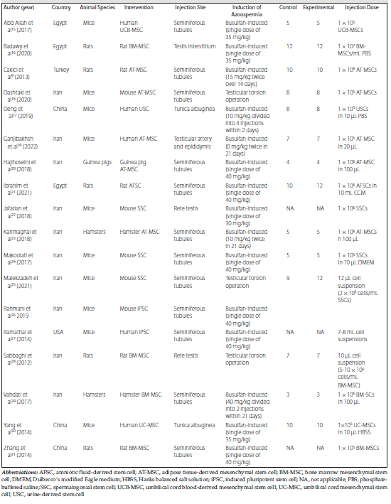
Table 1. Characteristics of 18 Selected Studies
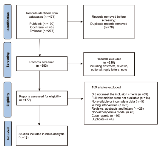
Figure 1. Flow Diagram Summarizing the Study Selection Process
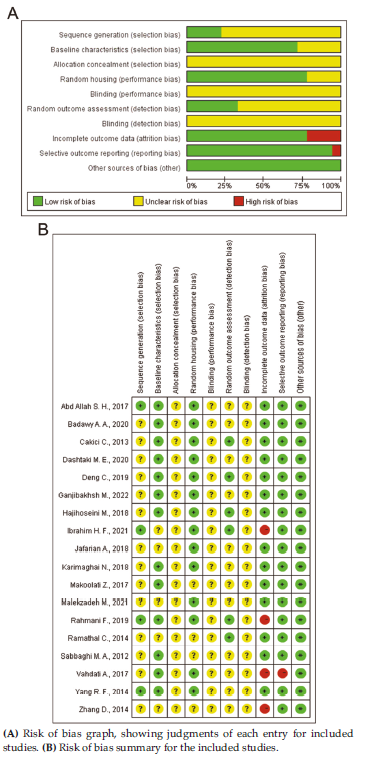
Figure 2. Risk of Bias Assessed With SYRCLE’s Risk of Bias Tool
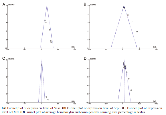
Figure 3. Funnel Plots of Comparing Results After Stem Cell Transplant on Azoospermia
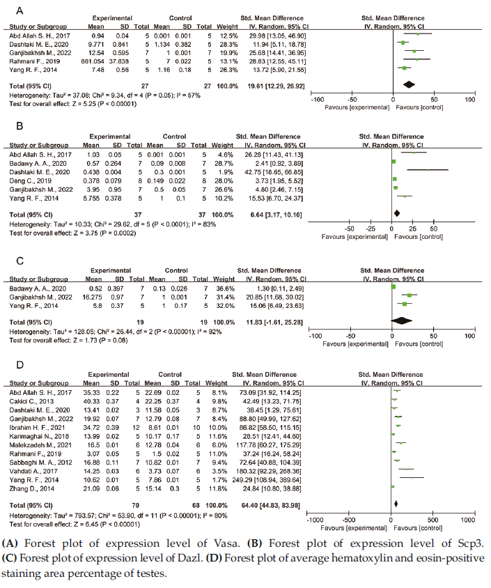
Figure 4. Forest Plots of Different Outcomes Related to Azoospermia Healing
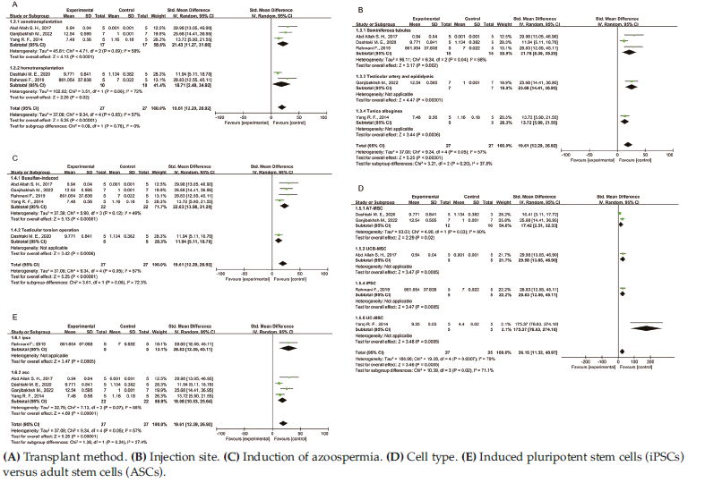
Figure 5. Forest Plots of Subgroup Analysis of Vasa Expression Level Based on Different Characteristics
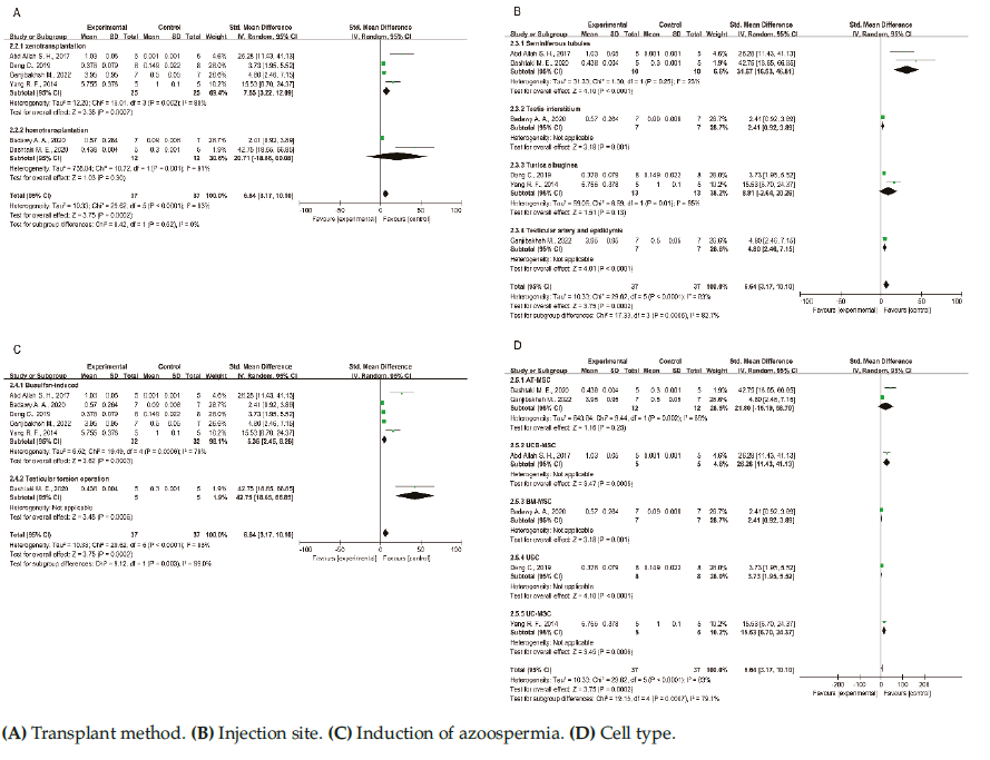
Figure 6. Forest Plots of Subgroup Analysis of Scp3 Expression Level Based on Different Characteristics
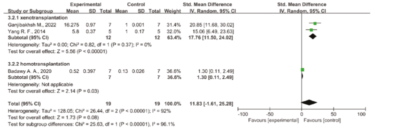
Figure 7. Forest Plot of Subgroup Analysis of Dazl Expression Level Based on Different Transplant Methods
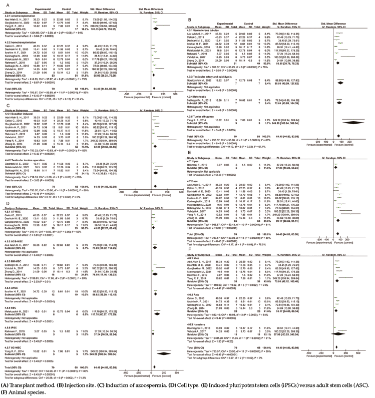
Figure 8. Forest Plots of Subgroup Analysis of Average Hematoxylin and Eosin-Positive Staining Area Percentage of Testes Based on Different Characteristics