Volume: 19 Issue: 6 June 2021
FULL TEXT
Cryptococcal infection has been documented in 2.8% of solid-organ transplant recipients, with the median time to disease onset being 21 months. Renal transplant recipients account for the majority of cases. Most patients present with central nervous system or disseminated disease, with only a minority having cutaneous manifestations. We present the case of a 47-year-old female renal transplant recipient who presented with refractory acute cellulitis 7 months after transplant. She had received thymoglobulin induction and was on a maintenance immunosuppressive regimen of tacrolimus, mycophenolic acid, and prednisone (5 mg/d). She did not respond to broad-spectrum antibacterial therapy for presumed bacterial cellulitis. Skin and soft tissue biopsies subsequently showed the presence of yeast; Cryptococcus neoformans was recovered in culture. Blood cultures, chest radiography, and cerebrospinal fluid sampling were negative, which resulted in a diagnosis of multifocal soft tissue cryptococcosis, a form of disseminated disease. Serum cryptococcal antigen testing was strongly positive (≥ 1:2560). The patient’s immunosuppression was reduced, and she received treatment with liposomal-amphotericin B and flucytosine for 2 weeks, which resulted in symptomatic improvement. This was followed by 1 year of consolidation and subsequent maintenance therapy with fluconazole. This case should increase awareness of the broader differential diagnosis of soft tissue infection in immunocompromised patients. Her case mimicked bacterial cellulitis, which delayed administration of effective therapy. Although our patient was initially diagnosed via biopsy, early clinical suspicion and serum cryptococcal antigen testing can lead to the correct diagnosis more rapidly. As transplant patients return to their community providers, heightened vigilance for unusual infections and presentations is warranted. The possibility of a cryptococcal cause for acute soft tissue infection should be considered, even in the absence of pulmonary or central nervous system involvement.
Key words : Cellulitis, Cryptococcus neoformans, Kidney transplant
Introduction
Cryptococcal infection is uncommon in solid-organ transplant recipients; most patients present with central nervous system (CNS) infection or disseminated disease. A minority of patients with disseminated disease develop cutaneous lesions. Our case highlights the importance of maintaining a high index of suspicion for atypical pathogens in immunocompromised patients who are not responding to empiric antibacterial therapy and highlights the need for a low threshold for biopsy to guide diagnosis and therapy.
Case Report
A 47-year-old female from a small city in southern Minnesota presented with right lower extremity pain, erythema, and swelling. She denied any current substance use, reported smoking cigarettes in the past (up to 1 pack per day), and denied significant environmental exposures; she had previously worked as a cashier.
The patient had a history of end-stage renal disease of unknown etiology and had received a second (3/6 antigen mismatched deceased donor) renal transplant 7 months earlier. Her first transplant, which occurred 11 years before her second transplant, was a living-related renal transplant that failed due to humoral and cellular rejection after 3 years. For her second transplant, she received thymoglobulin induction with a maintenance immunosuppressive regimen of tacrolimus, mycophenolic acid, and prednisone (5 mg/d). Four months after transplant, renal biopsy showed grade 1a acute cellular rejection and focal segmental glomerulosclerosis, which was treated with steroids, intravenous immunoglobulin, rituximab, plasmapheresis, and augmentation of tacrolimus dosing. On initial presentation, she reported being afebrile with 1 week of progressively worsening erythema, edema, and pain of the right lower extremity, which had not responded to oral cephalexin. She denied any history of trauma to the area.
Examination revealed edema and faint erythema of bilateral distal lower extremities, with a darker, more confluent 4 × 6-cm area of erythema and tenderness on the right calf. No crepitus was discovered. The patient was afebrile, and vital signs were stable. Laboratory testing revealed no leukocytosis; however, erythrocyte sedimentation rate and C-reactive protein were elevated to 63 mm/h and 110 mg/L, respectively. Because of severe patient discomfort and concern for possible necrotizing infection, a magnetic resonance imaging scan of the right lower extremity was performed. This revealed extensive edema along the deep fascial planes in the posterior compartment and within the gastrocnemius. She was diagnosed clinically with cellulitis and myositis. Because she had improved on vancomycin and meropenem therapy, bacterial infection was thought to be most likely, and she was dismissed from the hospital to complete a 2-week course of vancomycin and ertapenem (switched from meropenem for convenience as an outpatient). Before she completed therapy, she was seen in the outpatient clinic. She had remained afebrile, but reported worsening pain and increased erythema of the calf (Figure 1).
The patient was readmitted; laboratory tests showed no leukocytosis, erythrocyte sedimentation rate was 44 mm/h, and C-reactive protein level was 26.7 mg/L. A repeat magnetic resonance imaging scan was unchanged. A 5-mm punch biopsy of the right calf was performed, with samples sent for Gomori methenamine silver and mucicarmine stains (Figure 2) and fungal culture (Figure 3).
Due to the presence of yeast in tissue, histoplasmosis and cryptococcosis were considered. Histoplasma urine antigen was negative (0.0 ng/mL), as was serologic testing (complement fixation and immunodiffusion). However, serum cryptococcal antigen testing was strongly positive, with a titer of ≥ 1:2560. A tissue culture grew Cryptococcus neoformans, characterized by smooth mucoid colonies on inhibitory mold agar plates. Lumbar puncture and chest radiography showed no evidence of CNS or pulmonary disease, and routine blood cultures, as well as fungal blood cultures, were negative. On hospital day 4, a left inguinal soft tissue mass was noted, which was biopsied and showed numerous organisms consistent with Cryptococcus species, leading to the diagnosis of multifocal soft tissue cryptococcosis.
The patient’s immunosuppression was reduced, and she received treatment with liposomal-amphotericin B and flucytosine for 2 weeks, which resulted in symptomatic improvement. This was followed by consolidation and maintenance therapy with fluconazole.
Discussion
Cryptococcus neoformans is a ubiquitous encapsulated yeast found throughout the world. Human infection typically occurs after inhalation of the organism, which has been isolated from various sources in nature, notably including avian (eg, pigeon) excreta. Factors resulting in compromise of the cell-mediated immune system, such as a low CD4 count (with human immunodeficiency virus [HIV] or after transplant) or with steroid therapy, are important risk factors leading to disease development.1,2 Cryptococcal infection has been documented in 2.8% of solid-organ transplant recipients, with a median time to disease onset of 21 months.2,3 Our patient presented 7 months after her most recent renal transplant.
In a case study of 306 HIV-negative cryptococcosis patients in the United States, 18% were solid-organ transplant recipients, with 40% of these having renal transplants.1 Similarly, 50% of a cutaneous cryptococcosis cohort reported by the Cryptococcal Collaborative Transplant Study Group were renal transplant recipients.1,4 This number was higher in a more recent study from Australia, with 95% of their cryptococcal disease cohort with solid-organ transplants having had renal transplants. Only 17% of their transplant cohort had cutaneous manifestations, with 61% having pulmonary and 57% having CNS involvement.5
We searched Embase, Medline/PubMed, and Scopus for English language full-text primary reports of renal transplant recipients presenting with cutaneous manifestations of cryptococcal disease. In total, 37 cases with reports of time from transplant were identified. Most patients were male (81%), with median age at presentation of 51 years (interquartile range [IQR], 44.5-61.6 y). The median time to presentation with cellulitis was 55 months (IQR, 21.5-102 mo). Most patients presented with lower extremity involvement (28/37 patients; 75.7%), with bilateral involvement in 7 patients (18.9%) and unilateral involvement in 19 patients (51.4%). Eight patients (21.6%) had CNS disease, and 7 patients (18.9%) had pulmonary involvement.6-35
It has been hypothesized that cryptococcal disease results from either reactivation of quiescent latent infection or is due to primary infection.36 Given our patient’s presentation, it was postulated that her disease was likely due to reactivation. No recent exposures or cutaneous trauma was reported, and her immunosuppression had recently been augmented. Most organ transplant patients present with CNS or disseminated disease.3 However, only 15% of patients with disseminated disease develop cutaneous lesions, including papules, pustules, ulcers, cellulitis, plaques, granulomas, or abscesses.3,4 Our case was unusual as the patient only had multifocal soft tissue involvement, without evidence of CNS or pulmonary disease. Calcineurin inhibitors inhibit fungal calcineurin at 37°C but not at 24°C, which could predispose to cutaneous involvement.37 Alternatively, cutaneous lesions can arise from primary inoculation. Overall mortality ranges from 15% to 42%.1,2,4
Management recommendations vary based on disease severity and have been extrapolated from experiences in the HIV population.3,38 Immunosuppression should be reduced, when possible; preferentially steroids should be reduced first, as calcineurin inhibitors have direct anticryptococcal activity. This should be done cautiously to reduce the risk of immune reconstitution inflammatory syndrome and organ dysfunction. Antifungal therapy should consist of induction, consolidation, and maintenance phases, which can lead to resolution of disease and a low risk of relapse. Induction therapy for disseminated disease should include liposomal-amphotericin B and flucytosine for 2 weeks, followed by consolidation with fluconazole.
Our case highlights the importance of maintaining a high index of suspicion for atypical pathogens in immunocompromised patients who do not respond to empiric antibacterial therapy and highlights the need for a low threshold to consider tissue biopsy for diagnosis and to guide appropriate therapy.
References:
- Pappas PG, Perfect JR, Cloud GA, et al. Cryptococcosis in human immunodeficiency virus–negative patients in the era of effective azole therapy. Clin Infect Dis. 2001;33(5):690-699.
CrossRef - PubMed - Husain S, Wagener MM, Singh N. Cryptococcus neoformans infection in organ transplant recipients: variables influencing clinical characteristics and outcome. Emerg Infect Dis. 2001;7(3):375-381.
CrossRef - PubMed - Perfect JR, Dismukes WE, Dromer F, et al. Clinical practice guidelines for the management of cryptococcal disease: 2010 update by the Infectious Diseases Society of America. Clin Infect Dis. 2010;50(3):291-322.
CrossRef - PubMed - Sun HY, Alexander BD, Lortholary O, et al. Cutaneous cryptococcosis in solid organ transplant recipients. Med Mycol. 2010;48(6):785-791.
CrossRef - PubMed - Gassiep I, McDougall D, Douglas J, Francis R, Playford EG. Cryptococcal infections in solid organ transplant recipients over a 15-year period at a state transplant center. Transpl Infect Dis. 2017;19(1).
CrossRef - PubMed - Anderson DJ, Schmidt C, Goodman J, Pomeroy C. Cryptococcal disease presenting as cellulitis. Clin Infect Dis. 1992;14(3):666-672.
CrossRef - PubMed - Baer S, Baddley JW, Gnann JW, Pappas PG. Cryptococcal disease presenting as necrotizing cellulitis in transplant recipients: Case report. Transpl Infect Dis. 2009;11(4):353-358.
CrossRef - PubMed - Basaran O, Emiroglu R, Arikan U, Karakayali H, Haberal M. Cryptococcal necrotizing fasciitis with multiple sites of involvement in the lower extremities. Dermatol Surg. 2003;29(11):1158-1160.
PubMed - Bhowmik D, Dinda AK, Xess I, et al. Fungal panniculitis in renal transplant recipients. Transpl Infect Dis. 2008;10(4):286-289.
CrossRef - PubMed - Blocher KS, Weeks JA, Noble RC. Cutaneous cryptococcal infection presenting as vulvar lesion. Genitourin Med. 1987;63(5):341-343.
CrossRef - PubMed - Carlson KC, Mehlmauer M, Evans S, Chandrasoma P. Cryptococcal cellulitis in renal transplant recipients. J Am Acad Dermatol. 1987;17(3):469-472.
CrossRef - PubMed - Chakradeo K, Paul Chia YY, Liu C, Mudge DW, De Silva J. Disseminated cryptococcosis presenting initially as lower limb cellulitis in a renal transplant recipient - A case report. BMC Nephrol. 2018;19(1):18.
CrossRef - PubMed - Ding YG, Fang H. Edematous erythema, subcutaneous plaques, and severe pain in the lower extremities in an immunocompromised patient. JAMA. 2013;309(15):1632-1633.
CrossRef - PubMed - Gloster HM, Swerlick RA, Solomon AR. Cryptococcal cellulitis in a diabetic, kidney-transplant patient. J Am Acad Dermatol. 1994;30(6):1025-1026.
CrossRef - PubMed - Granier F, Kanitakis J, Hermier C, Zhu YY, Thivolet J. Localized cutaneous cryptococcosis successfully treated with ketoconazole. J Am Acad Dermatol. 1987;16(1 Pt 2):243-249.
CrossRef - PubMed - Gupta RK, Khan ZU, Nampoory MR, Mikhail MM, Johny KV. Cutaneous cryptococcosis in a diabetic renal transplant recipient. J Med Microbiol. 2004;53(5):445-449.
CrossRef - PubMed - Hall JC, Brewer JH, Crouch TT, Watson KR. Cryptococcal cellulitis with multiple sites of involvement. J Am Acad Dermatol. 1987;17(2):329-332.
CrossRef - PubMed - Hayashida MZ, Seque CA, Pasin VP, Enokihara MMSES, Porro AM. Disseminated cryptococcosis with skin lesions: Report of a case series. An Bras Dermatol. 2017;92(5 Suppl 1):69-72.
CrossRef - PubMed - Iacobellis FW, Jacobs MI, Cohen RP. Primary cutaneous cryptococcosis. Arch Dermatol. 1979;115(8):984-985.
CrossRef - PubMed - Jennings HS, Bradsher RW, Mcgee ZA, Johnson HK, Alford RH. Acute cryptococcal cellulitis in renal-transplant recipients. South Med J. 1981;74(9):1150-1153.
CrossRef - PubMed - Kimmatkar P, Raveendran N, Tanwar R, Leon D'souza AV, Beniwal P, Malhotra V. An unusual cause of lower limb ulcer in renal allograft recipient. Indian J Nephrol. 2016;26(4):309-310.
CrossRef - PubMed - Kothiwala SK, Prajapat M, Kuldeep CM, Jindal A. Cryptococcal panniculitis in a renal transplant recipient: Case report and review of literature. J Dermatol Case Rep. 2015;9(3):76-80.
CrossRef - PubMed - Kulkarni A, Sinha M, Anandh U. Primary cutaneous cryptococcosis due to Cryptococcous laurentii in a renal transplant recipient. Saudi J Kidney Dis Transpl. 2012;23(1):102-105.
PubMed - Marques S, Carmo R, Ferreira I, Bustorff M, Sampaio S, Pestana M. Cryptococcosis in renal transplant recipients: a single-center experience. Transplant Proc. 2016;48(7):2289-2293.
CrossRef - PubMed - Mayers DL, Martone WJ, Mandell GL. Cutaneous cryptococcosis mimicking gram-positive cellulitis in a renal transplant patient. South Med J. 1981;74(8):1032-1033.
CrossRef - PubMed - Moe K, Lotsikas-Baggili AJ, Kupiec-Banasikowska A, Kauffman CL. The cutaneous predilection of disseminated cryptococcal infection in organ transplant recipients. Arch Dermatol. 2005;141(7):913-914.
CrossRef - PubMed - Nampoory MR, Khan ZU, Johny KV, et al. Invasive fungal infections in renal transplant recipients. J Infect. 1996;33(2):95-101.
CrossRef - PubMed - Nelson GE, Neofytos D, Fischer M, Durand CM. A 70-year-old kidney transplant recipient presenting with persistent leg cellulitis. Clin Infect Dis. 2014;59(5):688, 745-744.
CrossRef - PubMed - Orsini J, Nowakowski J, Delaney V, Sakoulas G, Wormser GP. Cryptococcal infection presenting as cellulitis in a renal transplant recipient. Transpl Infect Dis. 2009;11(1):68-71.
CrossRef - PubMed - Pasqualotto AC, Bittar AE, de Quadros M, Severo LC. Cryptococcal cellulitis in a renal transplant patient. Nephrol Dial Transpl. 2005;20(9):2007-2008.
CrossRef - PubMed - Perfect JR, Durack DT, Gallis HA. Cryptococcemia. Medicine (Baltimore). 1983;62(2):98-109.
CrossRef - PubMed - Rerolle JP, Szelag JC, Diaconita M, Paraf F, Aldigier JC, Le Meur Y. Intracranial granuloma and skull osteolysis: complication of a primary cutaneous cryptococcosis in a kidney transplant recipient. Am J Kidney Dis. 2005;46(6):e113-e117.
CrossRef - PubMed - Shrader SK, Watts JC, Dancik JA, Band JD. Disseminated cryptococcosis presenting as cellulitis with necrotizing vasculitis. J Clin Microbiol. 1986;24(5):860-862.
PubMed - Vo-Cong MT, Persu A, Marot L, Goffin E. The transplanted patient who suffered from excruciating lower limb pain and developed nodular skin lesions. Nephrol Dial Transplant. 1999;14(12):2947-2949.
CrossRef - PubMed - Zorman JV, Zupanc TL, Parac Z, Cucek I. Primary cutaneous cryptococcosis in a renal transplant recipient: case report. Mycoses. 2010;53(6):535-537.
CrossRef - PubMed - Saha DC, Goldman DL, Shao X, et al. Serologic evidence for reactivation of cryptococcosis in solid-organ transplant recipients. Clin Vaccine Immunol. 2007;14(12):1550-1554.
CrossRef - PubMed - Cruz MC, Del Poeta M, Wang P, et al. Immunosuppressive and nonimmunosuppressive cyclosporine analogs are toxic to the opportunistic fungal pathogen cryptococcus neoformans via cyclophilin-dependent inhibition of calcineurin. Antimicrob Agents Chemother. 2000;44(1):143-149.
CrossRef - PubMed - Singh N, Lortholary O, Alexander BD, et al. Antifungal management practices and evolution of infection in organ transplant recipients with Cryptococcus neoformans infection. Transplantation. 2005;80(8):1033-1039.
CrossRef - PubMed

Volume : 19
Issue : 6
Pages : 609 - 612
DOI : 10.6002/ect.2017.0292
From the Division of Infectious Diseases, Department of Medicine, Mayo Clinic,
Rochester Minnesota 55905, USA
Acknowledgements: The authors have no sources of funding for this study and have
no conflicts of interest to declare. The authors thank Alina Bridges, MD, for
providing the fungal stain images for this report.
Corresponding author: Saira Ajmal, Mayo Clinic, Division of Infectious Diseases,
Department of Medicine, 200 First Street SW, Rochester, MN 55905, USA
Phone: +1 507 284 3309
E-mail: ajmal.saira@mayo.edu
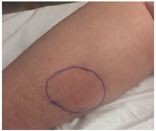
Figure 1. Right Calf Erythema
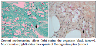
Figure 2. Skin Biopsy
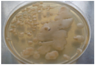
Figure 3. Fungal Culture (Inhibitory Mold Agar Plate) of Skin Biopsy