Volume: 19 Issue: 6 June 2021
FULL TEXT
Objectives: The Achilles tendon, which is composed of tendinous parts of gastrocnemius and soleus muscles, is the strongest and the largest tendon in the human body. Chronic renal disease can lead to reduced physical activity and exercise capacity. Spontaneous rupture of the Achilles tendon can occur in patients with chronic renal failure, with recurrent microtraumas, hypoxia, and chronic acidosis as predisposing factors. Here, we assessed and compared the elastographic findings in the Achilles tendon using acoustic radiation force impulse elastography in patients on chronic hemodialysis, in renal transplant patients, and in healthy volunteers.
Materials and Methods: Our study included 25 patients on chronic hemodialysis, 25 renal transplant patients, and 25 healthy individuals (control group). The thickness and shear wave velocity of the Achilles tendons were measured bilaterally by ultrasonography and acoustic radiation force impulse elastography.
Results: The mean shear wave velocity was 3.67 m/s in the right and 3.64 m/s in the left Achilles tendon in the hemodialysis group. In the renal transplant group, the mean shear wave velocity was 4.29 and 4.25 m/s for the right and left Achilles tendon, respectively. In the control group, the mean shear wave velocity was 6.68 and 6.59 m/s, respectively for the right and left Achilles tendon. A statistically significant difference in shear wave velocities was shown among the groups (P < .05).
Conclusions: Achilles tendons in patients with chronic renal failure and on hemodialysis were softer than in renal transplant patients and softer than in the control group. Chronic tendinopathy causes softening of the tendon. In the renal transplant group, stiffness of the Achilles tendon was increased versus the hemodialysis group but still softer than the control group, which could be explained as a positive clinical effect of renal transplant. Acoustic radiation force impulse elastography is an objective, easy, and noninvasive method to assess Achilles tendinopathy.
Key words : Chronic renal failure, Dialysis, Kidney transplantation, Tendinopathy
Introduction
The Achilles tendon, which is composed of the tendinous parts of the gastrocnemius and soleus muscles, is the strongest, largest, and thickest tendon in the human body.1 Achilles tendon injury is frequent both in athletes and in the general population due to sports-related trauma and overuse.2 A spontaneous Achilles tendon rupture, which is a rare complication, has been reported in patients with end-stage renal failure. Most of these patients have additional factors, such as hyperparathyroidism, diabetes mellitus, and use of fluoroquinolone antibiotics or corticosteroids.3 Some authors argue that uremic toxins cause tendon degeneration and spontaneous rupture.4,5 Metabolic acidosis, hypoxia, and hyperparathyroidism secondary to chronic renal failure may also lead to tendon degeneration.6 To prevent Achilles tendon rupture and chronification of tendon injury, early diagnosis and effective treatment of tendinopathy are important. Magnetic resonance imaging with excellent soft tissue contrast can be used to evaluate the Achilles tendon, but this method is expensive and time consuming. Conventional ultrasonography, which is not expensive or invasive, can be easily performed as an alternative imaging modality to evaluate tendon disorders. It has a high diagnostic accuracy for tendon ruptures, but its value in tendinopathy is still controversial.2,7,8
Ophir and associates9 first described sonoelastography, which is based on the deformation response of the tissue with compression and release by the transducer. Sonoelastography enables a prediction of tissue elasticity by measuring the tissue deformation parallel to the direction of the external force. Each tissue or lesion behaves differently due to its molecular structure. Hard tissues or lesions become less deformed, whereas soft tissues or lesions become more deformed. Qualitative information, presented as color maps, or semi-quantitative information, presented as size ratios or elasticity ratios, can be obtained.
Most studies regarding elastographic findings in the Achilles tendon have used the sonoelastography technique.1,6,10-12 Acoustic radiation force impulse (ARFI) elastography, another newly developed elastography method, uses sound waves to quantify tissue elasticity, thus allowing tissues or focal lesions to be evaluated without external compression. This technique measures the shear wave velocity (SWV) of the target region inside the region of interest (ROI) without external compression and can estimate minor differences inside the ROI. The target tissue is mechanically stimulated by short-duration acoustic pulses to produce small (1-10 mm) localized tissue displacements. The tissue displacement results in shear wave dissemination, which is tracked with a probe, and is measured as the SWV (in m/s).13 Acoustic radiation force impulse elastography technology also includes virtual touch tissue quantification, a new technique for the quantification of tissue elasticity by calculating the SWV.14,15 Fu and associates2 assessed the elastic properties of normal Achilles tendons with virtual touch tissue quantification and evaluated whether these properties changed with age and sex. Ruan and colleagues16 used ARFI to evaluate elasticity of healthy Achilles tendons and found that SWV increases with age.
In their study, Chen and associates17 used ARFI elastography to show significant softening of ruptured Achilles tendon.
In this study, we assessed the elastographic findings of the Achilles tendon in patients on chronic hemodialysis, in renal transplant patients, and in healthy volunteers using ARFI elastography. Our aim was to compare the ARFI elastography findings in the Achilles tendon among the 3 groups.
Materials and Methods
Study population
Our study included analysis of 150 Achilles tendons (50 Achilles tendons from 25
patients on chronic hemodialysis, 50 Achilles tendons from 25 renal transplant
recipients, and 50 Achilles tendons of 25 healthy volunteers [control group]).
All participants were asymptomatic, and none had a history of tendon injury.
Patients in the hemodialysis group were on hemodialysis 3 times per week for at
least 5 years regularly, and none had secondary hyperparathyroidism. Renal
transplant recipients had history of at least 5 years of hemodialysis and were
at a minimum of 5 years posttransplant. Their serum creatinine levels (0.8-1.2
mg/dL) were within normal levels. None of the healthy volunteers had a history
of systemic disease, surgery, or chronic drug use that could be associated with
tendinopathy. This study was performed in accordance with the ethical guidelines
of the declaration of Helsinki and was approved by the local ethical committee.
Informed, written consent was obtained from all participants.
Imaging
All participants underwent B-mode ultrasonography and ARFI elastography of the
right and left Achilles tendons. Sonographic examinations were performed with
the Siemens ACUSON S3000 diagnostic ultrasonographic system (Siemens Medical
Solutions, Erlangen, Germany). A Siemens 9L4 linear probe with a frequency range
of 4 to 9 MHz was used for both the conventional ultrasonographic and
elastographic examinations. All sonographic examinations were performed by a
radiologist with 10 years of experience in ultrasonography and 5 years of
experience in elastography. Each Achilles tendon was examined in axial and
longitudinal planes in the prone position with the foot hanging over the
examination bed in a relaxed position.17 The middle portion of the Achilles
tendon, which is nearly 2 to 6 cm proximal to the calcaneus insertion, was
chosen for both ultrasonographic and ARFI elastographic examinations. It is the
most affected part of the tendon because of the vascular supply and
biomechanical factors.2
The thickness of the tendon was determined by measuring the anteroposterior diameter from the middle third portion (2 to 6 cm above the insertion of the calcaneus) of the tendon. The ARFI elastography was performed by switching to the elastographic mode. Tendons were examined in the longitudinal plane, and the probe was held perpendicular to the tendon to avoid anisotropy.18 During elastography, the probe was placed in contact with the skin without pressure; because of the superficial localization of the Achilles tendon, more medical ultrasonic coupling gel was used. A fixed size of 5 × 6-mm ROI was placed in the middle third portion of Achilles tendon; after image stabilization, the probe was held steady for 3 to 5 seconds for framing. The image was then recorded and saved to the hard drive, and SWV values were calculated in meters per second and viewed on the screen.2,16 Four measurements were made for each tendon, and the mean value was calculated and used for statistical analyses. The SWV ranged from 0 to 9 m/s (Figures 1 to 3).
Statistical analyses
Statistical analyses were performed with SPSS software (SPSS: An IBM Company,
version 20, IBM Corporation, Armonk, NY, USA). P < .05 was considered
statistically significant. Analysis of variance was used to analyze the
thickness and SWV of Achilles tendons in the chronic hemodialysis, renal
transplant, and control groups. Subgroup comparisons were analyzed by
independent t test.
Results
The hemodialysis group consisted of 14 men and 11 women with a median age of 37 years (range, 22-52 y). The median duration of hemodialysis was 12 years (range, 5-20 y). The mean Achilles tendon thickness was 4.7 mm (range, 4-5.5 mm) for the right ankle and 4.7 mm (range, 3.6-5.5 mm) for the left ankle.
The renal transplant group consisted of 13 men and 12 women with median age of 38 years (range, 20-50 y). The median duration of hemodialysis was 8 years (range, 5-16 y), and the average time passed after transplant was 8 years (range, 5-20 y). The mean Achilles tendon thickness was 4.5 mm (range, 4.3-5.2 mm) for the right ankle and 4.6 mm (range, 4-5.3 mm) for the left ankle.
The control group consisted of 13 men and 12 women with median age of 38 years (range, 20-53 y). The mean Achilles tendon thickness was 6 mm (range, 5-6.5 mm) for the right ankle and 6 mm (range, 5-6.6 mm) for the left ankle.
Mean SWV results for the right and left Achilles tendons in the 3 groups are presented in Table 1. There were statistically significant differences among the SWV results for Achilles tendons of patients on chronic hemodialysis, in renal transplant recipients, and in healthy control participants. There was a statistically significant thinning shown in the right and left Achilles tendons of the hemodialysis and renal transplant groups (P < .05). No significant differences were shown between the right and left Achilles tendon SWV results in all 3 groups (P > .05). In addition, no significant differences were shown between men and women regarding SWV results within the 3 groups (P > .05).
Discussion
Acoustic radiation force impulse elastography has been widely used to assess both nodular lesions, such as breast masses, and thyroid nodules and diffuse diseases such as liver fibrosis and autoimmune thyroiditis.19-21 This technique has also been used to evaluate tendons, especially the Achilles tendon, which is the largest and thickest tendon in the human body. To the best of our knowledge, this is the first study that evaluated the elastic properties of the Achilles tendon in patients on chronic hemodialysis and in renal transplant recipients. The major findings of our study were that Achilles tendons in patients with chronic renal failure and in renal transplant recipients were softer than those in the control group (P < .0001). The stiffness of the Achilles tendons in renal transplant recipients was higher than in patients on chronic hemodialysis (P = .003) but still softer than in the control group (P < .0001).
Several studies have verified that an abnormal Achilles tendon becomes soft. In a study from Balaban and associates elastographic findings of Achilles tendons in asymptomatic volleyball players showed that they had softening and thickening of Achilles tendons. Turan and colleagues11 evaluated the Achilles tendons of patients with ankylosing spondylitis and reported that tendons became softer in these patients. De Zordo and associates22 found that the Achilles tendon was hard in asymptomatic healthy volunteers. With tendinopathy, collagen defects can cause softening of the tendon, which may also lead to ruptures.23
Chronic renal failure is a notable health problem that can cause serious complications. Physical inactivity and decreased exercise capacity are both related to chronic renal failure, and these can increase morbidity and mortality. Hypoxia, metabolic acidosis, vascular changes, and calcification may cause degeneration of the Achilles tendon.6,24 Spontaneous Achilles tendon rupture is a rare but an important complication.3,4 Although the mechanisms of Achilles tendon rupture in patients with chronic renal failure is not well known, use of corticosteroids, hyperparathyroidism, use of fluoroquinolone antibiotics, and amyloidosis have been reported as predisposing factors in the literature.3 Some authors have stated that uremic toxins can cause tendon degeneration and spontaneous rupture.4,5 In addition, a relation between length of time on hemodialysis and tendon rupture has been reported.25,26
Teber and associates6 studied 53 patients on chronic hemodialysis and 25 healthy control individuals with ultrasonography and sonoelastography and found softening and thinning of quadriceps tendons in the patient group, although no quadriceps tendon ruptures were observed. The group used sonoelastography, which is an operator-dependent method, and evaluated the quadriceps tendons with color mapping. In our study, we evaluated Achilles tendon with ARFI elastography, which is an operator-independent and quantitative elastography method. In our study, we found softening and thinning of Achilles tendon in both the hemodialysis group and the renal transplant group, as similarly reported in the previous study. We also did not find Achilles tendon rupture, which could be explained by regular hemodialysis and close follow-up of patients regarding hyperparathyroidism and acidosis. In our study, elastographic findings in renal transplant patients were different from the previous study. The Achilles tendons of renal transplant patients were harder than shown in patients with chronic renal failure but still softer than shown in the control group. This could be explained by the increased mobility and exercise capacity of patients after renal transplant. In addition, excretion of uremic toxins from the body by urine could be another reason.
Our study has several limitations. First, our sample size was small. Second, we did not perform a histological analysis. Because all of our patients were asymptomatic, an invasive procedure like a biopsy could not be performed. Third, only the middle part of the Achilles tendon was evaluated. However, most abnormalities have been reported in the middle third of the tendon.2,19 The middle third portion of the tendon has poor vascular supply.27
In conclusion, we found thinning and softening of the Achilles tendon in patients who were on chronic hemodialysis. Chronic tendinopathy causes softening and weakening of the tendon. In the renal transplant group, stiffness of the Achilles tendon was increased versus the hemodialysis group but still softer than the control group, which could be explained as a positive clinical effect of renal transplant. An ARFI elastography is an inexpensive, objective, noninvasive, and easy method that can be used in the assessment of Achilles tendinopathy.
References:
- Balaban M, Idilman IS, Ipek A, Ikiz SS, Bektaser B, Gumus M. Elastographic findings of Achilles tendons in asymptomatic professional male volleyball players. J Ultrasound Med. 2016;35(12):2623-2628.
CrossRef - PubMed - Fu S, Cui L, He X, Sun Y. Elastic characteristics of the normal Achilles tendon assessed by virtual touch imaging quantification shear wave elastography. J Ultrasound Med. 2016;35(9):1881-1887.
CrossRef - PubMed - Park JH, Kim SB, Shin HS, Jung GH, Jung YS, Rim H. Spontaneous and serial rupture of both Achilles tendons associated with secondary hyperparathyroidism in a patient receiving long-term hemodialysis. Int Urol Nephrol. 2013;45(2):587-590.
CrossRef - PubMed - Basic-Jukic N, Juric I, Racki S, Kes P. Spontaneous tendon ruptures in patients with end-stage renal disease. Kidney Blood Press Res. 2009;32(1):32-36.
CrossRef - PubMed - Hofmann GO, Weber T, Lob G. [Tendon rupture in chronic kidney insufficiency--"uremic tendonopathy"? A literature-supported documentation of 3 cases]. Chirurg. 1990;61(6):434-437.
PubMed - Teber MA, Ogur T, Bozkurt A, et al. Real-time sonoelastography of the quadriceps tendon in patients undergoing chronic hemodialysis. J Ultrasound Med. 2015;34(4):671-677.
CrossRef - PubMed - Paavola M, Paakkala T, Kannus P, Jarvinen M. Ultrasonography in the differential diagnosis of Achilles tendon injuries and related disorders. A comparison between pre-operative ultrasonography and surgical findings. Acta Radiol. 1998;39(6):612-619.
CrossRef - PubMed - Khan KM, Forster BB, Robinson J, et al. Are ultrasound and magnetic resonance imaging of value in assessment of Achilles tendon disorders? A two year prospective study. Br J Sports Med. 2003;37(2):149-153.
CrossRef - PubMed - Ophir J, Cespendes I, Ponnekonti H, Yazdi Y, Li X. Elastography: a quantitative method for imaging the elasticity of biological tissues. Ultrason Imaging. 1991;13(2):111-134.
CrossRef - PubMed - Agladioglu K, Akkaya N, Gungor HR, Akkaya S, Ok N, Ozcakar L. Effects of cigarette smoking on elastographic strain ratio measurements of patellar and Achilles tendons. J Ultrasound Med. 2016;35(11):2431-2438.
CrossRef - PubMed - Turan A, Tufan A, Mercan R, et al. Real-time sonoelastography of Achilles tendon in patients with ankylosing spondylitis. Skeletal Radiol. 2013;42(8):1113-1118.
CrossRef - PubMed - Turan A, Teber MA, Yakut ZI, Unlu HA, Hekimoglu B. Sonoelastographic assessment of the age-related changes of the Achilles tendon. Med Ultrason. 2015;17(1):58-61.
CrossRef - PubMed - Zhang H, Shi Q, Gu J, et al. Combined value of Virtual Touch tissue quantification and conventional sonographic features for differentiating benign and malignant thyroid nodules smaller than 10 mm. J Ultrasound Med. 2014;33(2):257-264.
CrossRef - PubMed - Golatta M, Schweitzer-Martin M, Harcos A, et al. Evaluation of virtual touch tissue imaging quantification, a new shear wave velocity imaging method, for breast lesion assessment by ultrasound. Biomed Res Int. 2014;2014:960262.
CrossRef - PubMed - Han R, Li F, Wang Y, Ying Z, Zhang Y. Virtual touch tissue quantification (VTQ) in the diagnosis of thyroid nodules with coexistent chronic autoimmune Hashimoto's thyroiditis: a preliminary study. Eur J Radiol. 2015;84(2):327-331.
CrossRef - PubMed - Ruan Z, Zhao B, Qi H, et al. Elasticity of healthy Achilles tendon decreases with the increase of age as determined by acoustic radiation force impulse imaging. Int J Clin Exp Med. 2015;8(1):1043-1050.
PubMed - Chen XM, Cui LG, He P, Shen WW, Qian YJ, Wang JR. Shear wave elastographic characterization of normal and torn Achilles tendons: a pilot study. J Ultrasound Med. 2013;32(3):449-455.
CrossRef - PubMed - Allison SJ, Nazarian LN. Musculoskeletal ultrasound: evaluation of ankle tendons and ligaments. AJR Am J Roentgenol. 2010;194(6):W514.
CrossRef - PubMed - Kohla MAS, Abbasy M, Abozeid M, et al. Assessment of liver fibrosis with acoustic radiation force impulse imaging versus liver histology in patients with chronic hepatitis C: a pilot study. Eur J Gastroenterol Hepatol. 2017;29(8):951-955.
CrossRef - PubMed - Friedrich-Rust M, Romenski O, Meyer G, et al. Acoustic radiation force impulse-imaging for the evaluation of the thyroid gland: a limited patient feasibility study. Ultrasonics. 2012;52(1):69-74.
CrossRef - PubMed - Magalhaes M, Belo-Oliveira P, Casalta-Lopes J, et al. Diagnostic value of ARFI (acoustic radiation force impulse) in differentiating benign from malignant breast lesions. Acad Radiol. 2017;24(1):45-52.
CrossRef - PubMed - De Zordo T, Fink C, Feuchtner GM, Smekal V, Reindl M, Klauser AS. Real-time sonoelastography findings in healthy Achilles tendons. AJR Am J Roentgenol. 2009;193(2):W134-138.
CrossRef - PubMed - Dillehay GL, Deschler T, Rogers LF, Neiman HL, Hendrix RW. The ultrasonographic characterization of tendons. Invest Radiol. 1984;19(4):338-341.
CrossRef - PubMed - Klauser AS, Faschingbauer R, Jaschke WR. Is sonoelastography of value in assessing tendons? Semin Musculoskelet Radiol. 2010;14(3):323-333.
CrossRef - PubMed - Ureten K, Ozturk MA, Ozbek M, Unverdi S. Spontaneous and simultaneous rupture of both Achilles tendons and pathological fracture of the femur neck in a patient receiving long-term hemodialysis. Int Urol Nephrol. 2008;40(4):1103-1106.
CrossRef - PubMed - Shiota E, Tsuchiya K, Yamaoka K, Kawano O. Spontaneous major tendon ruptures in patients receiving long-term hemodialysis. Clin Orthop Relat Res. 2002(394):236-242.
CrossRef - PubMed - Ahmed IM, Lagopoulos M, McConnell P, Soames RW, Sefton GK. Blood supply of the Achilles tendon. J Orthop Res. 1998;16(5):591-596.
CrossRef - PubMed

Volume : 19
Issue : 6
Pages : 534 - 538
DOI : 10.6002/ect.2018.0015
From the 1Department of Radiology, the 2Department of General Surgery, the
3Department of Family Physician, and the 4Department of Nephrology, Baskent
University Faculty of Medicine, Ankara, Turkey
Acknowledgements: The authors declare that they have no sources of funding for
this study and have no conflicts of interest to declare.
Corresponding author: Feride Kural Rahatli, Baskent University Faculty of
Medicine, Department of Radiology, 10 Sokak No. 45, 06490, Bahcelievler, Ankara,
Turkey
Phone: +90 312 2036868 1163
E-mail: drkural@hotmail.com
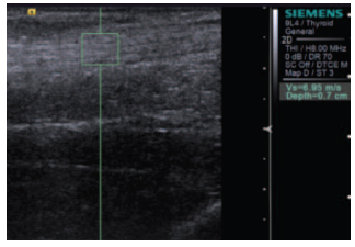
Figure 1. Acoustic Radiation Force Impulse Elastography of Achilles Tendon in a Healthy 35-Year-Old Woman (Shear Wave Velocity = 6.95 m/s)
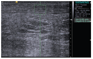
Figure 2. Acoustic Radiation Force Impulse Elastography of Achilles Tendon in a 33-Year-Old Woman on Chronic Hemodialysis for 9 Years (Shear WaveVelocity = 3.65 m/s)
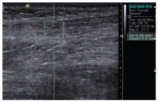
Figure 3. Acoustic Radiation Force Impulse Elastography of Achilles Tendon in a 30-Year-Old Female Renal Transplant Recipient at 6 Years Posttransplant (Shear Wave Velocity = 4.55 m/s)
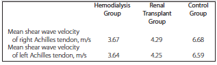
Table 1. Mean Shear Wave Velocity Results of Achilles Tendon in the Study Groups