Volume: 18 Issue: 1 February 2020
FULL TEXT
We describe a case of a 24-year-old female renal transplant recipient who, 10 years after receiving a deceased-donor kidney, presented with acute and massive increases in serum creatinine and proteinuria levels of 13 g over 24 hours. At a previous outpatient clinic visit, her baseline serum creatinine was noted to be 87 µmol/L; on admission, serum creatinine was 1377 µmol/L. Renal biopsy results were consistent with acute cellular rejection with severe interstitial lymphoplasmacytic infiltrates and edema with no evidence of glomerular pathology, including transplant glomerulopathy. The immunofluorescence test results were negative, and the ultrastructural features were consistent with podocytopathy with no immune deposits present. We believe that this is the first case of acute cellular rejection typified by severe interstitial lymphoplasmacytic infiltrates and edema with severe proteinuria secondary to minimal change disease (or podocytopathy).
Key words : Glomerular disorder, Kidney transplant, Podocytopathy, Proteinuria
Case Report
We report a case of a 24-year-old female patient who underwent deceased-donor renal transplant 10 years previously. The cause of her native kidney disease was unknown. The patient presented with sore throat, runny nose, fever, dry cough, shortness of breath, and fatigue for 1 week. There was no hematuria or change in urine amount or color. She was prescribed cefuroxime by her primary care physician for 5 days with no improvement, resulting in change to amoxicillin and clavulanate.
The patient was pale but alert and was not in distress. Physical examinations of the chest, cardiovascular system, and abdomen were normal. Her blood pressure was 149/114 mm Hg, her heart rate was 92 beats/min, her temperature was 36.9°C, and her peripheral oxygen saturation was 100%.
Serum creatinine level on admission was 1377 μmol/L, showing a large increase versus a baseline serum creatinine at an outpatient clinic visit 5 months previously of 87 μmol/L. Her estimated glomerular filtration rate dropped from 74 to 3 mL/min over that period.
With regard to blood test results, her hemoglobin was 98 g/L (baseline is 11.9 g/L), platelet count was 242 × 109/L, and white blood cell count was 7.3 × 10⁹/L. On admission, urinalysis revealed protein level of > 600 mg/L, red blood cell count of 1049 per high-power field, and white blood cell count of 859 per high-power field. Serum albumin level on admission was 31 g/L. During her prior outpatient clinic visit, there was no detectable protein or red blood cells in urine samples, and serum albumin level was 42 g/L.
The protein-to-creatine ratio was 11.24. Tests showed that hepatis B surface antigen, hepatitis B virus antibody, and hepatitis C virus tests were negative. Erythrocyte sedimentation rate was 77 mm/h and C-reactive protein was 28 mg/L (normal range < 8 mg/L). Antiglomerular basement antibody, perinuclear antineutrophil cytoplasmic antibody, cytoplasmic antineutrophil cytoplasmic antibody, antinuclear antibody, and anti-double-strand DNA tests were all negative. Tests also excluded any evidence of thrombotic microangiopathy or antiphospholipid syndrome. With the use of the Luminex HLA single antigen class I and II products (Luminex, Austin, TX, USA), no donor-specific antibodies were detected in the patient.
The patient was given methylprednisolone intravenously at 500 mg/day on the assumption that she may have rapidly progressive glomerulonephritis or antibiotic-induced acute interstitial nephritis or possibly rejection. She also received dialysis in preparation for renal biopsy, which was done 3 days after admission.
The biopsy showed heavy interstitial lymphoplasmacytic infiltrates with severe interstitial edema, tubulitis, severe tubular injury, and focal mild endarteritis (Figure 1). We also observed focal mild peritubular capillaritis. C4d staining in the cortical peritubular capillaries was negative. Light microscopy showed mild mesangial matrix expansion with no hypercellularity or mesangiolysis; the glomerular basement membranes were normal with no spike reaction, replication, or microaneurysmal dilatation. The glomeruli showed no significant changes and no proliferation, no glomerular basement thickening, and no glomerulitis. We also detected no inflammatory cell infiltrates, endocapillary hypercellularity, fibrinoid necrosis, crescents, or hyaline deposits. Polyomavirus immunostaining was negative.
Immunofluorescence showed 2 glomeruli (Figure 2). All antibodies (immunoglobulins A, G, and M, C3, C1q, C4, albumin, fibrinogen, and kappa and lambda light chains) were negative. Electron microscopy visualization of the ultrastructure of 1 glomerulus showed marked podocyte injury with vacuolization, microvillus transformation, and diffuse foot process effacement consistent with podocytopathy (Figure 3).
The patient was given methylprednisolone 500 mg/day for 3 days, and her oral prednisolone dose was increased to 50 mg/day. The patient showed no improved renal function with this regimen, and she was started on antithymocyte globulin on day 6 of admission at a dose of 1 mg/kg/day for 7 days.
A later outpatient follow-up confirmed that the patient has remained off dialysis but with very poor kidney function (serum creatinine level of 698 μmol/L, estimated glomerular filtration rate of 7 mL/min) and with persistence of her urinary proteinuria.
Discussion
Here, we described a case of severe acute cellular rejection in a transplanted kidney 10 years after transplant with the unusual association of severe proteinuria but in the absence of transplant glomerulopathy, immune deposits, or C4d positivity in biopsy tests. The only explanation we could find for the presence of this heavy proteinuria was the clear ultrastructural features consistent with podocytopathy.
Although we did not know the cause of chronic kidney disease in this patient, it is unlikely that it was recurrent focal segmental glomerulosclerosis (FSGS): we did not see a late presentation of proteinuria, there was no evidence of FSGS on light microscopy, and the ultrastructural features of podocytopathy were more consistent with minimal change disease versus FSGS or diffuse mesangial sclerosis. In any case, recurrent FSGS would not explain the severe acute rejection we found in our patient.1,2 We believe that this is the first case of acute cellular rejection leading to minimal change disease.
It is now well recognized that podocyte dysfunction can result from a variety of reasons, including genetic, idiopathic, or as reactive response to a variety of insults such as mechanical stress, medications, toxins, and viral infections. The stress in our patient that led to podocytopathy may be related to the severe form of acute rejection that we observed, which is typified by severe interstitial lymphoplasmacytic infiltrates and edema.
In their extensive paper describing the advances in the biology and genetics of podocytopathies, especially with regard to kidney transplant, Barisoni and associates mainly discussed FSGS recurrence and did not mention any case of rejection causing posttransplant podocytopathy.3 Similarly, in the review of podocytopathies from Wiggins,4 no examples of acute cellular rejection being associated with podocytopathy were provided. However, it was pointed out that any injury to the glomerulus, be it immune, toxic, infectious, or ischemic, can result in podocyte injury.4
Our case presented several unusual acute rejection histologic features: namely, the presence of severe interstitial edema and cellular infiltrates rich with plasma cells. Normally, severe edema is not seen in cellular rejection, and plasma cells normally account for < 5% of the infiltrating cells in acute cellular rejection.5
In another study that examined acute cellular rejection, the authors found that acute cellular rejection was characterized histologically by severe interstitial edema (defined as a cortical area in which at least 10 adjacent tubules were separated by intertubular spaces of more than 1 tubular diameter in width and filled with serous fluid devoid of mononuclear cells) and cellular infiltrates rich with plasma cells (defined as plasmocytes accounting for >10% of the graft infiltrating cells).6 In 826 biopsies, the investigators found that only 14 (1.7%) met those criteria. At biopsy, the patients in this group had a mean baseline serum creatinine level of 263 ± 21 μmol/L. This study also reported that rejection occurred in these patients after a median period of 187 days posttransplant, and all cases were steroid resistant.6 Graft survival was 40% at 1 year following rejection.6
Other studies estimated that the prevalence of this type of acute rejection histology was between 1.7% and 14%, and all such patients with this type of acute rejection histologic type have poor prognosis.7-10 Our patients also showed poor response to the antirejection therapy.
Presence of nephrotic-range proteinuria posttransplant is not common; when seen, it is usually associated with glomerular pathology (with recurrent or de novo glomerulonephritis or when transplant glomerulopathy is present or with the use of sirolimus). Rejection without evidence of transplant glomerulopathy is not associated with heavy proteinuria.
In a previous study of 613 recipients who had 1-year routine surveillance biopsy,11 only 3% had proteinuria of > 3 g over 24 hours. Patients found to have glomerular pathology at biopsy (56/613 patients) had a mean 24-hour urine output of 2.7 g. Patients found to have normal or other types of pathology on biopsy (acute rejection, fibrosis/atrophy, or other) had a mean 24-hour urine output of < 0.26 g (reported in 557/613 patients).11 In a review by Ponticelli and colleagues, the prevalence of proteinuria at 1 year after renal transplant was reported to range from 11% to 45%.12 However, only 8.7% to 9.1% of patients were found to have proteinuria of > 1 g over 24 hours.11,13
Conclusions
We believe that this is the first case of severe acute cellular rejection associated with podocytopathy leading to severe proteinuria. The acute rejection features in this case are unusual, showing severe interstitial lymphoplasmacytic infiltrates and severe interstitial edema. Previous studies of acute rejection have not mentioned podocytopathy with interstitial edema and cellular infiltrates rich with plasma cells.
References:
- Singh L, Singh G, Dinda AK. Understanding podocytopathy and its relevance to clinical nephrology. Indian J Nephrol. 2015;25(1):1-7.
CrossRef - PubMed - Barisoni L, Schnaper HW, Kopp JB. A proposed taxonomy for the podocytopathies: a reassessment of the primary nephrotic diseases. Clin J Am Soc Nephrol. 2007;2(3):529-542.
CrossRef - PubMed - Barisoni L, Schnaper HW, Kopp JB. Advances in the biology and genetics of the podocytopathies: implications for diagnosis and therapy. Arch Pathol Lab Med. 2009;133(2):201-216.
CrossRef - PubMed - Wiggins RC. The spectrum of podocytopathies: a unifying view of glomerular diseases. Kidney Int. 2007;71(12):1205-1214.
CrossRef - PubMed - Hancock WW, Thomson NM, Atkins RC. Composition of interstitial cellular infiltrate identified by monoclonal antibodies in renal biopsies of rejecting human renal allografts. Transplantation. 1983;35(5):458-463.
CrossRef - PubMed - Desvaux D, Le Gouvello S, Pastural M, et al. Acute renal allograft rejections with major interstitial oedema and plasma cell-rich infiltrates: high gamma-interferon expression and poor clinical outcome. Nephrol Dial Transplant. 2004;19(4):933-939.
CrossRef - PubMed - Meehan SM, Domer P, Josephson M, et al. The clinical and pathologic implications of plasmacytic infiltrates in percutaneous renal allograft biopsies. Hum Pathol. 2001;32(2):205-215.
CrossRef - PubMed - Gartner V, Eigentler TK, Viebahn R. Plasma cell-rich rejection processes in renal transplantation: morphology and prognostic relevance. Transplantation. 2006;81(7):986-991.
CrossRef - PubMed - Gupta R, Sharma A, Mahanta PJ, Agarwal SK, Dinda AK. Plasma cell-rich acute rejection of the renal allograft: A distinctive morphologic form of acute rejection? Indian J Nephrol. 2012;22(3):184-188.
CrossRef - PubMed - Dufek S, Khalil A, Mamode N, Sebire NJ, Marks SD. Plasma-cell-rich infiltrates in paediatric renal transplant biopsies are associated with increased risk of renal allograft failure. Pediatr Nephrol. 2017;32(4):679-684.
CrossRef - PubMed - Amer H, Fidler ME, Myslak M, et al. Proteinuria after kidney transplantation, relationship to allograft histology and survival. Am J Transplant. 2007;7(12):2748-2756.
CrossRef - PubMed - Ponticelli C, Graziani G. Proteinuria after kidney transplantation. Transpl Int. 2012;25(9):909-917.
CrossRef - PubMed - Tsampalieros A, Knoll GA. Evaluation and management of proteinuria after kidney transplantation. Transplantation. 2015;99(10):2049-2060.
CrossRef - PubMed

Volume : 18
Issue : 1
Pages : 106 - 109
DOI : 10.6002/ect.2019.0277
From the 1Department of Medicine and the 2Department of Pathology, King Saud Bin
Abdulaziz University for Health Sciences, Riyadh; and the 3King Abdulaziz
Medical City, Riyadh, Kingdom of Saudi Arabia
Acknowledgements: The authors have no sources of funding for this study and have
no conflicts of interest to declare.
Corresponding author: Abdulla Al Sayyari, King Saud Bin Abdulaziz University for
Health Sciences, Riyadh, King Abdulaziz Medical City, PO Box 22490, Riyadh
11426, Kingdom of Saudi Arabia
E-mail: aaalsayyari@gmail.com
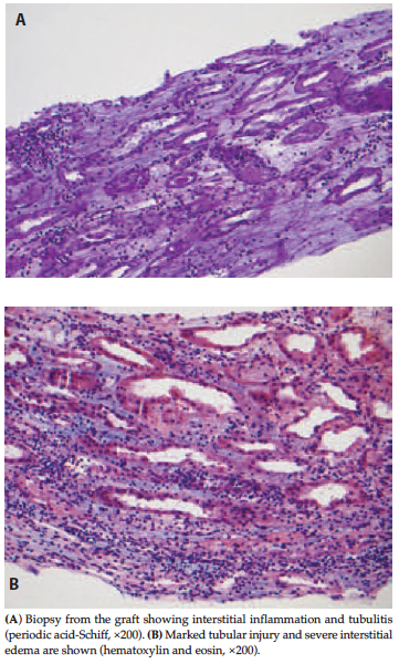
Figure 1. Biopsy Results
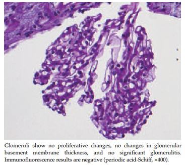
Figure 2. Immunofluorescence Results
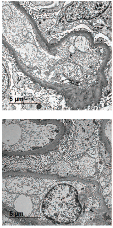
Figure 3. Ultrastructural Findings of Podocyte Injury: Vacuolization, Microvillous Transformation, and Diffuse Foot Process Effacement(transmission electron microscopy, ×12 000)