Volume: 17 Issue: 3 June 2019
FULL TEXT
Objectives: Nitrite as an alternative source of nitric oxide has been proposed, as it can mediate the protective response in the presence of ischemia or hypoxic conditions and inorganic nitrite can be reduced to nitric oxide by xanthine oxidoreductase. Here, we investigated whether pretreatment with sodium nitrite can attenuate liver damage in hepatic ischemia-reperfusion injury and identified the possible mechanism of nitrite reduction using 2-(4-carboxyphenyl)-4,5dihydro-4,4,5,5-tetramethyl-1H-imidazolyl-1-oxy-3oxide (C-PTIO), a nitric oxide scavenger, and allopurinol, a xanthine oxidoreductase inhibitor.
Materials and Methods: In experiment 1, 30 male Sprague-Dawley rats were divided into 5 groups: (1) sham-operated; (2) hepatic ischemia-reperfusion injury; and (3-5) sodium nitrite administered intra-peritoneally 30 minutes before ischemia at 2.5, 25, and 250 μmol/kg, respectively. In experiment 2, 24 male Sprague-Dawley rats were divided into 4 groups: (1) hepatic ischemia-reperfusion injury; (2) sodium nitrite + hepatic ischemia-reperfusion injury; (3) C-PTIO + sodium nitrite + hepatic ischemia-reperfusion injury; and (4) allopurinol + sodium nitrite + hepatic ischemia-reperfusion injury. Sodium nitrite (25 μmol/kg) was then administered 30 minutes before hepatic ischemia, and C-PTIO or allopurinol was administered 5 minutes before sodium nitrite admi-nistration. Blood aspartate aminotransferase, alanine aminotransferase, hepatic tissue malondialdehyde, histologic changes, and expression of mitogen-activated protein kinase family members were evaluated.
Results: Sodium nitrite limited serum elevation of alanine aminotransferase and aspartate aminotransferase induced by hepatic ischemia-reperfusion with a peak effect occurring at 25 μmol/kg sodium nitrite. Pre-treatment with allopurinol abolished the protective effect of sodium nitrite, and C-PTIO treatment attenuated the hepatoprotection of sodium nitrite in rats with hepatic ischemia-reperfusion injury. Liver malondialdehyde activity after ischemia-reperfusion decreased in sodium nitrite-treated rats. Sodium nitrite also prevented hepatic ischemia-reperfusion-induced c-Jun N-terminal kinase and extracellular signal-regulated kinase phosphorylation.
Conclusions: Exogenous sodium nitrite had protective effects against hepatic ischemia-reperfusion injury. Catalytic reduction to nitric oxide and attenuation of hepatic ischemia-reperfusion is dependent on xanthine oxidoreductase.
Key words : Liver transplantation, Nitric oxide, Xanthine oxidoreductase
Introduction
Hepatic ischemia-reperfusion (IR) injury can occur in many clinical conditions, such as liver trans-plantation, hepatic resection, trauma, and shock.1,2 Extensive research efforts have been focused on the investigation of various pathophysiologic components of hepatic IR injury. Among these components, nitric oxide (NO) has been known to have diverse effects on its mediation, which is dependent on the site of production, concentration, timing and duration of exposure, and the presence of reactive oxygen intermediates.3,4
Nitric oxide is synthesized from L-arginine by nitric oxide synthase (NOS) and depends on oxygen; there are 3 isoforms of NOS: endothelial NOS (eNOS), inducible NOS, and neuronal NOS.5 In settings where oxygen is lacking or IR injury occurs, however, conventional NO production is compro-mised because of constitutive eNOS dysfunction. Consequently NOS-independent generation of NO may be needed as the physiologic component. Recently, nitrite (NO2-) as an alternative source of NO has been proposed as a way to mediate the protective response in the presence of ischemia or hypoxia.6 Many studies have shown that nitrite exerts beneficial effects as an alternative source of NO in liver, heart, and kidney IR injury.7-9
Inorganic nitrite can be produced by the oxidation of NO at the physiologic state,10 whereas it can be reduced to NO in ischemic or hypoxic conditions.6 The NO released from nitrite is favored by various mechanisms, including xanthine oxidoreductase (XOR), deoxyhemoglobin, and components of the mitochondrial electron transport chain.11-13 Xanthine oxidoreductase has been recognized as the terminal enzyme of purine catabolism; however, interests in it have been directed toward its pathophysiologic significance, specifically, as a source of reactive oxygen species (ROS) production in IR injury.14 In addition to its accepted role related to ROS formation, XOR has risen in prominence in mediation of NO formation as catalyzing the reduction of nitrite to NO.6
In this study, we investigated whether pre-treatment with sodium nitrite (NaNO2) can attenuate liver damage in hepatic IR injury and also evaluated the possible mechanisms of nitrite reduction to NO using 2-(4-carboxyphenyl)-4,5dihydro-4,4,5,5-tetramethyl-1H-imidazolyl-1-oxy-3oxide (C-PTIO), an NO scavenger, and allopurinol, an XOR inhibitor. Moreover, to verify the intracellular signaling pathway of nitrite-mediated hepatoprotection in hepatic IR injury, we analyzed the activities of the mitogen-activated protein kinase (MAPK) pathway.
Materials and Methods
Animals
This study was approved by the Institutional Animal Care and Use Committee of
Kyungpook National University (Daegu, Korea), and all procedures were conducted
according to the institutional guidelines.
Experimental protocol
In experiment 1, 30 male Sprague-Dawley rats
(290-310 g) were divided into the following 5 groups (n = 6/group): (1)
sham-operated, (2) hepatic IR,
(3) NaNO2 2.5 μmol/kg + hepatic IR, (4) NaNO2
25 μmol/kg + hepatic IR, and (5) NaNO2 250 μmol/kg + hepatic IR. For these
groups, NaNO2 was dissolved in distilled water and administered
intraperitoneally (IP) with 1 mL of final solution at 30 minutes before
ischemia.
In experiment 2, 24 male Sprague-Dawley rats (290-310 g) were divided into the following 4 groups (n = 6/group): (1) hepatic IR, (2) NaNO2 25 μmol/kg + hepatic IR, (3) C-PTIO + NaNO2 25 μmol/kg + hepatic IR, and (4) allopurinol + NaNO2 25 μmol/kg + hepatic IR. Each drug (25 μmol/kg NaNO2, 2 mg/kg C-PTIO, and 50 mg/kg allopurinol) was dissolved in distilled water and administered with 1 mL of final solution. NaNO2 was administered 30 minutes before hepatic ischemia, and C-PTIO or allopurinol was administered 5 minutes before intraperitoneal administration of 25 μmol/kg NaNO2.
Rats were anesthetized by intraperitoneal injection of ketamine (60 mg/kg) and xylene (10 mg/kg). Vehicle in the sham and hepatic IR groups was distilled water. Each drug dosage was determined based on previous studies.15,16
Hepatic ischemia-reperfusion injury model
After rats were anesthetized, a midline laparotomy was performed. Ischemia in
left lateral and median lobes of liver was induced by clamping the portal vein
and left lateral branch of the hepatic artery using a microvascular clamp; this
procedure resulted in partial segmental (70%) hepatic ischemia. The duration of
ischemia was 30 minutes, after which the microvascular clamp was removed to
induce reperfusion. After 6 hours of reperfusion, blood samples were collected
from the heart to minimize hemolysis during sampling, and ischemic liver tissues
were retrieved for biochemical and histopathologic analysis.
Liver enzyme assessment
Levels of aspartate aminotransferase (AST) and alanine aminotransferase (ALT)
were analyzed by using a Vitros 250 device (Johnson & Johnson, Raritan, NJ,
USA), following the manufacturer’s protocol.
Hepatic tissue malondialdehyde level assessment
Malondialdehyde levels, a measure of lipid per-oxidation, were assayed
spectrophotometrically using thiobarbituric acid reactive substances.17 The
mixture consisted of cell lysate, 0.375% thiobarbituric acid (Alfa Aesar, Ward
Hill, MA, USA), 15% trichloroacetic acid (Sigma, St. Louis, MO, USA), and 0.25 N
HCl. The mixture was heated in boiling water for 15 minutes. After the mixture
was cooled, it was centrifuged for 10 minutes at 12 000 revolutions/min. The
absorbance was read spectrophotometrically at a
535-nm wavelength. Tissue protein concentration was estimated according to the
Bradford assay, with malondialdehyde content expressed as nmol/mg of protein.
Western blot analyses
Hepatic tissues were homogenized in radioim-munoprecipitation assay buffer
(Thermo Scientific, Rockford, IL, USA) and centrifuged at 12 500 revolutions/min
for 20 minutes at 4°C. The super-natants were removed, and extracted protein was
quantified using the Bradford assay (Thermo Scientific). The samples were mixed
with a loading buffer solution containing 60 mM Tris-HCl, 25% glycerol, 2%
sodium dodecyl sulfate, 14.4 mM
2-mercaptoethanol, and 0.1% bromophenol blue, separated in a 10% sodium dodecyl
sulfate-polyacrylamide gel, and transferred to a nitrocellulose membrane
(Whatman GmbH, Dassel, Germany). The membrane was blocked with 1% bovine serum
albumin in Tris-buffered saline containing 0.1% Tween 20 for 2 hours at room
temperature. After washes were completed, blots were incubated overnight at 4°C
with antibodies to c-Jun N-terminal kinase (JNK), extracellular signal-regulated
kinase (ERK), and p38 (diluted 1:1000; Santa Cruz, Santa Cruz, CA, USA) and
β-actin (diluted 1:5000; Cell Signaling Technology, Beverly, MA, USA).
Mem-branes were then incubated with secondary anti-mouse horseradish
peroxidase-conjugated antibody (diluted 1:1000; Cell Signaling Technology),
devel-oped using an enhanced chemiluminescence substrate kit (Advansta, Menlo
Park, CA, USA), and exposed onto medical radiography film. The signal intensity
was quantified using NIH Image J version 1.47 software.
Liver histopathology assessment
Liver tissue from the left lobe was fixed in 10% buffered formalin for 24 hours
and embedded in paraffin, and 5-μm sections were stained with hematoxylin and
eosin. The degree of liver injury was determined using light microscopy at ×200
in a blinded fashion. Five liver tissue samples from each group were used, and
10 fields per slide were evaluated using a scoring system for sinusoidal
congestion, cytoplasmic vacuolation, and liver necrosis. Results were analyzed
and graded on a scale of 0 to 3 for sinusoidal congestion (0, none; 1, mild with
10% to 40% hepatic tissue; 2, moderate with 40% to 70% hepatic tissue; 3, severe
with > 70% hepatic tissue), 0 to 3 for cytoplasmic vacuolation (0, none; 1, mild
with 10% to 40% hepatocytes; 2, moderate with 40% to 70% hepatocytes; 3, severe
with > 70% hepatocytes), or 0 or 1 for liver necrosis (0, none; < 50%
hepatocytes; 1, > 50% hepatocytes).18
Statistical analyses
All results are shown as means ± standard error. A one-way analysis of variance
test with a Bonferroni correction was performed. To compare all values, data
were analyzed using SPSS software version 23.0 (SPSS, Chicago, IL, USA).
Differences were consi-dered to be statistically significant at P < .05.
Results
Experiment 1
The effects of nitrite on hepatic IR injury were determined after
intraperitoneal injection of NaNO2 (2.5, 25, and 250 μmol/kg) 30 minutes before
ischemia. In the hepatic IR control group, serum ALT and AST levels were
significantly increased com-pared with the sham group (P < .0001 for ALT,
P < .0001 for AST). With 2.5, 25, and 250 μmol/kg NaNO2, elevation of serum
liver transaminase ALT and AST levels was limited (P < .0001 for ALT,
P < .01 for AST), with a peak effect occurring at
25 μmol/kg NaNO2. The level of ALT was lower
in the 25 μmol/kg NaNO2 group than in the
2.5 μmol/kg NaNO2 group, and the 250 μmol/kg NaNO2 group showed increased ALT
levels com-pared with the 25 μmol/kg NaNO2 group. The level of AST was lower in
the 25 μmol/kg NaNO2 group than in the 2.5 μmol/kg NaNO2 group (Figure 1, A and
B).
Experiment 2
To assess the possible mechanisms of nitrite-mediated hepatoprotection,
allopurinol and C-PTIO were administered 5 minutes before NaNO2 treatment.
Pretreatment with allopurinol abolished the protective effect of NaNO2 (P = .001
for ALT and P = .002 for AST), and treatment with C-PTIO attenuated the
hepatoprotection of NaNO2 in rats with hepatic IR injury (P = .002 for ALT and
P
= .001 for AST) (Figure 2, A and B).
For measurement of cellular lipid peroxidation caused by oxidative stress, liver malondialdehyde activity was assessed. With regard to malondialdehyde levels, we could not discern differences between the sham group and the hepatic IR groups; however, malondialdehyde activity after hepatic IR decreased significantly more in rats treated with 25 NaNO2 than in rats with hepatic IR only (P = .047) (Figure 3).
To verify the intracellular signaling pathway of nitrite-mediated hepatoprotection after hepatic IR injury, we analyzed MAPK. Phosphorylation levels of JNK, p38, and ERK were significantly increased after hepatic IR compared with that shown in the sham group (P = .02 for JNK, P = .004 for p38, and P= .002 for ERK). Expression levels of phos-phorylated JNK and phosphorylated ERK were lower in rats treated with 25 μmol/kg NaNO2 than in rats with hepatic IR only (P = .02 for JNK, P = .044 for p38, and P= .015 for ERK) (Figure 4).
We performed histologic analyses of liver to evaluate the degree of structural injury among experimental groups. There was no evidence of hepatic injury in rats that underwent sham operation; however, significant differences were observed in the number of incidences of sinusoidal congestion, cytoplasmic vacuolation, and necrosis in the hepatic IR group (P < .001). The 25 μmol/kg NaNO2 treatment group exhibited much less structural derangement in histology compared with those in hepatic IR group (P < .001). Allopurinol and C-PTIO pretreatment resulted in more severe histologic injury than NaNO2 treatment alone (P = .002 for allopurinol and P = .001 for C-PTIO) (Figure 5).
Discussion
This study showed that treatment with 25 μmol/kg NaNO2 conferred protective effects against hepatic IR injury, preserving hepatic function and limiting hepatocellular necrosis. Pretreatment with C-PTIO, an NO scavenger, in NaNO2-treated rats resulted in more severe liver damage than NaNO2 treatment alone, and administration of allopurinol, an XOR inhibitor, with NaNO2 abolished the hepato-protective effects of NaNO2. These results provided evidence that XOR acts as a functional nitrite reductase in the conversion of nitrite to NO. Moreover, we identified that JNK and ERK phos-phorylation induced by hepatic I/R injury was attenuated by NaNO2 treatment.
The synthesis of NO is catalyzed by the enzyme NOS. Nitric oxide production from constitutive eNOS is generally considered to play a physiologic role in IR injury. Previous studies have shown the protective effects of eNOS-induced NO in hepatic IR injury,19,20 and the beneficial effects of eNOS have been confirmed in studies using eNOS knockout animals, which revealed marked liver injury due to hepatic IR.21 However, during the IR period, the formation of eNOS-dependent NO can be com-promised due to endothelial cell injury or hypoxic conditions. Thus, eNOS-independent NO formation may be needed as a salvage pathway in physiologic endothelial function and integrity. Recently, the use of nitrite at physiologic concentrations has been suggested as a promediator of NO homeostasis.6 Nitrite can be reduced to NO under hypoxic and acidic conditions by enzymatic and nonenzymatic components,22 which may be helpful to maintain physiologic NO levels in hepatic IR injury.
Among various components related to nitrite reductase action, XOR is a molybdo-flavoenzyme that contains 4 redox centers: one molybdenum cofactor, 1 flavin adenine dinucleotide, and 2 Fe2S2 sites,23 which are found abundantly in liver, gut, and plasma with inflammation24 and which are upregulated during IR injury by processes involving phosphorylation.25 The well-established activity of XOR is catalyzing the hydroxylation of hypoxanthine to xanthine and xanthine to urate with generation of ROS during IR injury.26
Recently, XOR has been shown to catalyze the reduction of nitrite to NO at its molybdenum site under ischemic conditions, mediating beneficial outcomes against IR injury.16 Herein, we may assume that XOR-induced superoxide interacts with NO in the presence of nitrite, subsequently increasing peroxynitrite formation. However, Tripatara and associates16 hypothesized that the reduction of inorganic nitrite to NO may compete for electrons related to oxygen reduction at the flavin adenine dinucleotide site of XOR, resulting in reducing superoxide and peroxynitrite formation. Therefore, they showed the protective effects of nitrite as an alternative substrate against IR injury. In this study, concentrations of NaNO2 at both 2.5 and 25 μmol/kg resulted in similar protection, suggesting that even relatively small amounts of NaNO2 may have a protective effect. However, hepatoprotection was lost at higher dosing (250 μmol/kg), suggesting that this dose may have a noxious effect via by nitrite-derived NO production. The efficacy of NaNO2 with U-shaped pattern and not a linear dose-response pattern was similar to the results of a rat brain IR injury study from Jung and associates27 and to results of a mouse cardiac and liver IR injury study from Duranski and associates.28 In line with these findings, many studies have concluded that excessive NO may mediate pathogenesis of IR injury rather than beneficial outcomes.15,27,28
After we established an administration dose for beneficial effects of NaNO2, we tried to address the underlying mechanism. First, C-PTIO with NaNO2 was administered to investigate whether NO formation was induced from nitrite. The NO scavenger C-PTIO attenuated the protective role of NaNO2 against hepatic IR injury, suggesting that not NaNO2 per se but nitrite-derived NO generation mediates the protective effects. This result was consistent with results from other studies in the brain, lung, and kidney.27,28 Second, we used allopurinol, an XOR inhibitor, to determine whether XOR was responsible for reduction of nitrite to NO. It has been shown that XOR can generate ROS in IR injury, and studies have already shown the beneficial effects of allopurinol as inhibiting XOR in hepatic IR injury.29,30 However, in contrast to the negative role of XOR, its role as a nitrite reductase showed beneficial outcomes. Pickerodt and associates15 showed that the beneficial effect of NaNO2 for ventilation-induced lung injury was abolished by allopurinol, suggesting that the involvement of XOR-dependent nitrite-derived NO formation. In this study, allopurinol abolished the hepatoprotective effects of NaNO2, and this result may provide the evidence that the role of XOR as a nitrite reductase has greater capacity than its role in formation of ROS.
The MAPK signaling pathway, which plays an important role in intracellular signal transduction, is closely related to hepatic IR injury.31 Three distinct subgroups of MAPK (ERK, JNK, and p38) can be activated by a variety of pathophysiologic stresses, including oxidative stress, inflammatory cytokines, and heat shock.32 The phosphorylation of these MAPK family members can increase stress-induced cell damage and regulate the signaling events relevant to apoptosis.33 Therefore, tight regulation of MAPK phosphorylation may be needed to maintain physiologic cell function. In this study, NaNO2 suppressed the phosphorylation of ERK and JNK, which was consistent with the attenuation of malondialdehyde activity, a surrogate marker of lipid peroxidation caused by oxidative stress after hepatic IR, suggesting that the protective effects of NaNO2 against hepatic IR injury may be affected by weakened phosphorylation of the MAPK pathway.
This study has some limitations. First, reduction of nitrite to NO depends on the activity of XOR, but we could not exclude the possibility of other mechanisms underlying this conversion. Second, in terms of the role of XOR related to ROS generation or nitrite reduc-tion, we could not exactly document the degree of contribution of XOR during IR injury because we analyzed XOR-mediated nitrite reduction to NO indirectly using allopurinol and C-PTIO. Further research investigations with more precise methods such as direct real-time NO measurement are needed to confirm our results.
Conclusions
Exogenous NaNO2 protected hepatic IR injury. On the basis of our results, although the precise mechanisms underpinning XOR-dependent nitrite-derived NO formation remain unclear, we propose that any materials that contain or release nitrite may potentially affect therapeutic strategies in many conditions related to hepatic IR injury.
References:
- Douzinas EE, Livaditi O, Tasoulis MK, et al. Nitrosative and oxidative stresses contribute to post-ischemic liver injury following severe hemorrhagic shock: the role of hypoxemic resuscitation. PLoS One. 2012;7(3):e32968.
CrossRef - PubMed - Arkadopoulos N, Defterevos G, Nastos C, et al. Development of a porcine model of post-hepatectomy liver failure. J Surg Res. 2011;170(2):e233-242.
CrossRef - PubMed - Goligorsky MS, Brodsky SV, Noiri E. Nitric oxide in acute renal failure: NOS versus NOS. Kidney Int. 2002;61(3):855-861.
CrossRef - PubMed - Goligorsky MS, Brodsky SV, Noiri E. NO bioavailability, endothelial dysfunction, and acute renal failure: new insights into pathophysiology. Semin Nephrol. 2004;24(4):316-323.
CrossRef - PubMed - Zhang C, Hein TW, Wang W, Ren Y, Shipley RD, Kuo L. Activation of JNK and xanthine oxidase by TNF-alpha impairs nitric oxide-mediated dilation of coronary arterioles. J Mol Cell Cardiol. 2006;40(2):247-257.
CrossRef - PubMed - Li H, Samouilov A, Liu X, Zweier JL. Characterization of the magnitude and kinetics of xanthine oxidase-catalyzed nitrate reduction: evaluation of its role in nitrite and nitric oxide generation in anoxic tissues. Biochemistry. 2003;42(4):1150-1159.
CrossRef - PubMed - Lu P, Liu F, Yao Z, et al. Nitrite-derived nitric oxide by xanthine oxidoreductase protects the liver against ischemia-reperfusion injury. Hepatobiliary Pancreat Dis Int. 2005;4(3):350-355.
PubMed - Webb A, Bond R, McLean P, Uppal R, Benjamin N, Ahluwalia A. Reduction of nitrite to nitric oxide during ischemia protects against myocardial ischemia-reperfusion damage. Proc Natl Acad Sci U S A. 2004;101(37):13683-13688.
CrossRef - PubMed - Milsom AB, Patel NS, Mazzon E, et al. Role for endothelial nitric oxide synthase in nitrite-induced protection against renal ischemia-reperfusion injury in mice. Nitric Oxide. 2010;22(2):141-148.
CrossRef - PubMed - Lauer T, Preik M, Rassaf T, et al. Plasma nitrite rather than nitrate reflects regional endothelial nitric oxide synthase activity but lacks intrinsic vasodilator action. Proc Natl Acad Sci U S A. 2001;98(22):12814-12819.
CrossRef - PubMed - Zhang Z, Naughton DP, Blake DR, et al. Human xanthine oxidase converts nitrite ions into nitric oxide (NO). Biochem Soc Trans. 1997;25(3):524S.
CrossRef - PubMed - Kim-Shapiro DB, Schechter AN, Gladwin MT. Unraveling the reactions of nitric oxide, nitrite, and hemoglobin in physiology and therapeutics. Arterioscler Thromb Vasc Biol. 2006;26(4):697-705.
CrossRef - PubMed - Zweier JL, Wang P, Samouilov A, Kuppusamy P. Enzyme-independent formation of nitric oxide in biological tissues. Nat Med. 1995;1(8):804-809.
CrossRef - PubMed - Granger DN, Hollwarth ME, Parks DA. Ischemia-reperfusion injury: role of oxygen-derived free radicals. Acta Physiol Scand Suppl. 1986;548:47-63.
PubMed - Pickerodt PA, Emery MJ, Zarndt R, et al. Sodium nitrite mitigates ventilator-induced lung injury in rats. Anesthesiology. 2012;117(3):592-601.
CrossRef - PubMed - Tripatara P, Patel NS, Webb A, et al. Nitrite-derived nitric oxide protects the rat kidney against ischemia/reperfusion injury in vivo: role for xanthine oxidoreductase. J Am Soc Nephrol. 2007;18(2):570-580.
CrossRef - PubMed - Mihara M, Uchiyama M. Determination of malonaldehyde precursor in tissues by thiobarbituric acid test. Anal Biochem. 1978;86(1):271-278.
CrossRef - PubMed - Calabrese F, Valente M, Pettenazzo E, et al. The protective effects of L-arginine after liver ischaemia/reperfusion injury in a pig model. J Pathol. 1997;183(4):477-485.
CrossRef - PubMed - Duranski MR, Elrod JW, Calvert JW, Bryan NS, Feelisch M, Lefer DJ. Genetic overexpression of eNOS attenuates hepatic ischemia-reperfusion injury. Am J Physiol Heart Circ Physiol. 2006;291(6):H2980-2986.
CrossRef - PubMed - Katsumi H, Nishikawa M, Yamashita F, Hashida M. Prevention of hepatic ischemia/reperfusion injury by prolonged delivery of nitric oxide to the circulating blood in mice. Transplantation. 2008;85(2):264-269.
CrossRef - PubMed - Hines IN, Harada H, Flores S, Gao B, McCord JM, Grisham MB. Endothelial nitric oxide synthase protects the post-ischemic liver: potential interactions with superoxide. Biomed Pharmacother. 2005;59(4):183-189.
CrossRef - PubMed - Okamoto M, Tsuchiya K, Kanematsu Y, et al. Nitrite-derived nitric oxide formation following ischemia-reperfusion injury in kidney. Am J Physiol Renal Physiol. 2005;288(1):F182-187.
CrossRef - PubMed - Patton S, Keenan TW. The milk fat globule membrane. Biochim Biophys Acta. 1975;415(3):273-309.
CrossRef - PubMed - Parks DA, Granger DN. Xanthine oxidase: biochemistry, distribution and physiology. Acta Physiol Scand Suppl. 1986;548:87-99.
PubMed - Kayyali US, Donaldson C, Huang H, Abdelnour R, Hassoun PM. Phosphorylation of xanthine dehydrogenase/oxidase in hypoxia. J Biol Chem. 2001;276(17):14359-14365.
CrossRef - PubMed - Harrison R. Structure and function of xanthine oxidoreductase: where are we now? Free Radic Biol Med. 2002;33(6):774-797.
CrossRef - PubMed - Jung KH, Chu K, Ko SY, et al. Early intravenous infusion of sodium nitrite protects brain against in vivo ischemia-reperfusion injury. Stroke. 2006;37(11):2744-2750.
CrossRef - PubMed - Duranski MR, Greer JJ, Dejam A, et al. Cytoprotective effects of nitrite during in vivo ischemia-reperfusion of the heart and liver. J Clin Invest. 2005;115(5):1232-1240.
CrossRef - PubMed - Liu PG, He SQ, Zhang YH, Wu J. Protective effects of apocynin and allopurinol on ischemia/reperfusion-induced liver injury in mice. World J Gastroenterol. 2008;14(18):2832-2837.
CrossRef - PubMed - Yildirim S, Tok H, Koksal H, Erdem L, Baykan A. Allopurinol plus pentoxifilline in hepatic ischaemia/reperfusion injury. Asian J Surg. 2002;25(2):149-153.
CrossRef - PubMed - Mukhopadhyay P, Rajesh M, Horvath B, et al. Cannabidiol protects against hepatic ischemia/reperfusion injury by attenuating inflammatory signaling and response, oxidative/nitrative stress, and cell death. Free Radic Biol Med. 2011;50(10):1368-1381.
CrossRef - PubMed - Winter-Vann AM, Johnson GL. Integrated activation of MAP3Ks balances cell fate in response to stress. J Cell Biochem. 2007;102(4):848-858.
CrossRef - PubMed - Petrache I, Choi ME, Otterbein LE, et al. Mitogen-activated protein kinase pathway mediates hyperoxia-induced apoptosis in cultured macrophage cells. Am J Physiol. 1999;277(3):L589-595.
CrossRef - PubMed

Volume : 17
Issue : 3
Pages : 348 - 354
DOI : 10.6002/ect.2018.0169
From the 1Department of Anesthesiology and Pain Medicine, Yeungnam University
College of Medicine, Daegu, Korea; the 2Department of Anesthesiology and Pain
Medicine, School of Medicine, Kyungpook National University, Daegu, Korea; and
the 3Department of Pathology, Yeungnam University College of Medicine, Daegu,
Korea
Acknowledgements: This work was supported by Biomedical Research Institute
grant, Kyungpook National University Hospital, 2016. The authors have no
conflicts of interest.
Corresponding author: Dong Gun Lim, Department of Anesthesiology and Pain
Medicine, School of Medicine, Kyungpook National University, Daegu, Korea
Phone: +82 534205876
E-mail: dglim@knu.ac.kr
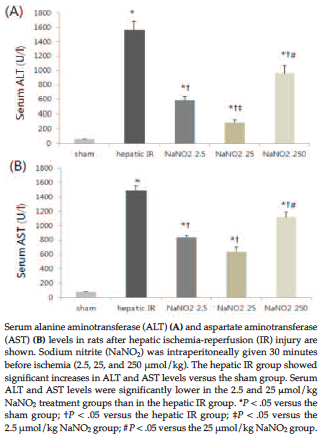
Figure 1. Dose-Dependent Effect of Sodium Nitrite on Hepatic Ischemia- Reperfusion Injury
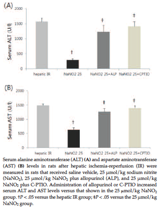
Figure 2. Sodium Nitrite-Mediated Hepatoprotection and Nitric Oxide Signaling Pathways
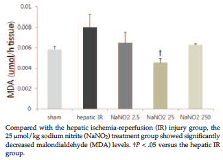
Figure 3. Renal Tissue Malondialdehyde Levels in the Experimental Groups
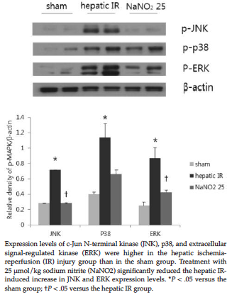
Figure 4. Western Blots of Phosphorylated Mitogen-Activated Protein Kinases of Liver Homogenates
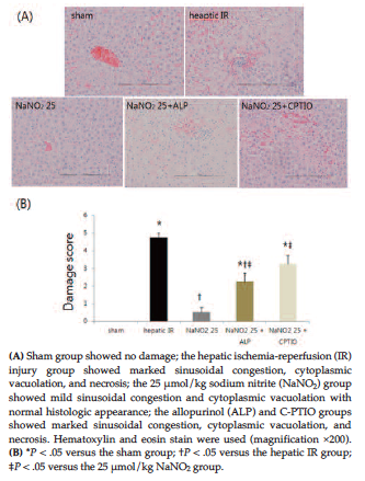
Figure 5. Histopathologic Study of Liver Tissue