Volume: 15 Issue: 2 April 2017
FULL TEXT
Diabetes mellitus is a disease with no definite cure. In recent years, stem cell transplant has led to treatment of various diseases including diabetes. We sought to report a type 1 diabetic patient with a brain mass, diagnosed as transitional meningioma, after a fetal hematopoietic stem cell transplant. A 57-year-old woman with type 1 diabetes who previously had undergone a fetal hematopoietic stem cell transplant, attended the clinic with a history of progressive bifrontal headaches accompanied by nausea, vomiting, and visual disturbances over the previous 8 months. Investigations revealed a 2-cm mass in the right temporal region. The patient underwent a craniotomy, and the lesion was removed and sent for pathological and genetic investigations. The results indicated transitional meningioma with the origin of transplanted fetal hematopoietic stem cells.
To our knowledge, this is the first report of transitional meningioma as a result of stem cell transplant. Despite all unanswered questions about the safety of stem cell transplant, this novel therapy provides hope for patients with type 1 diabetes.
Key words : Regenerative medicine, Benign tumor, Type 1 diabetes
Introduction
Curative therapy for diabetes mellitus can only be achieved by restoring functional insulin-producing pancreatic cells with either pancreas or islet-cell transplants. However, applying these procedures is vastly hindered by the shortage of donors. Stem cell therapy is an alternative curative treatment for diabetes without such limitation.1,2
Cell therapy has been demonstrated to be promising for treatment of several autoimmune diseases such as type 1 diabetes with the mechanism of engraftment and promoting β-cell regeneration through enhanced neovascularization.3,4 There are several studies that have demonstrated successfully the immunomodulatory effect of hematopoietic stem cells (HSCs). One of the promising sources of HSCs in fetal tissues is the fetal liver.5,6 However, there are serious concerns that embryonic-derived stem cells could proliferate excessively and thereby, causing solid tumors after transplant.7
We sought to present a type 1 diabetic patient who presented with a brain mass after having undergone a fetal hematopoietic stem cell transplant that was diagnosed later to be transitional meningioma.
Case Report
A 57-year-old woman with type 1 diabetes, who had undergone a fetal hematopoietic stem cell transplant was referred to our clinic with complaints of progressive headaches accompanied with nausea, vomiting, and visual disturbances over the previous 8 months. The patient had participated in another study evaluating fetal HSCs safety done by Ghodsi and associates and had received fetal hematopoietic stem cells intravenously 25 months before the onset of symptoms. It should be noted that the transplant procedure in the mentioned study was approved by the ethical board committee of Tehran University of Medical Sciences (Ethical Code: 0089) and all participants signed informed consent.8 All of the protocols conformed to the ethical guidelines of the 1975 Helsinki Declaration.
A physical examination before the transplant revealed no notable abnormalities. The blood pressure was 105/70 mm Hg, pulse rate was 78 beats/min, and oral temperature was 36.9°C. Complete blood cell count showed a hemoglobin of 13.9 g/dL, platelet count of 294 × 1000/μL, and white blood cell count of 4000/mm3, with 50% neutrophils. Fasting blood glucose and HbA1c levels were 146 mg/dL and 8.3%. Blood and urine cultures were negative for infection. Other laboratory tests such as liver function tests, blood urea nitrogen, and creatinine were within the normal ranges. Bone mass densitometry showed mild osteopenia. Her medications included glibenclamide (5 mg, 1 tablet per day), metformin (500 mg, 0.5 tablet per day), and lovastatin (20 mg, 1 tablet per day).
Cell preparation
Briefly, fetal liver-derived HSCs were isolated from legally and aborted
human fetuses aged 6 to 12 weeks after obtaining informed consent from the
parents (mother or both of the parents). To determine chromosomal abnormalities
and to identify the sex of the donated fetus, karyotyping was done for each
fetal sample. Whole fetal liver was placed in Hank’s balanced salt solution
without calcium and magnesium, Sigma, (Milwaukee, WI, USA) and dissociated and
homogenized mechanically. The cell suspension was filtered through nylon mesh to
undergo transplant; then isolated cells were cryopreserved using 5% dimethyl
sulfoxide in Hank’s balanced salt solution, Wak Chemie (Homburg, Germany) with a
programmable freezer, and were transferred to liquid nitrogen for long-term
storage. Before transplant, samples were thawed at 37°C, and cryoprotectant was
diluted by 5-mm normal saline before infusion. Total cell count in the prepared
suspension was approximately 35 to 55 × 106, twenty percent of which
was recognized as hematopoietic stem cells and were injected into the cubital
vein of the patients. No vehicles or any types of scaffolds were used for cell
delivery to patients. The suspension was checked before, during, and after
processing for aerobic, anaerobic, and fungal contamination and viral
infections. Rubella, herpes simplex virus, Cytomegalovirus, Chlamydia,
Mycoplasma hominis, Toxoplasma gondii, and Treponema pallidum were
checked using an enzyme-linked immunoassay. DNA/RNA extraction and real-time
polymerase chain reaction were done for checking viral contamination (hepatitis
B virus, hepatitis C virus, and human immunodeficiency virus). After evaluating
the results, cell samples were known qualified for the transplant and were
injected into the cubital vein of the patients.
Six months after transplant, laboratory evaluations revealed fasting blood glucose of 176 mg/dL, HbA1c of 7.7%, and C-peptide of 3.62 (ng/mL). One year after the transplant, fasting blood glucose, HbA1c and C-peptide levels were 139 mg/dL, 7.1%, and 2.9 (ng/mL).8
Twenty-five months after transplant, the patient was referred to the neurology clinic complaining of severe progressive bilateral frontal headache accompanied by nausea, vomiting and visual disturbances.
For further investigations, the patient was referred to an ophthalmologist and visual field defects were confirmed. Then, the patient was visited by a neurologist, and the computed tomography scan revealed a 2-cm enhancing lesion in the right medial frontal region of brain with surrounding edema (Figures 1A and B).
A computed tomography scan showed encephalomalacia in the left frontal lobe and a hyperdense calcific lesion in the right frontal region, suggestive of calcified meningioma. Supratentorial parenchyma and ventricular systems were normal.
Subsequently, the patient underwent an operation and the tumor was radically removed through a right frontal craniotomy and sent for pathological and genetic investigations. Five days after surgery, the patient had a normal neurologic examination and was discharged.
Pathological investigations showed a benign tumor composed of spindle and meningotheliomatous components with obvious psammoma bodies suggestive of transitional meningioma.
Transplanted fetal HSCs, patient’s peripheral blood cells, and tumor cells were compared with a PCR using highly polymorphic microsatellite markers and PCR with specific primers for amelogenin homologous gene located on X and Y chromosomes. Genomic DNA was extracted from the patient’s blood samples and from the tumor tissue using standard protocols.
Six highly polymorphic microsatellite markers (CA repeats) from different chromosomes (Table 1) were chosen from the Ge´ne´thon database9 and their corresponding primer sets were synthesized. The forward primer of each set was labeled with fluorescent dye (Cy5; Pharmacia Biotech; BioTek US, Winooski, VT, USA), but the reverse primer was not. Polymerase chain reaction amplification was performed in a 12.5 mL reaction mixture containing 50 ng of genomic DNA from blood or tissue, 1-3 mM MgCl2, 0.2 mM of each deoxynucleotide triphosphate, 0.5 U of DNA polymerase (AmpliTaq Gold; PE Biosystems), and 12.5 pmol of each primer. Polymerase chain reaction conditions consisted of an initial denaturation at 94°C for 10 minutes; 40 cycles of denaturation at 94°C for 30 seconds, annealing at 48°C to 58°C for 30 seconds, and extension at 72°C for 30 seconds; and final extension at 72°C for 10 minutes. Polymerase chain reaction products were dissolved by electrophoresis on 6% polyacrylamide DNA sequencing gel, by the use of an automated sequencer Pharmacia Biotech (Roosendaal, The Netherlands). Electrophoresis patterns were analyzed by computer software Pharmacia Biotech to determine the genotypes of the blood samples and the tissue. To confirm that the stem cells had originated from a different sex, specific primers for X and Y homologous of amelogenin genes were designed, PCR amplification was performed (as mentioned above), and PCR products were run in 1% agarose gel to visualize the amplified bands.
Amplified amelogenin alleles gene revealed that the tumor cells had originated from a female donor, similar to our patient. Analysis of DNA obtained from the blood and tumor cells with 6 microsatellite markers which are listed in Table 1, demonstrated that the allele size for 5 markers were different from the DNA originated from the patient’s blood sample and the DNA extracted from the tumor. The number of alleles detected in the tumor originated from nonhost cells indicated the presence of more than 1 cell population in the tumor. Although the possibility of genomic instability in tumor remains as an alternative cause for these differences, it is more likely that the tumor had not originated from recipient cells.
Discussion
Although stem-cell therapy has been the focus of interest in recent years, there are still serious concerns over its safety. Several types of tumors have been shown to be caused by stem cell transplant.7,10,11 To the best of our knowledge, this is the first report of a brain meningioma caused by fetal liver-derived HSCs transplant that developed in a 57-year-old woman with type 1 diabetes mellitus.
Meningioma is a slow growing tumor originally arising from the arachnoid. Besides headaches, nausea, and vomiting, meningioma may present with different visual disturbances based on the site of the tumor and its treatment of choice is usually surgical resection if the symptoms are present.12 In our patient, as the tumor had caused visual field defects as well as progressive headaches accompanied by nausea and vomiting which had affected her daily activities to a great extent, surgical management was considered and the mass was removed through frontal craniotomy. As pathological and genetic investigations confirmed the tumor was transitional meningioma, it can be concluded that the tumor originated from the transplanted fetal liver-derived HSCs.
It is noteworthy that the method of stem cell transplant in our study was different from that of other studies. For example, Thirabanjasak and associates, who reported angio myeloproliferative lesions after stem cell transplant, used direct injection of autologous stem cells. Similarly, in a study performed by Amariglio and associates, a brain tumor was reported after a stem cell transplant in a boy with ataxia telangiectasis, to whom the stem cells were injected intracerebellarly and intrathecally. As both mentioned trials used direct injection method, our study is the first one to report a tumor after intravenous injection of stem cells. Moreover, it must be noted that patients with ataxia telangiectasia are more susceptible to different types of tumors. In addition, as Prokhorava and associates showed that the rate of teratoma formation in mice is site dependent,13 the findings in our study might argue that the tumorigenicity of stem cells is not confined to their local administration. Therefore, it can be concluded that the results of our study can cast even more doubt on the safety of clinical application of HSCs for the treatment of type 1 diabetes.
Clinical application of stem cell technology has been the subject of intense speculation and controversy for several years. The case presented here demonstrates that the technology may result in grave consequences that should be studied and assessed thoroughly before they can be applied clinically in a large scale. Therefore, we suggest that more clinical trials with larger cohorts and long-term follow-up studies are of crucial importance to ensure the safety of stem cell transplant in the treatment of type 1 diabetes.
References:
- Ryan EA, Lakey JR, Paty BW, et al. Successful islet transplantation:
continued insulin reserve provides long-term glycemic control. Diabetes.
2002;51:2148-2157.
CrossRef - PubMed - Ryan EA, Paty BW, Senior PA, et al. Five-Year Follow-Up After Clinical
Islet Transplantation. Diabetes. 2005;54:2060-2069.
CrossRef - PubMed - Burt RK, Testor A, Craig R, Cohen B, Suffit R, Barr W. Hematopoietic
stem cell transplantation for autoimmune diseases: What have we learned?
Journal of Autoimmunity. 2008;30:116-120.
CrossRef - PubMed - Rosengren AH, Renström E. Autologous hematopoietic stem cell
transplantation in type 1-diabetes. Islets. 2009;1:81-83.
CrossRef - PubMed - O'Donoghue K, Fisk NM. Fetal stem cells. Best Pract Res Clin Obstet
Gynaecol. 2004;18:853-875.
CrossRef - PubMed - Petrenko YA, Jones D, Petrenko AY. Cryopreservation of human fetal liver
hematopoietic stem/progenitor cells using sucrose as an additive to the
cryoprotective medium. Cryobiology. 2008;57:195-200.
CrossRef - PubMed - Hess PG. Risk of tumorigenesis in first-in-human trials of embryonic
stem cell neural derivatives: Ethics in the face of long-term uncertainty.
Account Res. 2009;16:175-198.
CrossRef - PubMed - Ghodsi M, Heshmat R, Amoli M, et al. The effect of fetal liver-derived
cell suspension allotransplantation on patients with diabetes: first year of
follow-up. Acta Med Iran. 2012;50:541-546.
PubMed - Schuler G, Boguski M, Stewart E, et al. A gene map of the human genome.
Science. 1996;274:540-546.
CrossRef - PubMed - Amariglio N, Hirshberg A, Scheithauer BW, et al. Donor-derived brain
tumor following neural stem cell transplantation in an ataxia telangiectasia
patient. PLoS Med. 2009;6:e1000029.
CrossRef - PubMed - Thirabanjasak D, Tantiwongse K, Thorner PS. Angiomyeloproliferative
lesions following autologous stem cell therapy. J Am Soc Nephrol.
2010;21:1218-1222.
CrossRef - PubMed - Aminoff MJ. Brain's Diseases of the Nervous System. JAMA: The Journal of
the American Medical Association. 1994;272:1627-1628.
CrossRef - - Prokhorova TA, Harkness LM, Frandsen U, et al. Teratoma formation by
human embryonic stem cells is site dependent and enhanced by the presence of
Matrigel. Stem cells and development. 2009;18:47-54.
CrossRef - PubMed

Volume : 15
Issue : 2
Pages : 231 - 234
DOI : 10.6002/ect.2014.0243
From the 1Diabetes Research Center, Endocrinology and Metabolism
Clinical Sciences Institute; and the 2Endocrinology and Metabolism
Research Center, Endocrinology and Metabolism Clinical Sciences Institute,
Tehran University of Medical Sciences, Tehran, Iran
Acknowledgements: Ensieh Nasli-Esfahani participated in providing
conception and design, revised the study for important intellectual content, and
contributed to the statistical analysis; Mohammad Ghadami contributed to
administrative, technical, and material support, revising the study, and
searched for important intellectual content, data analysis and interpretation;
Peyvand Amini participated in data acquisition, analysis, and interpretation;
Somayeh Amiri participated in data acquisition; Maryam Ghodsi participated in
drafting the manuscript; Ali Tootee participated in drafting the manuscript; and
Bagher Larijani supervised the work, revised it for important intellectual
content, and gave the final approval of the version to be published. All authors
read and approved the final manuscript. The authors have no competing interests
and the study was funded by the Endocrinology and Metabolism Research Institute
of Tehran University of Medical Sciences.
Corresponding author: Bagher Larijani, Endocrinology and Metabolism
Research Center, 5th Floor, Shariati Hospital, North Karegar St. 1411413137,
Tehran, Iran
Phone: +98 21 8822 0037
Fax: +98 21 8822 0052
E-mail: emri@tums.ac.ir
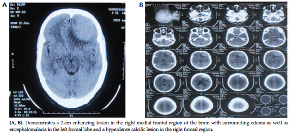
Figure 1. Computed Tomography Scan Before the Surgery
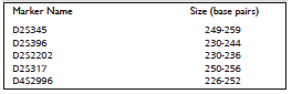
Table 1. Microsatellite Markers Used in This Study