Volume: 15 Issue: 2 April 2017
FULL TEXT
Chronic hepatitis in immunosuppressed patients because of infection with hepatitis E virus is increasingly recognized, but there is a paucity of knowledge about hepatitis E virus infection in solid-organ transplant recipients. Herein, we reported the first confirmatory case of hepatitis E virus genotype 4 infection in a 47-year-old woman who underwent a liver transplant recipient in Taiwan. The patient presented with unexplained hepatitis and severe jaundice. Hepatitis E virus RNA was present in serum and identified as the genotype 4 by reverse transcription polymerase chain reaction. Pathological findings revealed that prominent zone 3 canalicular and hepatocellular cholestasis with a few acidophilic bodies and giant cell transformation, which was typical of hepatitis E virus hepatitis. Even undergoing ribavirin treatment, she had worse cholestasis and recurrent urinary tract infections. She died from encephalopathy and sepsis 6 months after the initial presentation. So far, compared with genotype 3 hepatitis E virus hepatitis, genotype 4 hepatitis E virus infection in solid-organ transplant recipients has been reported less frequently in the literature, and that warrants further accumulation of experiences.
Key words : HEV, Liver recipient, Jaundice, Posttransplant infection
Introduction
Interest in hepatitis E virus (HEV) has increased in recent years because chronic infection, rather than a self-limited benign course, was not uncommonly observed in solid-organ recipients.1 Among these recipients with HEV infections, almost all cases were due to genotype 3 HEV (HEV3).2 Herein, we report the first confirmatory case of HEV genotype 4 (HEV4) infection in a previously stable liver transplant recipient, with a difficult initial diagnosis and a progressively detrimental course.
Case Report
A 47-year-old woman with hepatitis C-related liver cirrhosis and sustained virologic response after interferon and ribavirin treatment, received an uneventful living-related liver transplant 4 years earlier. She had acute flulike symptoms, spiking fever, and jaundice for 2 weeks. There was no uncooked food eating, travel, or animal contact history. First round of infection surveys, including HEV serology, were all negative. Empiric antibiotics, antifungal, and anticytomegaloviral agents were used for fever control with tapering tacrolimus, the immunosuppressant. She was referred to our transplant center because of prolonged and progressive jaundice 1 month after the first fever episode. An image study showed no biliary tree obstruction or stricture. Serum HEV RNA was positive and identified as the genotype 4 by reverse transcription polymerase chain reaction. Pathological examination of liver biopsy revealed prominent zone-3 canalicular and hepatocellular cholestasis, with a few acidophilic bodies and giant cell transformation, which was consistent with HEV hepatitis (Figure 1). No rejection sign was noted. Even under ribavirin 600 mg per day, she had worse cholestasis with serum bilirubin (blood concen-tration, 644.7 μmol/L) and intermittent urinary tract infections. She died of sepsis 6 months after the initial presentation of HEV hepatitis. Detailed clinical events, serum blood biochemistry, and serum levels of the immuno-suppressant because acute flulike symptoms are shown in Figure 2.
Discussion
Hepatitis E virus-4 infection, usually acquired by the consumption of undercooked pork, offal, sausages, or mussels,3 are prevalent commonly in Asia, including Taiwan.4 Kamar and associates demonstrated that more than 60% of solid-organ recipients infected with HEV developed chronic hepatitis, and 10% of them progressed to cirrhosis.5 Great quasispecies heterogeneity, a weak inflammatory response, and high serum concentrations of the chemokines involved in leukocyte recruitment to the liver in the acute phase were associated with persistent HEV infection in solid-organ transplant patients.6 Further, slow quasispecies diversification during the first year of infection was associated with rapidly developing liver fibrosis.6
The clinical features of our patient were different from the immunocompetent patients in that highly elevated serum bilirubin level occurred in the acute stage without marked elevation of liver enzymes, which hindered early diagnosis. The disease pattern of HEV4 infection may be of more frequent jaundice and with a more severe presentation compared with HEV3 in a French general population.7 Serological diagnosis of HEV is not reliable because sero-conversion may never occur in immunosuppressed patients1 and therefore, HEV viral load should be checked. Histology of HEV infection under immuno-suppression in the early phase is distinct from HEV infection in immunocompetent individuals.8 Molecular testing with a reverse transcription polymerase chain reaction assay designed for detecting the HEV open reading frame 2/3 gene region in liver biopsies is another powerful tool to evaluate liver transplant recipients with elevated transaminases of unknown origin.8
A dosage reduction of immunosuppressants is the first-line therapy that can result in a viral clearance in more than 30% of patients.5 Risk factors associated with failure of HEV clearance after acute infection included the degree of immunosuppression, the time between the last episode of acute rejection and HEV infection, time since transplant, low leucocyte count, low total-lymphocyte count, low T-cell count, and use of tacrolimus and thrombocytopenia.1,5 A recent, retrospective, multicenter study revealed that HEV clearance was observed in 95% of 59 solid-organ transplant recipients undergoing ribavirin treatment for a median of 3 months.9 The antiviral activity of ribavirin against wild-type genotype 1, 2, and 3 strains were confirmed in vitro, except genotype 4.10 The outcome of our patient under ribavirin monotherapy was not as good, which might be because of the delayed diagnosis or the varied therapeutic response of different HEV genotypes. Further study of HEV4 therapeutics is needed, especially in endemic areas.
In summary, our case report provides additional information on the clinical course of HEV4 infection in liver transplant recipients. We suggest you consider HEV infection in immunosuppressed patients with an unknown cause of viral hepatitis, not only in endemic HEV3 infection areas, but also in other places, such as Japan and China.
References:
- Kamar N, Selves J, Mansuy JM, et al. Hepatitis E virus and chronic
hepatitis in organ-transplant recipients. N Engl J Med. 2008;358(8):811-817.
CrossRef - PubMed - Unzueta A, Rakela J. Hepatitis E infection in liver transplant
recipients. Liver Transpl. 2014;20(1):15-24.
CrossRef - PubMed - Kamar N, Bendall R, Legrand-Abravanel F, et al. Hepatitis E. Lancet.
2012;379(9835):2477-2488. Erratum in: Lancet. 2012;380(9843):730.
CrossRef - PubMed - Lee JT, Shao PL, Chang LY, et al. Seroprevalence of Hepatitis E Virus
Infection among Swine Farmers and the General Population in Rural Taiwan.
PLoS One. 2013;8(6):e67180.
CrossRef - PubMed - Kamar N, Garrouste C, Haagsma EB, et al. Factors associated with chronic
hepatitis in patients with hepatitis E virus infection who have received
solid organ transplants. Gastroenterology. 2011;140(5):1481-1489.
CrossRef - PubMed - Lhomme S, Abravanel F, Dubois M, et al. Hepatitis E virus quasispecies
and the outcome of acute hepatitis E in solid-organ transplant patients. J
Virol. 2012;86(18):10006-10014.
CrossRef - PubMed - Jeblaoui A, Haim-Boukobza S, Marchadier E, Mokhtari C, Roque-Afonso AM.
Genotype 4 hepatitis E virus in France: an autochthonous infection with a
more severe presentation. Clin Infect Dis. 2013;57(4):e122-e126.
CrossRef - PubMed - Protzer U, Böhm F, Longerich T, et al. Molecular detection of hepatitis
E virus (HEV) in liver biopsies after liver transplantation. Mod Pathol.
2015;28(4):523-532.
CrossRef - PubMed - Kamar N, Izopet J, Tripon S, et al. Ribavirin for chronic hepatitis E
virus infection in transplant recipients. N Engl J Med.
2014;370(12):1111-1120.
CrossRef - PubMed - Debing Y, Emerson SU, Wang Y, et al. Ribavirin inhibits in vitro
hepatitis E virus replication through depletion of cellular GTP pools and is
moderately synergistic with alpha interferon. Antimicrob Agents Chemother.
2014;58(1):267-273.
CrossRef - PubMed

Volume : 15
Issue : 2
Pages : 228 - 230
DOI : 10.6002/ect.2015.0031
From the 1Departments of Surgery, 2Pathology, and
3Internal Medicine National Taiwan University Hospital and National Taiwan
University College of Medicine, Taipei, Taiwan
Acknowledgements: We thank Prof. Shiou-Hwei Yeh for laboratory support.
The authors declare that they have no sources of funding for this study, and
they have no conflicts of interest to declare.
Corresponding author: Cheng-Maw Ho, MD, Department of Surgery, National
Taiwan University Hospital, 7 Chung-Shan South Road, Taipei 100, Taiwan
Phone: +886 2 2 312 3456 65914
Fax: +886 2 2 356 8810
E-mail: miningho@ntu.edu.tw
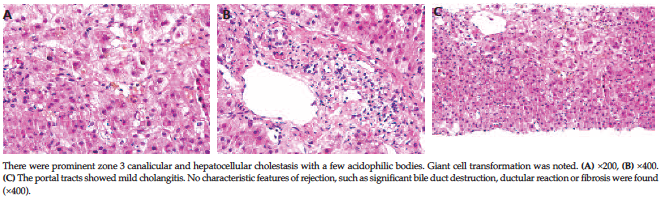
Figure 1. Pathologic Presentation of Hepatitis E Viral Genotype 4 Infection in a Liver Transplant Recipient
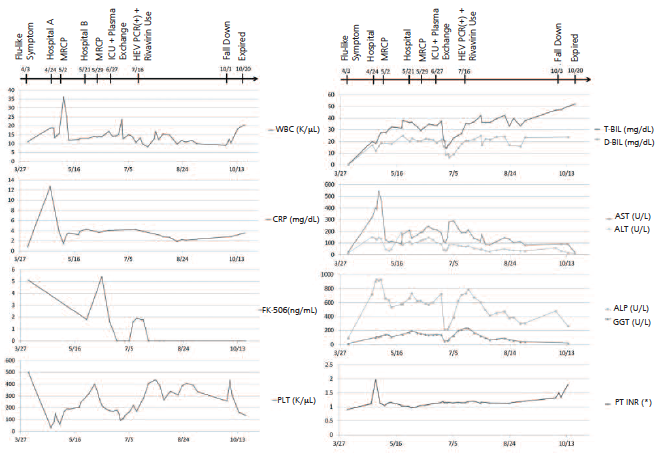
Figure 2. Clinical Events, Serum Blood Biochemistry, and Serum Levels of the Immunosuppressant Since the First Episode of Flulike Symptoms