Volume: 15 Issue: 2 April 2017
FULL TEXT
Infections are Achilles heel of kidney transplant recipients. Opportunistic fungal infections are increasingly recognized in these patients. We report a case of kidney transplant recipient with skin and soft tissue infection caused by Pyrenochaeta romeroi, a dematiaceous fungi. Infection by this organism is rare.
Key words : Fungal infection, Kidney transplant recipient, Opportunistic infection
Introduction
Phaeohyphomycosis is an opportunistic infection caused by dematiaceous fungi. Though ubiquitous, these organisms rarely cause human disease. However, in immunosuppressed organ transplant recipients, they are increasingly recognized as human pathogens. Here, we present a case of a kidney transplant patient with nodular lesions of the skin who had a diagnosis of phaeohyphomycosis caused by Pyrenochaeta romeroi.
Case Report
A 43-year-old farmer presented with painless nodular lesions over his left thigh, left calf, and right shin. Six months earlier, he underwent a kidney transplant with wife as the donor. For transplant, he received antithymocyte globulin for induction. Maintenance immunosuppression was accomplished with tacrolimus, mycophenolate mofetil, and prednisolone. He was also given valganciclovir prophylaxis for 100 days after the transplant. Except for new onset diabetes mellitus after the transplant, his posttransplant course was uneventful, and he had stable graft function.
As a farmer, he had a history of working on a farm and in a garden after the transplant. He had no history of fever or decreased urine output. On examination, the patient was hemodynamically stable. Multiple painless nodular lesions were seen over his left thigh, left calf, and right shin measuring about 1 to 1.5 cm in diameter. Systemic examination was unremarkable (Figure 1).
On investigation, he had hemoglobin of 15.6 g/dL, a total leucocyte count of 7250/mm3. His differential leucocyte count showed 89% neutrophils, 6% lymphocytes, and 5% monocytes. His platelet count was 1.53× 10⁹/L. Blood urea nitrogen was 4.9 mmol/L (14 mg/dL) and serum creatinine was 106.8 μmol/L (1.2 mg/dL). A urine examination was normal. An excision biopsy of the swollen left thigh showed granuloma with fungal hyphae. Aspirates from the lesions grew organisms belonging to the group called Phaeohyphomycosis. DNA sequencing showed that this organism was Pyrenochaeta romeroi. He was treated initially with itraconazole and terbinafine along with a reduction in his immunosuppression medications. During the course of the next 2 months, the lesions increased. New lesions were seen on his upper limbs and lower limbs (Figure 2). Some of the lesions developed ulcerations. Surgical debridement of the lesions was performed, and the patient was started on voriconazole. After starting this treatment, no new lesions were observed. The patient was continued on voriconazole.
Discussion
This patient had an unusual fungal infection caused by Pyrenochaeta romeroi. This dematiaceous fungus belongs to a group called Phaeohyphomycosis that has characteristic melanin like pigment in their cell wall.1 Pyrenochaeta is widely distributed in the soil or in association with wood and plant debris. This patient may have acquired this infection while farming.
Infection by this particular organism is rare. To the best of our knowledge, only 4 cases in a nontransplant setting and 3 cases in transplant recipients have been described to date.2-8 Nodulocystic lesion, subcutaneous abscess, and lesions mimicking plantar warts can be caused by this organism and have been described in kidney transplant recipients. Infection with Pyrenochaeta romeroi at multiple sites that cause such extensive lesions have not been described.
Treatment of this fungal infection can be difficult. No standard-of-care guidelines are available. The organism is usually resistant to fluconazole, ketoconazole, amphotericin B, and flucytosine. Voriconazole and posaconazole have been found to be effective in some cases.4,9 The combination of surgery and antifungal therapy can yield good results. Although not evidence based, a reduction of immunosuppression may quicken the patient’s recovery. One important consideration is drug interaction while using triazoles. Triazoles can increase the blood levels of tacrolimus. Careful dose modification with therapeutic drug monitoring is needed during treatment.
References:
- Revankar SG, Sutton DA.Melanized fungi in human disease.Clin Microbiol
Rev. 2010;23(4):884-928.
CrossRef - PubMed - Badali H, Chander J, Gulati N, et al. Subcutaneous phaeohyphomycotic
cyst caused by Pyrenochaeta romeroi. Med Mycol. 2010;48(5):763-768.
CrossRef - PubMed - Girard C, Dereure O, Rispail P, Durand L, Guilhou JJ. Subcutaneous
phaeohyphomycosis due to Pyrenochaeta romeroi in a patient with leprosy.
Acta Derm Venereol. 2004;84(2):154-155.
CrossRef - PubMed - Cerar D, Malallah YM, Howard SJ, Bowyer P, Denning DW. Isolation,
identification and susceptibility of Pyrenochaeta romeroi in a case of
eumycetoma of the foot in the UK. Int J Antimicrob Agents.
2009;34(6):617-618.
CrossRef - PubMed - Khan Z, Ahmad S, Kapila K, et al. Pyrenochaeta romeroi: a causative
agent of phaeohyphomycotic cyst. J Med Microbiol. 2011;60 (Pt 6):842-846.
CrossRef - PubMed - Mathuram Thiyagarajan U, Bagul A, Nicholson ML. A nodulo-cystic
eumycetoma caused by Pyrenochaeta romeroi in a renal transplant recipient: A
case report. J Med Case Rep. 2011;5:460.
CrossRef - PubMed - Ocampo MA, Kanitakis J, Bienvenu AL, Chauvet C, Euvrard S.
Phaeohyphomycosis caused by Pyrenochaeta romeroi mimicking a plantar wart in
a kidney transplant recipient. Transpl Infect Dis. 2012;14(6):E173-E174.
CrossRef - PubMed - Chan YY, Tan AL, Tan BH. Subcutaneous abscess due to Pyrenochaeta
romeroi in a renal transplant recipient. Singapore Med J.
2014;55(4):e64-e66.
CrossRef - PubMed - Ogawa MM, Galante NZ, Godoy P, et al. Treatment of subcutaneous
phaeohyphomycosis and prospective follow-up of 17 kidney transplant
recipients. J Am Acad Dermatol. 2009;61(6):977-985.
CrossRef - PubMed

Volume : 15
Issue : 2
Pages : 226 - 227
DOI : 10.6002/ect.2014.0283
From the 1Department of Nephrology, Father Muller Medical College,
Mangalore, Karnataka; the 2Department of Nephrology, Seth GS Medical
College and KEM Hospital, Mumbai, Maharashtra; and the 3Department of
Microbiology, Seth GS Medical College and KEM Hospital, Mumbai, Maharashtra,
India
Acknowledgements: The authors would like to acknowledge the help of Dr.
Jacques Meis (The Netherlands) for DNA analysis of the organism. The authors
declare that they have no sources of funding for this study, and they have no
conflicts of interest to declare.
Corresponding author: Manjunath Kulkarni, Associate Professor, Department
of Nephrology, Father Muller Medical College, Kankanady, Mangalore 575002,
Karnataka, India
Phone: +91 77 9506 9393
Fax: +08 2 4243 6352
E-mail: drmjkulkarni@gmail.com
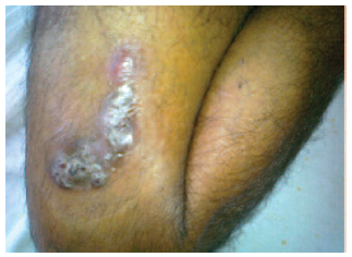
Figure 1. Nodular Lesion of Thigh
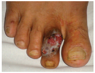
Figure 2. Ulcerative Nodule on the Second Toe