Volume: 15 Issue: 2 April 2017
FULL TEXT
Objectives: The aim of this study was to evaluate the incidence, possible risk factors, clinical presentation, and follow-up of patients with a gastrointestinal perforation after liver transplant.
Materials and Methods: We did a retrospective chart review of all patients who presented with a gastrointestinal perforation after liver transplant at our liver transplantation center between December 2009 and June 2011.
Results: In total, we performed 271 liver transplants during this period. Nine patients (3.3%), 5 pediatric and 4 adult patients (median age, 21.3 ± 16.2 y; range, 1-55 y), developed a gastrointestinal perforation after liver transplant. Six of the patients had living donors, and 3 had deceased-donor transplants. Four patients underwent prior abdominal surgery for unrelated reasons. The mean time between liver transplant and diagnosis of the gastrointestinal perforation was 12.9 ± 9.3 days (range, 4-30 d), and the mean hospitalization length was 50 ± 29.3 days (range, 18-102 d). Perforations were located in the stomach (n=1), jejunum (n=3), ileum (n=2), jejunum and ileum (n=1), and colon (n=2). Seven patients were managed by ostomies, and 2 by primary repair. Despite administration of proper antibiotic therapy and fluid resuscitation to all patients, 2 adults died of septic shock: 1 was caused by perforation and 1 was caused by anastomotic leakage after colostomy closure.
Conclusions: A gastrointestinal perforation after a liver transplant is a rare but mortal complication. Considering delayed wound healing owing to immunosuppression, potentially larger ischemic tissue around the perforation site owing to cautery burns and the atypical clinical course that may be further masked by bile leakage, ostomy treatment should be preferred to primary repair. A loop ostomy for small and large bowel perforations after the liver transplant decreases mortality and morbidity.
Key words : Gastrointestinal perforation, Complication, Liver transplant
Introduction
Enormous progress has been achieved since the first liver transplant (LT) was performed in humans in 1963. Since then, LT has established therapeutic effectiveness for patients with end-stage liver disease. However, it is not possible to completely prevent complications after LT.1, 2 While some complications are caused by using high-dose steroids and immunosuppressants, others develop as a result of surgery length, thromboembolic events, or opportunistic infections. Given that the liver is highly susceptible to iatrogenic injury during transplant, circulatory dysfunction may occur upon portal clamping, and damage to the gastrointestinal mucosal barrier can be induced by high-dose steroids, posttransplant complications are observed most commonly in gastrointestinal organs.1-14
Gastrointestinal complications are common among patients who undergo solid-organ transplant and range from mild to moderate conditions, such as diarrhea or nausea, to more severe, life-threatening complications such as digestive bleeding and gut perforation.5 Gastrointestinal perforation (GIP), which is potentially lethal, may occur at any point in the gastrointestinal tract, including the stomach, jejunum, ileum, and colon.1 Gastrointestinal perforation is more common in children than in adults and causes surgical morbidity and mortality. A delayed diagnosis can be life-threatening; however, early diagnosis is difficult because the large doses of steroids and immunosuppressants often given to LT patients may produce atypical clinical features.1 The cause of GIP after LT is unclear; possible risk factors include previous laparotomy, prolonged surgery, subsequent laparotomy, portal vein thromboembolism, treatment with high-dose steroids, and cytomegalovirus (CMV) infection.2-4 Here, we report the results of GIP after LT in adult and pediatric patients and analyze the associated risk factors, with specific emphasis on ostomies.
Materials and Methods
In total, 271 cases (231 adults and 40 children) underwent LT at the Liver Transplantation Center of Malatya Inonu University Medical Faculty between December 2009 and June 2011. During the postoperative follow-up, GIP occurred in 9 patients (4 adults and 5 children). A retrospective evaluation of the medical records of these 9 patients included the following: sex, age, primary disease, surgical history prior to transplant, immunosuppressant and other drugs used after transplant, blood biochemistry analysis results, radiologic findings, details of the perforation, Model for End-Stage Liver Disease score, Child-Turcotte-Pugh score, Pediatric End-Stage Liver Disease score, surgical approach, postoperative follow-up, conversion of immunosuppressive drugs, stoma care, hydration status, and culture results. The clinicopathological characteristics, operative history, surgical techniques, and postsurgical outcomes of the GIP cases are summarized in Tables 1 and 2 for adult and pediatric patients.
Six of the 9 cases underwent living-donor liver transplant (LDLT), and deceased-donor liver transplant was performed in 3 cases. Of the 6 LDLT cases, 3 received a right lobe, and 3 received a left lobe lateral segment. Diligent scrutiny of the case histories revealed that 2 pediatric patients had biliary atresia and underwent a Kasai type portoenterostomy (at 4 and 6 weeks after birth). Another pediatric patient, with Budd-Chiari syndrome, had been operated on for a hydatid cyst 1 year earlier. One adult patient had a cholecystectomy 10 years earlier. One deceased-donor LT patient had an ABO mismatch. Decompensate cirrhosis was prominent in 6 cases: 1 Wilson cirrhosis, 2 biliary atresia, 1 hepatitis B virus (HBV) infection, 1 hepatitis D virus infection, and 1 Budd-Chiari syndrome. Acute fulminant hepatic failure occurred in 3 patients (2 HBV infections, 1 acute phosphorus intoxication). Four pediatric cases were closed with a Bogota bag owing to a large liver size.
All cases received hydrocortisone (diminishing from 0.5 g/d to 0.25 mg/kg/d in adults, and diminishing from 100 mg/d to 0.25 mg/kg/d in children; stopped at postoperative month 6). In addition to steroids, cyclosporine (5-10 mg/kg/d) was preferred for pediatric patients. Tacrolimus (0.01-0.05 mg/kg/d) and mycophenolate mofetil were added to the steroid treatment in adult patients. Sirolimus was begun after cessation of calcineurin inhibitors in patients with creatinine clearance > 132.6 µmol/L.7 In the 7 patients with a stoma, follow-up of the daily volume of stoma output and 24-hour urinary output was calculated. Blood testing was analyzed on alternate days for cases with impaired renal function. Patients with a high stoma output volume or those with impaired kidney function received hydration and electrolyte supplementation.
Results
The records of 9 patients (3 females and 6 males; 3 adult and 6 pediatric cases; age range, 1-55 y; mean age, 21.3 ± 16.2 y) who experienced GIP after LT were retrospectively investigated. Mean portal venous clamping time was 37.5 minutes (range, 25-65 min). Mean cold ischemia time was 63 minutes (range, 50-75 min) for living-donor LT and 11.5 hours (range, 11-12 h) for deceased-donor LT. Mean operation time for LT was 9.22 hours (range, 8-11.5 h), with a mean intraoperative blood loss of 893 mL (range, 150-2000 mL). At the time of GIP diagnosis, mean white blood cell count was 12.8 ± 9.2 × 10⁹/L (range, 2.2-30 × 109/L). Mean time between LT and the diagnosis of a bowel perforation was 12.9 ± 9.3 days (range, 4-30 d), and mean hospitalization was 50 ± 29.3 days (range, 18-102 d).
The 3 adult cases with GIP complained of sudden subtle or moderate abdominal pain and abdominal tenderness without rebound tenderness. The decision to operate was made when abdominal ultrasonography revealed free fluid in the abdomen. One adult case was diagnosed based on bile mixed with food particles in the drainage fluid. Body temperature ranged from 37.5°C -39°C. Three cases were diagnosed as spontaneous, and 1 was an iatrogenic perforation. The adult patient with a peptic ulcer perforation (1 × 1.5 cm) at the antrum was treated with a Graham reconstruction, but died of sepsis on postoperative day 18. Another patient had an intraoperative cautery wound at the hepatic flexure, which was treated initially by primary closure. A Hartmann colostomy was performed on postoperative day 13 owing to leakage. The colostomy was closed after 2 months, but an anastomotic leakage and pericolonic abscess occurred. Despite an emergency colostomy, the patient died of multiple organ dysfunction and sepsis. During follow-up, no complications were encountered in the other 2 adult cases who received a loop jejunostomy. In both patients, the jejunostomy was closed without complications at postoperative week 6.
Three of the 6 pediatric patients developed nausea, vomiting, fever, and signs of an acute abdomen, which prompted ultrasonographic examination. The other 3 patients were diagnosed based on gastric content in the transparent Bogota bag. Four pediatric patients had a loop ileostomy during repair, with no significant postoperative problems. One pediatric case with an ileal perforation was diagnosed with another perforation at the jejunum, and a jejunostomy was added to the loop ileostomy. Only 1 case, who had an anastomotic biliary stricture treated with internal and external catheterization, was hospitalized until closure of the ostomy. No perforation-associated deaths were recorded among the 6 children, who were closely followed and received proper replacement therapy.
Drain cultures and an antibiogram were performed routinely in all patients with GIP. Enterococci were detected in 2 adult patients. No patients developed CMV or acute cellular rejection before the event.
The stoma output volume was 500-6000 cc in the 7 patients with an ostomy. Different dosages of diphenoxylate hydrochloride were administered in 4 cases with high stomal output. In 2 adult cases with a jejunostomy, creatinine levels remained between 212.1 and 291.7 µmol/L despite fluid resuscitation and electrolyte replacement. Sirolimus treatment was started, and cyclosporine (case 2) and tacrolimus (case 3) were stopped because of their nephrotoxic effects. Creatinine levels returned to normal in both cases during follow-up. Owing to a jejunostomy output of 4000-6000 cc, one pediatric patient was followed in the hospital until the ostomy was closed; kidney functions were maintained within normal limits.
Discussion
Since the first successful liver transplant was performed by Starzl in 1967, improved surgical techniques, antirejection treatment, and postoperative management have made LT the treatment of choice for pediatric and adult patients with end-stage liver disease or acute hepatic failure.2, 3 However, complications can still occur after LT. Gastrointestinal perforation after LT is a cause of surgical morbidity, and a delayed diagnosis may create a life-threatening situation. The cause of GIP after LT is unclear, but contributing factors include previous abdominal surgery (particularly the Kasai procedure for biliary atresia in children), serosal injury or devascularization of the bowel wall, prolonged LT procedure, retransplant, transfusions, posttransplant intra-abdominal bleeding requiring reoperation, early portal vein thrombosis, high-dose steroid therapy, poor nutritional status, and CMV infection.2-4, 9-11
The incidence of GIP after LT is 1% to 5.3% in adults and 8.3% to 14% in children.1, 3, 4, 9-11 The higher incidence in children is most likely attributable to tight adhesions of the liver and formation of intestinal loops during portoenterostomy before LT. When we look at the our literature analysis, the incidence ratio of GIP for adult and pediatric cases are 3.2% and 2.5%-35% (Table 3). Further, 115 (95%) of the 121 perforation cases were pediatric, whereas the remaining 6 were adult cases.1-4, 8-12 We documented that most of the pediatric GIP cases were patients who had undergone a portoenterostomy owing to biliary atresia. Thus, a portoenterostomy procedure performed before LT in children appears to be a serious risk factor.
Liver transplant is often accompanied by steroid therapy to decrease inflammation by suppressing macrophages, lymphocytes, and other inflammatory factors. As steroids delay wound healing and increase the risk for peptic ulceration owing to decreased gastric mucus secretion and increased gastric acid and pepsin production, patients receiving steroid treatment may be predisposed to perforations.1, 3 However, the claim that steroids are a risk factor for perforation is controversial; some studies have reported steroids as a risk factor, whereas others have not. Given the frequent administration of steroids in LT, the low rate of perforation would argue against steroids as a risk factor.9
One cause of perforation may be unnoticed mucosal abrasions resulting from extensive use of cautery, as in 1 of our adult patients. Postoperative bleeding necessitating reoperation developed in 1 pediatric and 1 adult case. Other possible causes such as postoperative early portal vein thrombosis, prolonged portal vein clamping, poor nutritional status, and CMV infection were not observed in our series. Gastrointestinal perforation is usually identified between 7 and 186 days after a transplant, but the diagnosis remains difficult because immunosuppression tends to alter clinical symptoms and signs. A high degree of suspicion is needed, as a delay in diagnosis is associated with significant morbidity and mortality.8-12 Despite developments in LT surgery, mortality owing to sepsis secondary to GIP is as high as 30% to 50%, and is 30% to 78% after reperforation surgery.9, 11 However, some studies have reported 0% mortality despite a high perforation rate.10, 12, 13
The signs and symptoms of perforation are usually overt in adults, but careful examination of the Bogota bag may be necessary to detect the only sign of perforation in a younger patient. Ultrasonography is usually sufficient to diagnose free liquid in the abdomen, and a microbiological confirmation of enterococci in the aspirate or drain material indicates a perforation. In our series, all immunosuppressive agents were stopped and proper antibiotics with major supportive therapy were given before and after the repair. Immune suppressants were not resumed before 2 to 5 days after repair. No acute rejection occurred using this methodology.
Gastrointestinal perforation after LT may occur in any part of the gastrointestinal tract. Overall, the highest incidence occurs in the Roux loop, ileum, colon, jejunum, duodenum, and stomach.9-12
We conducted a systematic literature search of the PubMed and Google Scholar databases using the search term "gut or gastrointestinal perforation and liver transplant," and investigated the full-texts and/or summaries of about 31 articles published between 1979 and 2011. Overall, GIPs in different locations were reported after 200 (5.7%) of 3512 LT operations. Most of the GIP cases were pediatric, and a sizable number of these perforations were related to biliary atresia. Nine of these studies referenced the topic "gastrointestinal perforations occurring after transplant." Table 3 summarizes the larger-scale studies in chronologic order.
Sanada and associates2 reported that GIPs in 4 of 148 pediatric patients undergoing LT, and all of these cases had a history of several surgeries performed owing to biliary atresia. Dehghani and associates3 reported that GIP developed in 4 of 72 pediatric patients undergoing LT; 3 of the 4 had previously undergone the Kasai operation for biliary atresia, and 1 had received high-dose steroid treatment. Xiong and associates1 reported GIPs in 6 of 187 patients who underwent OLT, and 4 of these patients had a history of previous operations. Beierle and associates4 described 158 OLTs performed in 128 pediatric patients, with GIPs occurring in 10 of the them. The authors proposed that CMV infection, steroid use, and a history of the Roux-en-Y operation could not be regarded as risk factors, based on their observations that CMV infection was detected in many of the patients, and that perforation occurred in only 6.4% of the cases despite steroid administration to all patients.
Melendez and associates10 found GIP as a complication in 13 of 194 patients who underwent OLT; the cause in 10 of the patients was biliary atresia, and another 10 patients had undergone previous surgery. Soubrane and associates9 reported perforation in 10 of 51 pediatric patients undergoing OLT; these patients had a history of the Kasai operation for biliary atresia. The authors suggested that factors such as splanchnic congestion, prolonged portal venous clamp time, portal vein thrombosis, and repeated trauma to the bowel caused by reoperation may lead to perforations. Yamanaka and associates12 reported on 119 OLT operations in 105 pediatric cases, and 37 of the cases developed GIP. They documented that 22 of the 37 patients had undergone no previous abdominal surgery, whereas 15 had a history of more than 1 surgical procedure. The authors suggested that CMV infection and steroid use were not directly related to the development of GIP, but that a history of surgical operations constituted a risk factor.
Shaked and associates11 documented GIPs in 24 of 246 pediatric patients who underwent orthotopic LT, and 22 of these patients had undergone previous liver-related surgeries. The authors reported difficult dissections owing to previous surgeries, and the duration of the transplant operation as the most important risk factors predicting the development of GIP. Marujo and associates8 found GIPs in 12 of 500 patients undergoing LT, and all were pediatric cases. Cytomegalovirus-related gastroenteritis was documented in only 1 case.
Creating an intestinal stoma is one of the most effective methods for preventing intra-abdominal sepsis in cases of GIP and anastomosis leakage. Although a loop ileostomy is the most commonly preferred method, jejunostomy and colostomy also may be performed for proximally and distally located leakages.
The most frequent complication in proximally located intestinal stomas is dehydration related to high stomal output. Dehydration rates after ileostomy are 0.8% to 20%. Dehydration is responsible for renal failure in 50% of cases who develop renal failure, and high stomal output is the dehydration culprit in 44% of cases.6 These ratios are highly significant in patients undergoing LT and receiving immunosuppressant agents, as renal function is either already impaired or on the verge of impairment in a sizable number of patients undergoing LT. Additionally, calcineurin inhibitors, including cyclosporine and tacrolimus, and prophylactic antibiotics used after transplant are toxic to the kidneys. Therefore, hydration is significant, even in uncomplicated transplant patients. Accordingly, transplant patients with GIP and a stoma should be monitored closely for any possible disturbances in stomal output, electrolyte balance, and fluid intake and output. The levels of blood urea nitrogen, creatinine, blood tacrolimus, and blood cyclosporine should be checked daily or on alternate days. Sirolimus treatment was initiated alternatively in 2 of the present cases with ongoing deterioration of kidney function despite appropriate fluid and electrolyte replacement. Kidney function test results returned to normal in both patients during follow-up. We recommend close monitoring of immunosuppressive drug levels in cases that become dehydrated and suggest a change to nonnephrotoxic medications when possible.7
The treatment of choice should be an ostomy, rather than primary repair, because this method decreases mortality, as seen in our pediatric patients. Loop ostomy is preferred for ileal or jejunal perforations, regardless of the level of injury. A protective ileostomy, rather than a Hartmann pouch, should be used for colonic perforations. The extent of ischemic injury owing to colonic cautery damage may be greater than the perforation itself; thus, a loop colostomy or loop ileostomy with primary repair is preferred. Diminished intraluminal pressure minimizes the risk for anastomotic leakage and related complications.
In conclusion, consensus has been reached regarding previous abdominal surgery, long duration of LT, posttransplant intra-abdominal bleeding requiring reoperation, splanchnic congestion, and portal vein thrombosis as the most-important risk factors for developing of GIP after LT. Nevertheless, debate persists over steroid use and CMV infection as risk factors. Primary repair and a protective ileal ostomy for colonic injuries, and a loop ostomy for perforations at all other sites are preferred in transplant patients with GIP.
References:
- Xiong J, You S, He XS. Gut perforation after orthotopic liver transplantation
in adults. World J Gastroenterol. 2007;13(14):2125-2128.
PubMed - Sanada Y, Mizuta K, Wakiya T, et al. Bowel perforation after pediatric living
donor liver transplantation. Pediatr Surg Int. 2011;27(1):23-27.
CrossRef - PubMed - Dehghani SM, Nikeghbalian S, Kazemi K, et al. Outcome of bowel perforation
after pediatric liver transplantation. Pediatr Transplant. 2008;12(2):146-149.
CrossRef - PubMed - Beierle EA, Nicolette LA, Billmire DF, Vinocur CD, Weintraub WH, Dunn SP.
Gastrointestinal perforation after pediatric orthotopic liver transplantation. J
Pediatr Surg. 1998;33(2):240-242.
CrossRef - PubMed - Herrero JI, Benlloch S, Bernardos A, et al. Gastrointestinal complications in
liver transplant recipients: MITOS study. Transplant Proc. 2007;39(7):2311-2313.
CrossRef - PubMed - Beck-Kaltenbach N, Voigt K, Rumstadt B. Renal impairment caused by temporary
loop ileostomy. Int J Colorectal Dis. 2011;26(5):623-626.
CrossRef - PubMed - Rogers CC, Johnson SR, Mandelbrot DA, et al. Timing of sirolimus conversion
influences recovery of renal function in liver transplant recipients. Clin
Transplant. 2009;23(6):887-896.
CrossRef - PubMed - Marujo WC, Stratta RJ, Langnas AN, Wood RP, Markin RS, Shaw BW Jr. Syndrome
of multiple bowel perforations in liver transplant recipients. Am J Surg.
1991;162(6):594-598.
CrossRef - PubMed - Soubrane O, el Meteini M, Devictor D, Bernard O, Houssin D. Risk and
prognostic factors of gut perforation after orthotopic liver transplantation for
biliary atresia. Liver Transpl Surg. 1995;1(1):2-9.
CrossRef - PubMed - Vilca Melendez H, Vougas V, Muiesan P, et al. Bowel perforation after
paediatric orthotopic liver transplantation. Transpl Int. 1998;11(4):301-304.
CrossRef - PubMed - Shaked A, Vargas J, Csete ME, et al. Diagnosis and treatment of bowel
perforation following pediatric orthotopic liver transplantation. Arch Surg.
1993;128(9):994-998; discussion 998-999.
CrossRef - PubMed - Yamanaka J, Lynch SV, Ong TH, Balderson GA, Strong RW. Posttransplant
gastrointestinal perforation in pediatric liver transplantation. J Pediatr Surg.
1994;29(5):635-638.
CrossRef - PubMed - Peng T, Peng MH, Li LQ, et al. Intestinal perforation after combined
liver-kidney transplantation for a case of congenital polycystic disease. World
J Gastroenterol. 2004;10(18):2769-2771.
PubMed - Koep LJ, Peters TG, Starzl TE. Major colonic complications of hepatic
transplantation. Dis Colon Rectum. 1979;22(4):218-220.
CrossRef - PubMed

Volume : 15
Issue : 2
Pages : 189 - 195
DOI : 10.6002/ect.2012.0061
From the Department of Surgery, Inonu University Faculty of Medicine, Division
of Liver Transplantation, 44280 Malatya, Turkey
Acknowledgements: The authors declare that they have no conflicts of interest, Mehmet Yilmaz and Sezai Yilmaz performed the surgical procedure; Sami Akbulut
and Mehmet Yilmaz contributed to the writing of the article and review of the
literature and undertaking a comprehensive literature search; Sami Akbulut and
Sezai Yilmaz contributed to the design and manuscript preparation.
Corresponding author: Sami Akbulut, Department of Surgery, Division of Liver
Transplantation, Inonu University Faculty of Medicine, 44280, Malatya, Turkey
Phone: +90 422 3410660
Fax: +90 422 3410036
E-mail:
akbulutsami@gmail.com
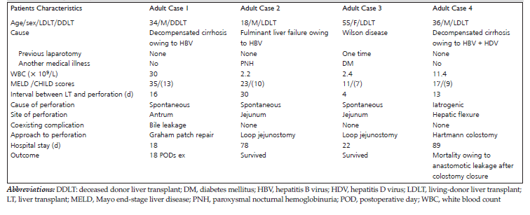
Table 1. Clinicopathologic Characteristics of Adult Patients With Gastrointestinal Perforation After Liver Transplant
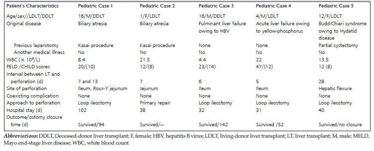
Table 2. Clinicopathologic Characteristics of Pediatric Patients With Gastrointestinal Perforation After Liver Transplant
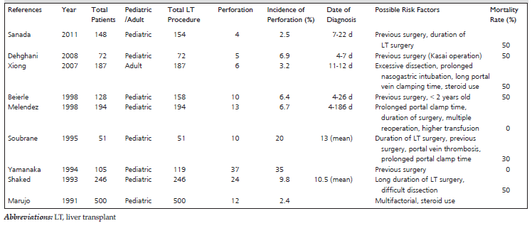
Table 3. Summary of 9 Studies Related to Gastrointestinal Perforation After Liver Transplant: A Short Literature Review