Volume: 14 Issue: 3 June 2016
FULL TEXT
Objectives: Our objective was to evaluate the effects of contrast-enhanced ultrasonography in monitoring microcirculation after rat liver ischemia-reperfusion injury.
Materials and Methods: Male Wistar rats (n = 36) were divided into sham-operated and ischemia-reperfusion groups. Rats in the ischemia-reperfusion groups underwent normothermic liver ischemia for 15 minutes followed by 1, 6, or 24 hours of reperfusion. At different time points, contrast-enhanced ultrasonography was performed to determine peak intensity in monitoring hepatic microcirculation. In addition, serum levels of alanine aminotransferase, aspartate aminotransferase, tumor necrosis factor α, and interleukin 1β levels were measured. Histopathologic changes were also observed.
Results: One hour after reperfusion, peak intensity values decreased, and serum levels of alanine aminotransferase, tumor necrosis factor α, and interleukin 1β increased significantly in the ischemia-reperfusion group compared with the sham-operated group. Histology results showed mild injury. Six hours after reperfusion, peak intensity values decreased continuously, serum levels of alanine aminotransferase, tumor necrosis factor α, and interleukin 1β decreased, and aspartate aminotransferase levels increased. Histology results showed severe injury compared with 1 hour after reperfusion. Twenty-four hours after reperfusion, peak intensity values increased, alanine aminotransferase and aspartate aminotransferase levels decreased, and histology results showed moderate injury compared with 6 hours after reperfusion. Peak intensity values were negatively correlated to alanine aminotransferase (P < .05; γ = -0.38) and aspartate aminotransferase (P < .01; γ = -0.78) levels.
Conclusions: Microcirculation dysfunction after liver ischemia-reperfusion injury can be monitored by contrast-enhanced ultrasonography. The perfusion of contrast agents negatively correlates to the severity of injuries.
Key words : Ultrasonography, Contrast media, Liver circulation, Injury, Ischemia-reperfusion
Introduction
Ischemia-reperfusion injury results from a prolonged ischemic insult followed by restoration of blood perfusion. It affects all oxygen-dependent cells that rely on an uninterrupted blood supply. These aerobic cells require mitochondrial oxidative phosphorylation for their energy supply. Consequently, all aerobically metabolizing tissues and organs are potential targets of ischemia-reperfusion injury.1,2
Regarding the liver, hepatic ischemia-reperfusion injury is commonly a consequence of prolonged portal triad clamping followed by reperfusion during surgical resection, performed as an elective preplanned procedure or as an emergency maneuver, to control excessive bleeding from the cut hepatic surface.2,3
Many techniques have been previously used for the study of hepatic microcirculation, such as the use of gamma-emitting radionuclide-labeled microspheres,4,5 laser Doppler flowmetry,6 and computed tomography.7 However, as a method in monitoring microcirculation in many other organs and tissues, contrast-enhanced ultrasonography (CEUS) has not still been mentioned in this area.8 In this study, we evaluated the effects of CEUS in monitoring hepatic microcirculation after rat liver ischemia-reperfusion injury.
Materials and Methods
Animals
Animal studies were approved by the institutional animal care committee of China Medical University. Inbred male Wistar rats, weighing 200 to 250 g, were used for experiments (Animal Center, China Medical University, Shenyang, China). Under monitored, pathogen-free conditions, at 22°C to 24°C, the rats were fed commercial pellets and water. Twelve hours before the procedure, only water was provided. Each experiment started at the same time of day to avoid the effects of circadian rhythm.
Operative procedure
Animals were anesthetized intraperitoneally with 10% chloral hydrate (3.5 mL/kg). The animals were placed in a supine position on a heating pad for maintaining a body temperature of 37.5°C to 38.5°C, monitored by a rectal thermometer. A polyethylene catheter was inserted into the femoral artery to monitor mean arterial blood pressure during each experiment. A polyethylene catheter, with a 3-way pipe connection, was inserted into the femoral vein for pretreatment with saline or ultrasonographic contrast agent (SonoVue, Bracco Suisse SA, Manno, Switzerland) infusion. In addition, an intraoperative saline infusion (2 mL) was also administered to compensate for intraoperative fluid loss and for collection of blood samples.
A median laparotomy was performed, and the liver was exposed. Warm ischemia was induced by the Pringle maneuver.9 The common portal vein, hepatic artery, and bile duct in the hepatoduodenal ligament were all clamped using a vascular clip. After an ischemic interval of 15 minutes, the clip was removed and reperfusion was started. After different durations of reperfusion, the microcirculation of hepatic parenchyma was monitored using CEUS. During the ischemia-reperfusion period, the animal's abdomen was covered with a plastic wrap to prevent fluid loss. After 45 minutes (15 min of ischemia and 30 min of reperfusion), the abdomen was closed. At the end of the 1, 6, or 24 hours of reperfusion, CEUS was performed and blood samples were taken from the inferior vena cava (about 2 mL). Blood samples were centrifuged (3000 revolutions/min for 10 min at room temperature), and the serum was stored at -80°C until further analyses. After completion of the procedure, animals were killed by exsanguination via the inferior vena cava, samples were taken for histologic analyses, and samples were placed in 4% formalin.
Experimental groups
Two groups of animals (n = 18 animals/group) were used. In the sham-operated group, rats underwent the surgical procedures described above except for the liver ischemia-reperfusion portion of the procedure. Laparotomy was performed, and the other previously mentioned procedures, including histologic analyses, use of CEUS, and collection of blood samples, were completed. In the ischemia-reperfusion group, rats underwent the surgical procedures described above. The groups were further divided into those that had 1-hour, 6-hour, and 24-hour ischemia-reperfusion injury.
Contrast-enhanced ultrasonography
To monitor hepatic microcirculation, we used CEUS with a Siemens Acuson Sequoia 512 (Siemens AG, Munich, Germany) using an endocavitary probe
(EV-8C4 with contrast pulse sequencing mode, Siemens). The contrast agents were dissolved in 5 mL of saline per 59 mg, with bolus injection of agents through the side port of the 3-way pipe connection (dose of 0.3 mL/kg). Saline was then injected immediately through the direct port, with a total injection volume of 1.5 mL. At the same time, the timer in the CEUS machine was started, with videos recorded for later analyses of time-intensity curves.
Time-intensity curves
Time-intensity curves were acquired automatically with the autotracking contrast quantification (ACQ) software (Siemens AG, Munich, Germany) in the CEUS machine. Regions of interest were selected in the whole parenchyma except for the hepatic vein. Peak intensity was also calculated automatically by the software.
Measurement of serum alanine aminotransferase and aspartate aminotransferase levels
Rat serum was analyzed within 24 hours by a laboratory for clinical chemistry results. Measurements of serum alanine aminotransferase (ALT) and aspartate aminotransferase (AST) levels were performed by standard spectrophotometry using an automated clinical chemistry analyzer (Hitachi 7600-210, Hitachi Medical Corporation, Tokyo, Japan).
Measurement of serum tumor necrosis factor α and interleukin 1β levels
Measurements of serum tumor necrosis factor α (TNF-α) and interleukin 1β (IL-1β) levels were performed using an ELISA method with a TNF-α and IL-1β immunoassay kit (Boster Bio-Engineering Limited Co., Wuhan, China) according to the manufacturer's instructions. The immunoassay was standardized using mouse TNF-α or IL-1β standards. A color reaction developed with a substrate containing equal volumes of stabilized hydrogen peroxide and tetramethylbenzidine. The color reaction was stopped after 20 minutes by the addition of hydrochloric acid. The absorbance of each sample was measured at 450 nm. From each data point (optical absorbance value), a value was calculated as a percentage of the average optical absorbance.
Histopathologic examination
The excised liver was fixed in 4% neutral buffered formalin for 24 hours, dehydrated, and embedded in paraffin. We used 3-μm thick sections for hematoxylin and eosin staining.
The following histopathologic changes were considered: sinusoidal width, hepatocellular swelling (vacuolization), desquamation of endothelial cells, and leukocyte infiltration. The pathologist was not informed of the applied treatment. All sections were studied using light microscopy (Nikon TS100 microscope, Tokyo, Japan).
Data collection and statistical analyses
Values are expressed as means ± standard deviation, with "n" representing the number of animals. Data were analyzed by 1-way analysis of variance and Pearson correlation analysis using SPSS software (SPSS: An IBM Company, version 13.0, IBM Corporation, Armonk, NY, USA). P < .05 was considered as statistically significant.
Results
Alanine aminotransferase and aspartate aminotransferase levels in serum
Serum ALT levels were significantly higher in the ischemia-reperfusion group than in the sham-operated group (P < .01). In the ischemia-reperfusion group, serum ALT levels were higher in the 1-hour group than in the 6-hour (P < .01) and 24-hour groups (P < .01), but higher in the 6-hour versus the 24-hour group (P < .05; Table 1).
In the ischemia-reperfusion groups, serum AST levels were significantly higher in the 6-hour (P < .01) and 24-hour (P < .05) groups than in the sham-operated group. However, there was no significant difference between the ischemia-reperfusion and sham-operated groups at 1 hour (P > .05). In addition, serum AST levels in the 6-hour group were higher than in the 1-hour (P < .01) and 24-hour ischemia-reperfusion groups (P < .01), which was higher in the 24-hour group than in the 1-hour group (P < .05; Table 2).
Tumor necrosis factor α and interleukin 1β contents in serum
In the ischemia-reperfusion groups, serum TNF-α contents were significantly higher in 1-hour group than in the sham-operated group (P < .01). However, there was no difference between the 6-hour and
24-hour groups and the sham-operated group. Serum TNF-α levels were significantly higher in the 1-hour ischemia-reperfusion group than in the 6-hour and 24-hour groups (P < .01; Table 3).
Serum IL-1β levels were significantly higher in the ischemia-reperfusion groups than in the sham-operated group. When we analyzed only the ischemia-reperfusion groups, serum IL-1β contents were significantly higher in 1-hour group than in the 6- and 24-hour groups (P < .01; Table 4).
Peak intensity values by contrast-enhanced ultrasonography
Table 5 and Figure 1 show CEUS changes with duration of reperfusion after ischemia of 15 minutes. Details of contrast agent perfusion in 6 animals per ischemia-reperfusion group and the peak intensity curve recordings are also shown. The ischemia-reperfusion groups showed lower peak intensity values than the sham-operated group. Furthermore, in the ischemia-reperfusion groups, peak intensity was lowest in the 6-hour group, with results for the 24-hour group lower than for the 1-hour group.
With Pearson correlation analyses, peak intensity negatively correlated to ALT (P < .05; γ = -0.38) and AST (P < .01; γ = -0.78). There was no significant correlation between peak intensity and TNF-α and peak intensity and IL-1β (P > .05).
Histopathologic analyses
No obvious injury was observed in the sham-operated group. In the ischemia-reperfusion groups, after 1 h of reperfusion, mild injury appeared, manifested as hepatocellular swelling, follicular fatty degeneration, thickness of hepatic plate, narrowing of sinus, and little desquamation of endothelial cells. After 6 hours of reperfusion, severe injury appeared, manifested as disappearance of hepatic sinus, severe hepatocellular vacuolization, and severe leukocyte infiltration at the portal area. After 24 hours of reperfusion, moderate-to-severe injury appeared, minor compared with the 6-hour group, manifested as more clear hepatic sinus structure, moderate hepatocellular swelling, and leukocyte infiltration at the portal area (Figure 2).
Discussion
Since 1908, when first described by Pringle,10 temporary clamping of the liver branch has been regularly used to shut down hepatic blood inflow to reduce excessive bleeding during liver surgery. At the same time, ischemia-reperfusion injury occurs. The injury is a result of the interaction between several different complex mechanisms. In these complex mechanisms, microcirculation plays an important role. It has been demonstrated that microcirculatory disorders are determinants of organ failure.2,8 The disorders include a dysregulation of the vasomotor control with a deterioration of the endothelin-nitric oxide balance, arterial and sinusoidal constriction, and shutdown of the microcirculation and an overwhelming inflammatory response with microvascular leukocyte accumulation, platelet adherence, and Kupffer cell activation. Within the sequelae of events, proinflammatory mediators such as TNF-α and IL-1β are key players, causing the microvascular dysfunction and perfusion failure.8
Many imaging techniques have been previously used to study the hepatic microcirculation, such as use of gamma-emitting radionuclide-labeled microspheres,4,5 laser Doppler flowmetry,6 and computed tomography.7 However, CEUS has still not been reported in the monitoring of liver microcirculation after ischemia-reperfusion injury, although it is a real-time and radiation-free method versus the other techniques.
Ultrasonographic contrast agents are nontoxic substances consisting of tiny microbubbles smaller than red blood cells and serve as true intravascular nondiffusible indicators. Compared with computed tomography contrast agents, ultrasonographic contrast agents are blood pool agents that remain in the intravascular compartment and do not leak into the organ tissue.11 The distribution and flow of microbubbles can be imaged in real time by using ultrasonography machines with a contrast-specific mode, which can effectively separate echo signals of contrast agent microbubbles from signals derived from tissue. Therefore, a specific contrast image is obtained12 in which the distribution of microbubble signals reflect the distribution of blood in vessels and capillaries of parenchymal tissue. Contrast-enhanced ultrasonography allows highly sensitive images of blood flow and perfusion in structures with real-time and spatial resolution at bedside through a low mechanical index (< 0.2) and the harmonic oscillation of microbubbles,11 which can be used in almost any clinical setting.13,14
In our study, we used microbubbles to monitor perfusion changes after liver ischemia-reperfusion injury. After 15 minutes of ischemia and 1 hour of reperfusion, the peak intensity decreased compared with that shown in the sham-operated group, reflecting that hepatic perfusion was reduced and microcirculation dysfunction occurred. This was confirmed by the narrowed hepatic sinus and mild injury shown in histologic analyses. After 6 hours of reperfusion, hepatic perfusion was further reduced. At the same time, the hepatic sinus became narrower and severe histologic injury occurred. After 24 hours of reperfusion, peak intensity increased compared with results at 6 hours, and microcirculation dysfunction partly recovered, as demonstrated by the wider hepatic sinus and milder injury shown in the histologic analyses.
Two important indexes in monitoring hepatic injury are ALT and AST, with ALT mainly located in the cytoplasm and AST mainly in the mitochondria. When mild to moderate injury occurs, the permeability of the cell membrane increases, leading to increased ALT levels in the blood. When severe injury occurs, AST levels in blood are significantly increased. In our study, serum ALT increased significantly after 1 hour of reperfusion and then decreased; AST increased at 6 hours and then decreased after that, indicating that mild injury happened after 1 hour of reperfusion, getting worse after 6 hours, and resumed recovered after 24 hours. These results also corresponded to histologic changes.
Two important proinflammatory mediators,
TNF-α and IL-1β, are both produced by Kupffer cells.15 Tumor necrosis factor α can induce microcirculation dysfunction by swelling endothelial cells and interacting with neutrophils and endotheliums.16 Interleukin 1β can stimulate the production of TNF-α, while inducing the aggregation of neutrophils by up-regulating expression of intercellular adhesion molecule 1.17 In our study, TNF-α and IL-1β levels significantly increased after 1 hour of reperfusion when hepatic injury was not so severe. However, when injury became quite severe at 6 hours, TNF-α and IL-1β levels decreased. Therefore, at the early times of reperfusion, TNF-α and IL-1β are produced quantitatively, and, as the procedure continues,
the proinflammatory mediators are exhausted, accompanied by more severe injury.
In summary, CEUS could be used in monitoring hepatic microcirculation after liver ischemia-reperfusion injury, which corresponds to alteration of histology and serum enzymes. Further studies are needed to observe the effects of drug administration in improving hepatic microcirculation after liver ischemia-reperfusion injury, especially in humans. After all, CEUS has been certified safe in humans.
References:
- de Groot H, Rauen U. Ischemia-reperfusion injury: processes in pathogenetic networks: a review. Transplant Proc. 2007;39(2):481-484.
CrossRef - PubMed - Abu-Amara M, Yang SY, Tapuria N, Fuller B, Davidson B, Seifalian A. Liver ischemia/reperfusion injury: processes in inflammatory networks--a review. Liver Transpl. 2010;16(9):1016-1032.
CrossRef - PubMed - Kim YI. Ischemia-reperfusion injury of the human liver during hepatic resection. J Hepatobiliary Pancreat Surg. 2003;10(3):195-199.
CrossRef - PubMed - Conzen PF, Vollmar B, Habazettl H, Frink EJ, Peter K, Messmer K. Systemic and regional hemodynamics of isoflurane and sevoflurane in rats. Anesth Analg. 1992;74(1):79-88.
CrossRef - PubMed - Vollmar B, Glasz J, Leiderer R, Post S, Menger MD. Hepatic microcirculatory perfusion failure is a determinant of liver dysfunction in warm ischemia-reperfusion. Am J Pathol. 1994;145(6):1421-1431.
PubMed - Szijártó A, Hahn O, Batmunkh E, et al. Short-termalanyl-glutamine dipeptide pretreatment in liver ischemia-reperfusion model: Effects on microcirculation and antioxidant status in rats. Clin Nutr. 2007;26(5):640-648.
CrossRef - PubMed - Guo CW, Shen SD, Zhang Y, Yi XL, Liang C, Luo W. Perfusion computed tomography evaluation of partial hepatic ischemia reperfusion in a rabbit model. Acad Radiol. 2011;18(10):1311-1317.
CrossRef - PubMed - Vollmar B, Menger MD. The hepatic microcirculation: mechanistic contributions and therapeutic targets in liver injury and repair. Physiol Rev. 2009;89(4):1269-1339.
CrossRef - PubMed - Karaman K, Tirnaksiz MB, Ulusu N, et al. Effects of dexamethasone on ischemia reperfusion injury following pringle maneuver. Hepatogastroenterology. 2011;58(106):465-471.
PubMed - Pringle JH. Notes of the arrest of hepatic hemorrhage due to trauma. Ann Surg. 1908;48(4):541-549.
CrossRef - PubMed - Greis C. Ultrasound contrast agents as markers of vascularity and microcirculation. Clin Hemorheol Microcirc. 2009;43(1-2):1-9.
PubMed - Greis C. Technology overview: SonoVue (Bracco, Milan). Eur Radiol. 2004;14(Suppl 8):P11-P15.
CrossRef - PubMed - Wilson SR, Burns PN. Microbubble-enhanced US in body imaging: what role? Radiology. 2010;257(1):24-39.
CrossRef - PubMed - Greis C. Quantitative evaluation of microvascular blood flow by contrast-enhanced ultrasound (CEUS). Clin Hemorheol Microcirc. 2011;49(1-4):137-149.
PubMed - Llacuna L, Marí M, Lluis JM, García-Ruiz C, Fernández-Checa JC, Morales A. Reactive oxygen species mediate liver injury through parenchymal nuclear factor-kappaB inactivation in prolonged ischemia/reperfusion. Am J Pathol. 2009;174(5):1776-1785.
CrossRef - PubMed - Shibuya H, Ohkohchi N, Tsukamoto S, Satomi S. Tumour necrosis factor-induced superoxide-mediated neutrophil accumulation in cold ischemia/reperfusion in rat liver. Hepatology. 1997;26(1):113-120.
CrossRef - PubMed - Shito M, Wakabayashi G, Ueda M, et al. Interleukin receptor blockade reduces tumour necrosis factor production, tissue injury and mortality after hepatic ischemia-reperfusion in the rat. Transplantation. 1997;63(1):143-148.
CrossRef - PubMed

Volume : 14
Issue : 3
Pages : 323 - 328
DOI : 10.6002/ect.2015.0246
From the 1Department of Ultrasound and the 2Department of Transplantation, The First Hospital of China Medical University, Shenyang, China; and the 3Department of Neurobiology, China Medical University, Shenyang, China
Acknowledgements: The authors have no conflicts of interest to declare. Funding was received from The National Natural Science Fund of China (No. 81100923). We acknowledge Xin Wen, Qian-Qian Zhang, and Xiao-Shu Li for their assistance in the experiments.
Corresponding author: Xiu-Bin Fang, Department of Neurobiology, China Medical University, 110001, Shenyang, Liaoning, China
Phone: +86 242 325 2582
E-mail: fxbcmu@yahoo.com
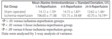
Table 1. Serum Alanine Aminotransferase Levels in Sham-Operated and Ischemia-Reperfusion Rats
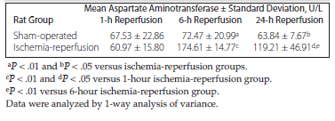
Table 2. Serum Aspartate Aminotransferase Levels in Sham-Operated and Ischemia-Reperfusion Rats
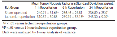
Table 3. Serum Tumor Necrosis Factor α Levels in Sham-Operated and Ischemia-Reperfusion Rats
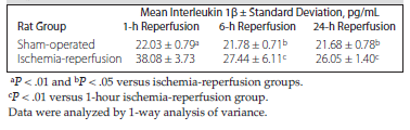
Table 4. Serum Interleukin 1β Levels in Sham-Operated and Ischemia-Reperfusion Rats
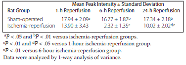
Table 5. Peak Intensity of Contrast-Enhanced Ultrasonography in Sham-Operated and Ischemia-Reperfusion Rats
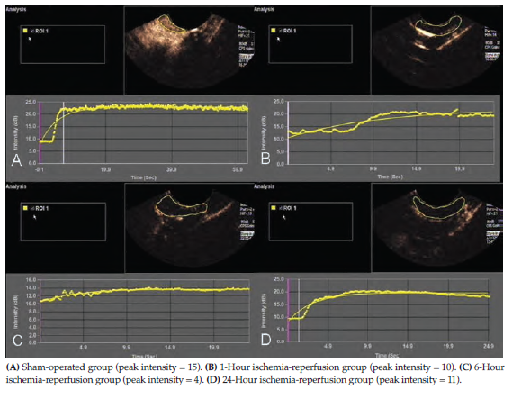
Figure 1. Peak Intensity Values in Liver by Contrast-Enhanced Ultrasonography
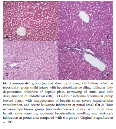
Figure 2. Histopathologic Changes of Liver by Hematoxylin and Eosin Staining