Volume: 14 Issue: 3 June 2016
FULL TEXT
Both autologous and allogeneic hematopoietic stem cell transplants are important therapeutic options for several benign and malignant disorders. Pulmonary complications, although they have become less frequent, remain a significant cause of morbidity and mortality after hematopoietic stem cell transplant. These complications range from bacterial, fungal, and viral pulmonary infections to noninfectious conditions such as diffuse alveolar hemorrhage and idiopathic pneumonia syndrome. Bronchiolitis obliterans syndrome is the primary chronic pulmonary complication, and treatment of this condition remains challenging. This report highlights the advances in the diagnosis and management of the major pulmonary complications after hematopoietic stem cell transplant. It also underscores the need for prospective and multicenter research to have a better understanding of the mechanisms behind these complications and to obtain more effective diagnostic tool and therapeutic options.
Key words : Adverse effects, Lung diseases/etiology, Lung diseases/diagnosis, Respiratory tract infections/etiology, Graft versus host disease
Introduction
Hematopoietic stem cell transplant (HSCT) is increasingly being used for treatment of a wide range of benign and malignant diseases. About 50 000 to 60 000 HSCTs are done annually.1 Hematopoietic stem cell transplant can be classified as autologous when the stem cells are harvested from the same patient and reinfused after high-dose chemotherapy, whereas, in allogeneic HSCT, the stem cells are donated by another person. Allogeneic HSCT can be related or unrelated and matched or mismatched. The hematopoietic stem cells might be derived from the bone marrow, umbilical cord, or peripheral blood.
Hematopoietic stem cell transplant can be a life-saving treatment of a wide range of neoplastic and nonneoplastic diseases; however, it is still limited by the development of serious complications. Advances in the preparative regimen using a nonmyeloablative preparative regimen, reduced-intensity conditioning transplants, and posttransplant supportive care have contributed to improved overall survival. Despite these advances, pulmonary complications still occur in about 37% of patient after HSCT2 and are associated with significant morbidity and mortality. In 1 study, the hazard ratio for death due to pulmonary complications was 30.1 However, there is evidence that the overall mortality and pulmonary complications after HSCT are improving. A recent review of allogeneic HSCT patients in a large transplant center from 1993 to 2007 showed that the risk of nonrelapse mortality decreased by 60% and overall mortality decreased by 41%. In addition, the risk of acute respiratory failure dropped by 36%, with risk of gram-negative bacterial and invasive mold infections decreasing by 39% and 51%.3 In this study, we provide an update of the main pulmonary complications (both infectious and noninfectious) after HSCT, including an overview of the diagnostic approach and treatment of these complications.
Noninfectious Complications After Hematopoietic Stem Cell Transplant
Advances in the prophylactic regimen against infections after HSCT, as well
as the diagnosis and treatment of the infectious complications after HSCT, have
led to their decreased incidence. However, the incidence of noninfectious
complications has not changed, which increases the significance of these
conditions.4,5 Noninfectious pulmonary complications after HSCT range
from acute to subacute causes of acute respiratory failure, such as diffuse
alveolar hemorrhage and idiopathic pneumonia syndrome, to chronic progressive
small airway disease like bronchiolitis obliterans syndrome (BOS),4
and include vascular complications like pulmonary veno-occlusive disease and
cytolytic thrombi. Table 1 provides a comparison of the primary noninfectious
causes of acute respiratory failure after HSCT. Although some of those entities,
such as pulmonary alveolar proteinosis, are reversible, others are obstinate and
associated with poor prognosis.5
Periengraftment Respiratory Distress Syndrome
Previously referred to as engraftment syndrome, periengraftment respiratory
distress syndrome is characterized by the development of fever with no
identifiable infectious cause, erythrodermatous rash not attributable to
medications, and noncardiogenic pulmonary edema. Other features might include
hepatic dysfunction, renal insufficiency, weight gain, and transient
encephalopathy. It is more common in autologous HSCT recipients and usually
occurs within 96 hours of engraftment, coinciding with neutrophil recovery. The
median onset time is 11 days, with an incidence of 7% to 11%.4-6
The incidence and severity of this syndrome have increased as a result of use of granulocyte colony-stimulating factor, which regulates the production of cytokines (tumor necrosis factor α, interleukin 1B, and interleukin 8). These mediators in turn increase neutrophil influx into the lungs and alveolar permeability and are likely implicated in acute lung injury.7
Patients with periengraftment respiratory distress syndrome have a favorable prognosis, and outcomes of most patients improve with supportive therapy. Consideration should be given to stopping granulocyte colony-stimulating factor. Patients with more severe cases associated with acute respiratory failure respond dramatically to systemic corticosteroids.5
Diffuse Alveolar Hemorrhage
Diffuse alveolar hemorrhage (DAH) is an important cause of acute respiratory
failure that occurs in 2% to 14% of recipients, with similar incidence in both
autologous and allogeneic HSCT recipients.8 Diffuse alveolar
hemorrhage is most commonly observed within the first month after HSCT (a median
of 23 days), often during the pre-engraftment phase; however, later onset is
encountered in up to 42% of cases.5 Risk factors for DAH after HSCT
include age > 40 years, total body irradiation, high-dose cyclophosphamide, HSCT
for solid tumors, the presence of high fevers, severe mucositis, leukocyte
recovery, and renal insufficiency.9 There is no relation between DAH
and type of preparatory regimen, whether it is reduced intensity or
myeloablative.10 Postmortem studies have shown that most patients
with DAH had a pattern of diffuse alveolar damage.6,8,9 The fact that
many cases occur around the time of engraftment suggests that neutrophil influx
into the lung may accentuate the injury and precipitate alveolar hemorrhage. The
hallmark of diagnosis of DAH is a finding of progressively bloodier return from
bronchoalveolar lavage (BAL) from at least 3 separate subsegments (or 20%
hemosiderin-laden macrophage) in the absence of an identifiable respiratory
tract infection. The following diagnostic criteria for DAH have been suggested:
(1) evidence of widespread alveolar injury manifested by multilobar pulmonary
infiltrates, symptoms, and signs of pneumonia and abnormal physiology with
hypoxemia and widened alveolar-arterial gradient; (2) BAL showing progressively
bloodier return from at least 3 separate subsegmental bronchi or > 20%
hemosiderin-laden macrophages on examination of the BAL fluid or, if a surgical
lung biopsy is performed, > 30% of the alveolar surface of the examined lung
tissue is covered by blood; and (3) absence of infection compatible with the
diagnosis.8
There are no prospective randomized trials that have addressed the treatment of DAH. Retrospective studies and case reports have suggested that early diagnosis of DAH and high-dose treatment with corticosteroids (500-1000 mg/d of methylprednisolone for 3-4 d followed by tapered dosage over 2-4 wk) may improve survival rate.6 In addition, aminocaproic acid and recombinant factor VIIa have been tried in some studies with variable results.11 However, a recent report showed no difference in outcome with methylprednisolone at low doses (≤ 250 mg/d), medium doses (250-1000 mg/d), or high doses (≥ 1000 mg/d) with or without the addition of aminocaproic acid.12 Mortality rates in patients with DAH range from 74% to 100%.8,9 However, mortality as low as 38% has been reported.13 Mortality is usually due to superimposed multiorgan failure or sepsis. Autologous HSCT, onset within the first 30 days of transplant, and lack of mechanical ventilation are associated with favorable outcome.6
Idiopathic Pneumonia Syndrome
Idiopathic pneumonia syndrome (IPS) is another subacute cause of acute
respiratory failure after HSCT. The definition of IPS has been recently updated
by the American Thoracic Society. It is diagnosed when there is (1) evidence of
widespread alveolar injury (with multilobar infiltrates on chest imaging,
symptoms and signs of pneumonia, and evidence of abnormal pulmonary physiology
such as hypoxemia with high alveolar-arterial gradient or new or increased
restrictive pulmonary function test abnormality); (2) absence of active lower
respiratory tract infection, such as bacteria, Legionella, viruses, and Fungi by
performing bronchoscopy with BAL and negative appropriate serologic studies for
viruses and fungi; and (3) absence of cardiac dysfunction, acute renal failure,
or iatrogenic fluid overload as cause of pulmonary dysfunction.14 The
recent American Thoracic Society definition of IPS suggested that this condition
includes a variety of noninfectious pulmonary complications after HSCT such as
acute respiratory distress syndrome, delayed pulmonary toxicity syndrome,
periengraftment respiratory distress syndrome, DAH, and cryptogenic organizing
pneumonia.14
The incidence rate of IPS ranges from 2% to 17%. It usually occurs in the first 120 days after HSCT, and the median time of onset is 21 to 49 days after transplant.5 Idiopathic pneumonia syndrome is more common after allogeneic HSCT. Risk factors for developing IPS after HSCT include older age, lower pretransplant performance status, transplant for a malignancy other than leukemia, high-intensity conditioning regimens, total body irradiation, high-grade acute graft-versus-host disease (GVHD), prior HSCT, and methotrexate-based GVHD prophylaxis.15-17
The clinical course of IPS is typically rapid, with most patients progressing to respiratory failure requiring mechanical ventilation. Mortality rate is around 74% and is primarily related to progressive respiratory failure. Poor prognostic factors include the need for mechanical ventilation and higher serum creatinine levels at the onset of IPS.15 Beyond supportive therapy, there is no proven therapy for IPS. High-dose corticosteroid therapy appears to be of minimal benefit. The administration of etanercept, a soluble tumor necrosis factor α binding protein, was promising in a few studies.18,19 However, in a recent randomized placebo controlled trial, 34 patients with IPS after allogeneic HSCT were randomized to 2 mg/kg/day of methylprednisolone with etanercept (0.4 mg/kg twice weekly for 4 wk). The 28-day mortality rate was not different between the 2 groups, and there were no differences in other outcomes. However, in this trial, the overall survival rate exceeded 60%, which is better than historic results.19 Etanercept may be considered in patients with severe refractory IPS.
Bronchiolitis Obliterans Syndrome
Bronchiolitis obliterans syndrome is the primary late noninfectious
pulmonary complication after allogeneic HSCT. It is reported to be more common
after peripheral blood HSCT.5 It is also more frequent after
conventional myeloablative than after nonmyeloablative HSCT and with
busulfan-based preparative regimens.5 It usually presents after the
first 100 days after HSCT.20 It is considered the primary pulmonary
manifestation of chronic GVHD. Risk factors for BOS include the presence of
chronic GVHD, older age of the recipient (> 20 y), the presence of airflow
obstruction (forced expiratory volume in 1 s/forced vital capacity < 0.7) before
HSCT, and respiratory viral infections in the first 100 days after HSCT.21,22
In a review of 1145 allogeneic HSCT recipients, 5.5% developed BOS. The
prevalence was 14% in patients with chronic GVHD. The median time between HSCT
and diagnosis of BOS was 439 days (range, 274-1690 d). Factors that were
significantly associated with BOS on multivariate analysis were baseline forced
expiratory volume in 1 second-to-forced vital capacity ratio, nonwhite race,
chronic GVHD, and immunoglobulin G level. Patients with BOS had a 1.62-fold
increase in mortality after diagnosis.23 In another recent review of 1854
allogeneic HSCT recipients, BOS was diagnosed in 4.8% of patients, with 97% of
these having chronic GVHD. Patients with BOS were compared with 2 matched groups
of patients with chronic GVHD and no BOS and another with no GVHD and no BOS. A
busulfan-based regimen even in the setting of reduced intensity conditioning,
use of an unrelated donor or female donor, pretransplant lung disease, acute
GVHD, Cytomegalovirus (CMV)-positive status, and high-risk transplant were
associated with increased risk of BOS.24 On the other hand, antithymocyte
globulin (ATG) was protective against GVHD and BOS. The study highlights the
importance of busulfan as a risk factor for chronic GVHD and BOS. This could be
due to direct pulmonary toxicity by the medication leading to exposure of
antigens and donor alloreactivity contributing to the development of BOS. Table
2 provides a summary of the risk factors that could lead to BOS after HSCT.
The presentation of BOS is usually insidious and may be preceded by upper respiratory tract symptoms. The main symptoms are dry cough, dyspnea, wheezing, and sinusitis. Approximately 20% of patients are asymptomatic, and the diagnosis is suspected based on pulmonary function test findings showing new-onset airflow obstruction.4 Most patients have a slow progressive airflow obstruction, with episodes of acute exacerbations. In the minority of patients, the airflow obstruction progresses rapidly into respiratory failure within a few months. In the advanced stages of BOS, patients are physically limited due to severe obstructive airway disease and may require home oxygen therapy. Some patients may develop features of bronchiectasis with recurrent respiratory tract infections.5 The most common radiologic sign of BOS on high-resolution computed tomography of the chest is the presence of air trapping during the expiratory phase of imaging. These views show areas of hypoattenuation that correspond to obstructed airways interspaced with areas of “ground-glass” appearance corresponding to the pulmonary lobules with patent airways giving a “mosaic” appearance. High-resolution computed tomography of the chest is recommended in the work-up of these patients to evaluate for the above features but also to rule out other pulmonary complications, such as infectious causes and cryptogenic organizing pneumonia or IPS, which are usually associated with pulmonary infiltrates. Spirometry is the main test used to diagnose and follow-up patients with BOS after HSCT. It usually shows evidence of new airflow obstruction with reduction in forced expiratory volume in the first second and the forced expiratory volume in 1 second-to-forced vital capacity ratio.
The following criteria were recently established by a special National Institutes of Health workshop for the diagnosis of BOS after HSCT25: (1) forced expiratory volume in 1 second-to-forced vital capacity ratio < 0.7 and forced expiratory volume in 1 second < 75% of predicted; (2) evidence of air trapping or small airway thickening or bronchiectasis on high-resolution chest computed tomography (with inspiratory and expiratory cuts), residual volume > 120%, or pathologic confirmation of constrictive bronchiolitis; and (3) absence of infection in the respiratory tract, documented by investigations targeting clinical symptoms, such as radiologic studies (radiographs or computed tomographic scans) or microbiologic cultures (sinus aspiration, upper respiratory tract viral screen, sputum culture, or BAL).
Pathologic confirmation of bronchiolitis obliterans can be achieved by surgical lung biopsy and occasionally by transbronchial biopsy. However, these are rarely necessary in clinical practice. A recent study confirmed that pathologic bronchiolitis obliterans diagnosis was not superior to BOS criteria in predicting decreased pulmonary function beyond the time of biopsy.26
Treatment of BOS is based primarily on expert opinion and aims at stabilization of the pulmonary function. Clinical evidence is limited to small and uncontrolled trials. Generally, treatment includes high-dose corticosteroids (usually prednisone at 1-1.5 mg/kg/d) to be tapered gradually over 6 to 12 months, reinstitution or augmentation of immunosuppressive agents such as tacrolimus and cyclosporine, macrolides such as low doses of azithromycin or clarithromycin, inhaled corticosteroids or bronchodilators, and montelukast.27-29 Treatment with budesonide/formoterol improved forced expiratory volume in 1 second compared with placebo in a small prospective study of patients with mild to severe BOS after HSCT.30 Patients should receive antireflux measures and prophylactic therapy against Pneumocystis jiroveci, Fungi, and CMV. Lung transplant has been successful in selected patients.30 Other treatments for BOS have been reported in small series or are currently under investigation, including leukotriene receptor antagonists, extracorporeal photodynamic therapy, antitumor necrosis factor α monoclonal antibodies (such as infliximab), imatinib mesylate, alefacept, daclizumab, and rituximab.31-34 In advanced cases, patients may benefit from supplemental oxygen therapy and pulmonary rehabilitation referral.35
The response to therapy is variable, and the best results of treatment are to maintain lung function and prevent further deterioration. Factors that have been shown to correlate with better outcomes include lymphocytic bronchiolitis versus constrictive bronchiolitis on surgical lung biopsy.36 In addition, in 1 study, the presence of high concentrations of neutrophils and interleukin 8 in BAL predicted better response to azithromycin.37
Cryptogenic Organizing Pneumonia
Cryptogenic organizing pneumonia has been previously called bronchiolitis
obliterans-organizing pneumonia and is frequently confused with BOS, although
they are separate entities. Cryptogenic organizing pneumonia complicates both
autologous and allogeneic HSCT but is more common in the latter, with an
incidence of 0.9% to 10.3%. Onset time ranges from 5 to 2800 days with a median
of 108 days.6 In a recent review of 9550 allogeneic HSCT recipients,38
cryptogenic organizing pneumonia was diagnosed in 2% of patients. HLA disparity,
female-to-male HSCT, and peripheral blood stem cell transplant were
significantly associated with an increased risk of cryptogenic organizing
pneumonia. On the other hand, busulfan-based myeloablative conditioning or
fludarabine-based reduced-intensity conditioning in comparison with a total body
irradiation-based regimen and in vivo T-cell depletion were associated with a
lower risk. The 3-year nonrelapse mortality rate and overall survival rate in
the cryptogenic organizing pneumonia group were 29.0% (95% confidence interval,
20.2%-38.3%) and 60.5% (95% confidence interval, 49.9%-69.5%).38 The
clinical presentation is usually acute or subacute with 2 weeks of fever,
nonproductive cough, and dyspnea. Radiologically, findings include patchy
(multifocal, diffuse, or focal) consolidations that tend to be peripheral and/or
peribronchovascular, ground glass opacities, or nodular lesions. In contrast to
BOS, pulmonary function tests typically show mild to moderate restrictive
pattern and characteristically low diffusing capacity and usually no airflow
obstruction. Bronchoscopy is useful in these patients to exclude other
conditions such as infections. Bronchoalveolar lavage fluid reveals
lymphocytosis with low CD4-to-CD8 ratio.39 Histologic confirmation
is usually necessary and can be made by transbronchial biopsies; however, many
patients require surgical lung biopsy by video-assisted thoracoscopy.
Pathologically, cryptogenic organizing pneumonia is characterized by patchy
distribution of plugs of granulation tissue that fill the lumens of the distal
airways extending to the alveolar ducts and spaces associated with chronic
interstitial inflammation. Contrary to BOS, patients with cryptogenic
organizing pneumonia after HSCT have more favorable response to systemic
corticosteroid therapy tapered over 3 to 6 months. Most patients achieve
resolution or stability of the disease.40
Infectious Complications After Hematopoietic Stem Cell Transplant
Despite the advances in prophylaxis and treatment of respiratory infections,
infections after HSCT remain a cause of death in more than 40% of HSCT
recipients.4,5 Infectious complications are more common in patients
with allogeneic HSCT because of the prolonged immune-suppressive therapy and
GVHD.
Bacterial pneumonia
Hematopoietic stem cell transplant recipients are at highest risk for
bacterial pneumonia within the first 30 days after transplant, with incidence
rates reported to be 20% to 50%.4 Bacterial pneumonia is more common
in allogeneic transplants with the risk significantly increased depending on the
presence of previous GVHD, the degree and duration of neutropenia, the type of
conditioning regimen (being more common with administration of myeloablative
regimen), and the presence of indwelling catheters.5,41,42
Gram-negative organisms such as Pseudomonas are the usual causatives during the pre-engraftment phase and are associated with a higher risk of septic shock and acute respiratory distress syndrome. During this phase, patients may present with neutropenic fever. Chest radiographs may be negative in about half of the cases. High-resolution computed tomography of the chest is usually recommended in febrile neutropenic patients due to their higher sensitivity in detecting pneumonic infiltrates.5
Bacterial pneumonia is less common in the postengraftment period, and the incidence of bacterial pneumonia is much lower after 100 days of HSCT except in patients with chronic GVHD where it is usually due to encapsulated organisms such as Streptococcus pneumoniae and Haemophilus influenzae.
The most recent guidelines from the Infectious Diseases Society of America recommend prophylaxis with a fluoroquinolone in high-risk patients with expected durations of prolonged and profound neutropenia (absolute neutrophil count < 100 cells/mm3 for > 7 d),41 although it might increase the risk of fluoroquinolone resistance.43 Patients should receive pneumococcal vaccine at 12 and 24 months after HSCT and the Haemophilus influenzae type b vaccine at 12, 14, and 24 months. Prophylaxis with intravenous immunoglobulin may be considered for HSCT recipients with unrelated HSCT with profound hypogammaglobulinemia (immunoglobulin level < 400 mg/dL) in the first 100 days after transplant.44
Nocardia pneumonia
Nocardia pneumonia is uncommon but should be considered in patients not
responding to initial antibiotic therapy. The median time of onset is 210 days
after HSCT. Risk factors include use of pentamidine instead of
trimethoprim-sulfamethoxazole for Pneumocystis jiroveci prophylaxis and chronic
GVHD. The usual radiologic presentation is pulmonary nodules with or without
infiltrates. The first-line treatment is with trimethoprim-sulfamethoxazole,
usually requiring prolonged duration of therapy and has a good prognosis.5,45
Mycobacterial diseases
The incidence of Mycobacterium tuberculosis infection is low, 0.1% to 5.5% in
nonendemic areas, but is of more of concern in endemic areas or in patients from
endemic areas.46 The incidence increases by 3-fold in patients who
have received allogeneic HSCT compared with those who have had autologous
transplant, for which the incidence is similar to the general population.5
The infection usually occurs more than 90 days after HSCT, with median time
being 324 days after transplant. The clinical presentation and treatment are
similar to the general population with consideration given to drug interactions.
According to a recent report, the diagnosis of M tuberculosis is made by
cultures of respiratory samples (55% of cases), acid fast bacilli stain (25% of
cases), and histology (20% of cases).47 It should be noted that HSCT
recipients should be treated for latent M tuberculosis if they have more than
5-mm induration on tuberculin skin test or have M tuberculosis contact even with
a negative tuberculin skin test.5,46,47
Nontuberculous mycobacterial infections (such as Mycobacterium avium complex, Mycobacterium kansasii, and Mycobacterium chelonae) are rare in HSCT recipients and are more common in patients with chronic GVHD involving the lungs. Clinical presentation and treatment are similar to the general population.48
Invasive fungal infections
Invasive fungal infections are serious problem after HSCT, especially with
allogeneic transplant. In a recent multicenter study involving 16 200 HSCT
transplant patients, invasive Aspergillosis was the most common fungal infection
(43%), followed by invasive Candidiasis (28%) and Mucormycosis (8%).49
Risk factors for fungal pneumonia include prolonged neutropenia,
immunosuppressive therapies to prevent or treat GVHD, the widespread use of
broad-spectrum antibiotics, and a high ferritin level of > 1550 ng/mL.50
Invasive pulmonary Aspergillosis (IPA) is more common after allogeneic HSCT, especially in those with chronic GVHD, with reported incidence of 5% to 30% in allogeneic and 1% to 5% in autologous HSCT.4,5 Invasive pulmonary Aspergillosis becomes increasingly more significant during the late postengraftment phase during treatment of chronic GVHD. The incidence of IPA in the neutropenic phase is now decreased due to the routine use of stem cells instead of bone marrow for transplant, the use of nonmyeloablative regimens, the use of colony-stimulating factors during neutropenia, and the widespread use of antifungal agents for prophylaxis.5 In a recent multicenter study, the median time for IPA infection was 99 days after HSCT.49 Risk factors include unrelated allogeneic HSCT, the use of high-dose corticosteroids, cyclosporine, antitumor necrosis factor α agents, and concurrent CMV infection.5,50,51 Radiologic features on high-resolution computed tomography of the chest include ill-defined consolidations and ground glass opacities, nodules with halo sign and air-crescent sign, and pleural effusions. A negative sputum sample does not rule out the infection, and usually a bronchoscopy with BAL is needed. Elevated BAL galactomannan with cut-off > 0.5 has 73% sensitivity and about 85% specificity in diagnosing IPA in patients with HSCT.52 In a study of 67 patients with hematologic malignancies (45% were HSCT recipients), the sensitivity and specificity of BAL galactomannan were 73% and 89%, with positive and negative predictive values of 73% and 83%. Bronchoalveolar lavage galactomannan performed better than BAL cytology (0%), BAL culture (27%), transbronchial biopsies (40%), and serum galactomannan (67%). Bronchoalveolar lavage galactomannan was positive in all patients who had invasive fungal infection and were on active antifungal therapy for < 3 days.53 Serum galactomannan can be falsely elevated in patients with gastrointestinal GVHD and those receiving beta lactams such as piperacillin-tazobactam.54 Elevated serum 1,3-beta-D-glucan assay levels may be useful in the diagnosis but is not well defined in patients with IPA. The treatment of choice for IPA is voriconazole, which is a triazole.55 Echinocandins either alone or in combination with posaconazole are used as a second-line treatment in patients who no longer respond to voriconazole.56 Surgical resection is reserved to selected patients, such as those with massive hemoptysis.5 Prophylaxis against IPA is important and can be achieved with voriconazole, posaconazole, or itraconazole. Recent guidelines recommend that allogeneic HSCT recipients receive prophylaxis through the neutropenic period and for at least 75 days after transplant or until cessation of immunosuppressive therapy.42
The incidence of invasive Candidiasis has decreased due to the widespread use of prophylactic antifungal therapy after HSCT; however, there is a shift in the Candida species from Candida albicans to more resistant species such as Candida glabrata and Candida krusei.5
Mucormycosis infections, including those involving Mucor species and Rhizopus species, are rare, with the most recent data indicating the incidence might be rising with the use of voriconazole prophylaxis. Treatment includes amphotericin B and surgical resection.5
The incidence of Pneumocystis jiroveci infection is almost negligible in the era of prophylaxis with trimethoprim-sulfamethoxazole and usually occurs in patients who are on other types of prophylaxis, such as atovaquone or pentamidine. Infection usually happens in month 2 to 6 after HSCT.4
Viral pneumonia
The incidence of CMV pneumonia has decreased significantly after the use of
prophylactic and preemptive antiviral therapy in high-risk patients. Currently,
in allogeneic HSCT recipients, the incidence of CMV pneumonia is 10% to 30%,
whereas, in autologous recipients; it is extremely rare (1%-9%). The onset of
CMV pneumonia in allogeneic HSCT is now delayed to after the first 100 days
(median of 169 d).5 Diagnosis depends on the presence of clinical and
radiologic features in combination with detection of CMV in blood and BAL
through CMV DNA quantitative polymerase chain reaction, shell assay, and viral
cultures. Patients present clinically with rapid onset of fever, nonproductive
cough, dyspnea, and hypoxemia, with progression to acute respiratory failure
within 2 weeks. Radiologically, the most common findings are an interstitial
pattern with tiny pulmonary nodules and patchy areas of consolidation.57
The definitive diagnosis is made histologically with presence of
intracytoplasmic inclusion bodies seen on lung biopsies. The treatment of choice
is ganciclovir with or without CMV immunoglobulins. Although ganciclovir is
effective for CMV prophylaxis, the myelosuppressive toxicity has limited its use
as routine prophylaxis. Preemptive treatment of CMV with ganciclovir or
valganciclovir is an alternative approach and is usually based on plasma pp65
antigen or polymerase chain reaction testing.58
Community respiratory viruses also include respiratory syncytial virus, parainfluenza, influenza, and adenovirus. The incidence is close to 30% in patients with HSCT, with more common incidence after allogeneic transplant. Respiratory syncytial virus pneumonia is the most common. Mortality from untreated respiratory syncytial virus pneumonia is high.5 In a recent retrospective cohort of 82 patients with respiratory syncytial virus pneumonia, the 30-day and 100-day mortality rates were 32% and 43%. The increased mortality was associated with bone marrow as a source of HSCT and baseline need for oxygen supplementation (> 2 L/min). Aerosolized ribavirin and respiratory syncytial virus-specific immunoglobulins might be beneficial.59
Parainfluenza and influenza viruses have a similar incidence in both autologous and allogeneic transplant recipients. The mortality is much lower than for respiratory syncytial virus. Treatment is supportive. The use of oseltamivir within 48 hours of onset of symptoms reduces progression to pneumonia.4,5 In addition to higher risk of lower respiratory tract infection by these viruses, there is an increased risk of coinfection or superinfection with bacteria and fungi.60
Approach to the Diagnosis of Pulmonary Complications After Hematopoietic
Stem Cell Transplant
There are no standard guidelines for the evaluation of pulmonary complications
after HSCT. Most studies in the literature are based on retrospective analysis
of single center studies. There are general considerations when approaching HSCT
patients with pulmonary symptoms and/or infiltrates. These include the immune
status, such as presence and duration of neutropenia, immunosuppressive therapy,
the dose and duration of corticosteroid therapy, and immunoglobulin deficiency,
all important in predicting the cause of the respiratory infection. Recently,
tobacco smoking has been identified as a potential risk factor for respiratory
infections after HSCT.61 The type of HSCT (allogeneic vs autologous)
and the presence of GVHD are also significant considerations in the spectrum of
respiratory problems. Another consideration is that pulmonary complications
after HSCT tend to follow a predictable timeline, as illustrated in Figure 1.
Therefore, it is prudent to consider the timing of the respiratory event during
the approach to diagnosis and management. In addition, the type of prophylactic
antimicrobial agent is also useful in excluding certain infections such as
trimethoprim-sulfamethoxazole in cases of P jiroveci. Prior hospitalizations for
respiratory problems and previous infections, such as CMV, may shed some light
on the cause of pulmonary infiltrates. Respiratory infections may lack the
classic signs and symptoms, and presentation may be subtle or nonspecific. These
observations could be related to presence of neutropenia or medications that may
affect the inflammatory response such as corticosteroids. Not all pulmonary
infiltrates are due to infections. Commonly, pulmonary complications after HSCT
are due to noninfectious complications such as pulmonary edema, engraftment
syndrome, DAH, IPS, cryptogenic organizing pneumonia, or sometimes secondary
malignancy. Radiologic findings are helpful but not specific. A classic example
is the presence of the halo sign in patients with IPA. It appears primarily in
neutropenic patients and early in the course of infection. The absence of this
radiologic sign does not exclude IPA, whereas its presence does not always mean
IPA. Pseudomonas aeruginosa, Nocardia species, and Mucormycosis also may cause
halo infiltrate.62,63 In determining whether to proceed with an
invasive procedure such as bronchoscopy or surgical lung biopsy, it is important
to decide whether such an intervention is likely to change the care or outcome
of the patient, as will be discussed further below. The best outcome of the
diagnostic approach to pulmonary infiltrates is when it is carried out
expeditiously by a multidisciplinary team that includes specialists from
pulmonary, bone marrow transplant, infectious diseases, and radiology
departments and, when indicated, from thoracic surgery.
Although sputum cultures have several limitations with low sensitivities and false positive results, they may be helpful in certain circumstances, such as Mycobacterial infection, P jiroveci, Legionella, Nocardia, and fungal infections. The detection of Aspergillus spp. may be difficult to interpret because it may colonize the upper respiratory tract, especially in patients with chronic lung disease. However, in an HSCT patient, it should not be ignored, and further studies are generally indicated to exclude IPA.
The use of chest radiography may be helpful; however, most patients with respiratory symptoms after HSCT require chest computed tomography with high-resolution images to evaluate for infectious and noninfectious causes that may have distinctive features. Radiologic signs that are better detected by chest computed tomography include pulmonary nodules, the halo and air crescent signs, “ground-glass” infiltrates, changes that are suggestive of BOS and cryptogenic organizing pneumonia, and mediastinal lymphadenopathy. Furthermore, chest computed tomography assists in better yield if invasive procedures are indicated. There is evidence that early use of chest computed tomography for evaluation of pulmonary disease was associated with earlier diagnosis of serious infections such as IPA and better outcomes of patients.63
Serologic studies have recently emerged as useful tools in the evaluation of infectious pulmonary complications after HSCT. Serum and BAL galactomannan antigens are very useful, in the appropriate setting, for diagnosis of IPA as outlined above. In addition, serum 1,3-beta-D-glucan analysis is another supportive test that could show the presence of fungal infection. Bronchoalveolar lavage 1,3-beta-D-glucan has not been studied yet, and polymerase chain reaction assay for Aspergillus DNA in BAL is currently under evaluation.64 Serum CMV polymerase chain reaction is helpful in diagnosis and monitoring of patients with CMV infection.
Fiberoptic bronchoscopy and BAL are the major investigative and least invasive tools for obtaining lower respiratory tract specimens. The overall diagnostic yield of bronchoscopy with BAL in HSCT patients ranges between 42% and 65%.65 The highest yield is in allogeneic HSCT patients, when there is presence of focal versus diffuse infiltrates, and when the procedure is done within 24 hours of presentation or before start of antimicrobial therapy. It is worth noting that BAL is still useful within 3 days of initiation of therapy.66,67 The lowest diagnostic yield of BAL is in critically ill patients with acute respiratory failure and in the presence of acute GVHD and neutropenia.68,69 Changes in the management in HSCT following bronchoscopy with BAL vary in the literature from 20% to 70%.65 Changes in the outcome based on the procedure are even more variable and inconsistent.
Transbronchial biopsies are commonly restricted by severe thrombocytopenia and critical respiratory status. The additional diagnostic yield is around 10%.70,71 Transbronchial biopsies are more useful in the diagnosis of noninfectious pulmonary complications after HSCT such as cryptogenic organizing pneumonia.
Surgical lung biopsy, preferably by video-assisted thoracoscopic surgery, is the diagnostic criterion standard in the evaluation of pulmonary infiltrates after HSCT. The outcome of patients is improved when a specific diagnosis is made. However, commonly, this procedure is limited by the relatively high surgical morbidity and mortality rates in this patient population.72 Chellapandian and associates recently compared the diagnostic yield of BAL with that of lung biopsy.73 Although yield was similar for both procedures, BAL diagnosed more infectious causes, whereas lung biopsies led to more noninfectious ones. Not surprisingly, complications and procedure-related mortality were higher in patients who underwent lung biopsies.
Figure 2 provides a suggested approach to evaluation of pulmonary infiltrates in HSCT recipients.
Conclusions
Hematopoietic stem cell transplant is a therapeutic option for a variety of diseases, and indications for this procedure and other types of stem cell transplant are likely to expand in the future. There have been significant advances in the conditioning regimens and supportive measures. However, pulmonary complications remain a major problem after HSCT. The available evidence for risk factors, diagnostic approach, and treatment options is primarily based on retrospective, single center studies. Prospective, observational, and interventional multicenter trials are needed to better diagnose and treat these complications.
References:
- Afessa B, Abdulai RM, Kremers WK, Hogan WJ, Litzow MR, Peters SG. Risk
factors and outcome of pulmonary complications after autologous
hematopoietic stem cell transplant. Chest. 2012;141(2):442-450.
CrossRef - PubMed - Ghalie R, Szidon JP, Thompson L, Nawas YN, Dolce A, Kaizer H. Evaluation
of pulmonary complications after bone marrow transplantation: the role of
pretransplant pulmonary function tests. Bone Marrow Transplant.
1992;10(4):359-365.
PubMed - Gooley TA, Chien JW, Pergam SA, et al. Reduced mortality after
allogeneic hematopoietic-cell transplantation. N Engl J Med.
2010;363(22):2091-2101.
CrossRef - PubMed - Chi AK, Soubani AO, White AC, Miller KB. An update on pulmonary
complications of hematopoietic stem cell transplantation. Chest.
2013;144(6):1913-1922.
CrossRef - PubMed - Zaza Dit Yafawi J, Shehada E, Soubani, AO. Pulmonary complications following hematopoietic stem cell transplantation. In: Montgomery WG, Burton HI, eds. Hematopoietic Stem Cells: New Research. New York, NY: Nova Publishers; 2012:1-49.
- Soubani AO, Pandya CM. The spectrum of noninfectious pulmonary
complications following hematopoietic stem cell transplantation. Hematol
Oncol Stem Cell Ther. 2010;3(3):143-157.
CrossRef - PubMed - Karlin L, Darmon M, Thiery G, et al. Respiratory status deterioration
during G-CSF-induced neutropenia recovery. Bone Marrow Transplant.
2005;36(3):245-250.
CrossRef - PubMed - Afessa B, Tefferi A, Litzow MR, Krowka MJ, Wylam ME, Peters SG. Diffuse
alveolar hemorrhage in hematopoietic stem cell transplant recipients. Am J
Respir Crit Care Med. 2002;166(5):641-645.
CrossRef - PubMed - Robbins RA, Linder J, Stahl MG, et al. Diffuse alveolar hemorrhage in
autologous bone marrow transplant recipients. Am J Med. 1989;87(5):511-518.
CrossRef - PubMed - Majhail NS, Parks K, Defor TE, Weisdorf DJ. Alveolar hemorrhage
following allogeneic hematopoietic cell transplantation using reduced-intensity
conditioning. Bone Marrow Transplant. 2006;38(11):765-768.
CrossRef - PubMed - Elinoff JM, Bagci U, Moriyama B, et al. Recombinant human factor VIIa
for alveolar hemorrhage following allogeneic stem cell transplantation. Biol
Blood Marrow Transplant. 2014;20(7):969-978.
CrossRef - PubMed - Rathi NK, Tanner AR, Dinh A, et al. Low-, medium- and high-dose steroids
with or without aminocaproic acid in adult hematopoietic SCT patients with
diffuse alveolar hemorrhage. Bone Marrow Transplant. 2015;50(3):420-426.
CrossRef - PubMed - Solh M, Arat M, Cao Q, Majhail NS, Weisdorf D. Late-onset noninfectious
pulmonary complications in adult allogeneic hematopoietic cell transplant
recipients. Transplantation. 2011;91(7):798-803.
CrossRef - PubMed - Panoskaltsis-Mortari A, Griese M, Madtes DK, et al. An official American
Thoracic Society research statement: noninfectious lung injury after
hematopoietic stem cell transplantation: idiopathic pneumonia syndrome. Am J
Respir Crit Care Med. 2011;183(9):1262-1279.
CrossRef - PubMed - Fukuda T, Hackman RC, Guthrie KA, et al. Risks and outcomes of
idiopathic pneumonia syndrome after nonmyeloablative and conventional
conditioning regimens for allogeneic hematopoietic stem cell transplantation.
Blood. 2003;102(8):2777-2785.
CrossRef - PubMed - Sano H, Kobayashi R, Iguchi A, et al. Risk factor analysis of idiopathic
pneumonia syndrome after allogeneic hematopoietic SCT in children. Bone
Marrow Transplant. 2014;49(1):38-41.
CrossRef - PubMed - Sakaguchi H, Takahashi Y, Watanabe N, et al. Incidence, clinical
features, and risk factors of idiopathic pneumonia syndrome following
hematopoietic stem cell transplantation in children. Pediatr Blood Cancer.
2012;58(5):780-784.
CrossRef - PubMed - Yanik GA, Ho VT, Levine JE, et al. The impact of soluble tumor necrosis
factor receptor etanercept on the treatment of idiopathic pneumonia syndrome
after allogeneic hematopoietic stem cell transplantation. Blood.
2008;112(8):3073-3081.
CrossRef - PubMed - Yanik G, Hellerstedt B, Custer J, et al. Etanercept (Enbrel)
administration for idiopathic pneumonia syndrome after allogeneic
hematopoietic stem cell transplantation. Biol Blood Marrow Transplant.
2002;8(7):395-400.
CrossRef - PubMed - Yanik GA, Horowitz MM, Weisdorf DJ, et al. Randomized, double-blind,
placebo-controlled trial of soluble tumor necrosis factor receptor: enbrel (Etanercept)
for the treatment of idiopathic pneumonia syndrome after allogeneic stem
cell transplantation: blood and marrow transplant clinical trials network
protocol. Biol Blood Marrow Transplant. 2014;20(6):858-864.
CrossRef - PubMed - Yoshihara S, Tateishi U, Ando T, et al. Lower incidence of Bronchiolitis
obliterans in allogeneic hematopoietic stem cell transplantation with
reduced-intensity conditioning compared with myeloablative conditioning.
Bone Marrow Transplant. 2005;35(12):1195-1200.
CrossRef - PubMed - Schwarer AP, Hughes JM, Trotman-Dickenson B, Krausz T, Goldman JM. A
chronic pulmonary syndrome associated with graft-versus-host disease after
allogeneic marrow transplantation. Transplantation. 1992;54(6):1002-1008.
CrossRef - PubMed - Chien JW, Duncan S, Williams KM, Pavletic SZ. Bronchiolitis obliterans
syndrome after allogeneic hematopoietic stem cell transplantation-an
increasingly recognized manifestation of chronic graft-versus-host disease.
Biol Blood Marrow Transplant. 2010;16(1 Suppl):S106-114.
CrossRef - PubMed - Gazourian L, Rogers AJ, Ibanga R, et al. Factors associated with
bronchiolitis obliterans syndrome and chronic graft-versus-host disease
after allogeneic hematopoietic cell transplantation. Am J Hematol.
2014;89(4):404-409.
CrossRef - PubMed - Filipovich AH, Weisdorf D, Pavletic S, et al. National Institutes of
Health consensus development project on criteria for clinical trials in
chronic graft-versus-host disease: I. Diagnosis and staging working group
report. Biol Blood Marrow Transplant. 2005;11(12):945-956.
CrossRef - PubMed - Uhlving HH, Andersen CB, Christensen IJ, et al. Biopsy-verified
bronchiolitis obliterans and other noninfectious lung pathologies after
allogeneic hematopoietic stem cell transplantation. Biol Blood Marrow
Transplant. 2015;21(3):531-538.
CrossRef - PubMed - Dudek AZ, Mahaseth H, DeFor TE, Weisdorf DJ. Bronchiolitis obliterans in
chronic graft-versus-host disease: analysis of risk factors and treatment
outcomes. Biol Blood Marrow Transplant. 2003;9(10):657-666.
CrossRef - PubMed - Lam DC, Lam B, Wong MK, et al. Effects of azithromycin in bronchiolitis
obliterans syndrome after hematopoietic SCT--a randomized double-blinded
placebo-controlled study. Bone Marrow Transplant. 2011;46(12):1551-1556.
CrossRef - PubMed - Norman BC, Jacobsohn DA, Williams KM, et al. Fluticasone, azithromycin
and montelukast therapy in reducing corticosteroid exposure in bronchiolitis
obliterans syndrome after allogeneic hematopoietic SCT: a case series of
eight patients. Bone Marrow Transplant. 2011;46(10):1369-1373.
CrossRef - PubMed - Bergeron A, Chevret S, Chagnon K, et al. Budesonide/Formoterol for
bronchiolitis obliterans after hematopoietic stem cell transplantation. Am J
Respir Crit Care Med. 2015;191(11):1242-1249.
CrossRef - PubMed - Soubani AO, Kingah P, Alshabani K, Muma G, Haq A. Lung transplantation
following hematopoietic stem cell transplantation: report of two cases and
systematic review of literature. Clin Transplant. 2014;28(7):776-782.
CrossRef - PubMed - Shapira MY, Abdul-Hai A, Resnick IB, et al. Alefacept treatment for
refractory chronic extensive GVHD. Bone Marrow Transplant.
2009;43(4):339-343.
CrossRef - PubMed - Sengsayadeth SM, Srivastava S, Jagasia M, Savani BN. Time to explore
preventive and novel therapies for bronchiolitis obliterans syndrome after
allogeneic hematopoietic stem cell transplantation. Biol Blood Marrow
Transplant. 2012;18(10):1479-1487.
CrossRef - PubMed - Lucid CE, Savani BN, Engelhardt BG, et al. Extracorporeal photopheresis
in patients with refractory bronchiolitis obliterans developing after allo-SCT.
Bone Marrow Transplant. 2011;46(3):426-429.
CrossRef - PubMed - Tran J, Norder EE, Diaz PT, et al. Pulmonary rehabilitation for
bronchiolitis obliterans syndrome after hematopoietic stem cell
transplantation. Biol Blood Marrow Transplant. 2012;18(8):1250-1254.
CrossRef - PubMed - Holbro A, Lehmann T, Girsberger S, et al. Lung histology predicts
outcome of bronchiolitis obliterans syndrome after hematopoietic stem cell
transplantation. Biol Blood Marrow Transplant. 2013;19(6):973-980.
CrossRef - PubMed - Vanaudenaerde BM, Meyts I, Vos R, et al. A dichotomy in bronchiolitis
obliterans syndrome after lung transplantation revealed by azithromycin
therapy. Eur Respir J. 2008;32(4):832-843.
CrossRef - PubMed - Nakasone H, Onizuka M, Suzuki N, et al. Pre-transplant risk factors for
cryptogenic organizing pneumonia/bronchiolitis obliterans organizing
pneumonia after hematopoietic cell transplantation. Bone Marrow Transplant.
2013;48(10):1317-1323.
CrossRef - PubMed - Yoshihara S, Yanik G, Cooke KR, Mineishi S. Bronchiolitis obliterans
syndrome (BOS), bronchiolitis obliterans organizing pneumonia (BOOP), and
other late-onset noninfectious pulmonary complications following allogeneic
hematopoietic stem cell transplantation. Biol Blood Marrow Transplant.
2007;13(7):749-759.
CrossRef - PubMed - Pandya CM, Soubani AO. Bronchiolitis obliterans following hematopoietic
stem cell transplantation: a clinical update. Clin Transplant.
2010;24(3):291-306.
CrossRef - PubMed - Poutsiaka DD, Price LL, Ucuzian A, Chan GW, Miller KB, Snydman DR. Blood
stream infection after hematopoietic stem cell transplantation is associated
with increased mortality. Bone Marrow Transplant. 2007;40(1):63-70.
CrossRef - PubMed - Freifeld AG, Bow EJ, Sepkowitz KA, et al. Clinical practice guideline
for the use of antimicrobial agents in neutropenic patients with cancer:
2010 update by the infectious diseases society of america. Clin Infect Dis.
2011;52(4):e56-93.
CrossRef - PubMed - Therriault BL, Wilson JW, Barreto JN, Estes LL. Characterization of
bacterial infections in allogeneic hematopoietic stem cell transplant
recipients who received prophylactic levofloxacin with either penicillin or
doxycycline. Mayo Clin Proc. 2010;85(8):711-718.
CrossRef - PubMed - Sullivan KM, Dykewicz CA, Longworth DL, et al. Preventing opportunistic
infections after hematopoietic stem cell transplantation: the Centers for
Disease Control and Prevention, Infectious Diseases Society of America, and
American Society for Blood and Marrow Transplantation Practice Guidelines
and beyond. Hematology Am Soc Hematol Educ Program. 2001:392-421.
CrossRef - PubMed - van Burik JA, Hackman RC, Nadeem SQ, et al. Nocardiosis after bone
marrow transplantation: a retrospective study. Clin Infect Dis.
1997;24(6):1154-1160.
CrossRef - PubMed - Russo RL, Dulley FL, Suganuma L, Franca IL, Yasuda MA, Costa SF.
Tuberculosis in hematopoietic stem cell transplant patients: case report and
review of the literature. Int J Infect Dis. 2010;14 Suppl 3:e187-191.
CrossRef - PubMed - Akan H, Arslan O, Akan OA. Tuberculosis in stem cell transplant patients.
J Hosp Infect. 2006;62(4):421-426.
CrossRef - PubMed - Al-Anazi KA, Al-Jasser AM, Al-Anazi WK. Infections caused by non-tuberculous
mycobacteria in recipients of hematopoietic stem cell transplantation. Front
Oncol. 2014;4:311.
CrossRef - PubMed - Kontoyiannis DP, Marr KA, Park BJ, et al. Prospective surveillance for
invasive fungal infections in hematopoietic stem cell transplant recipients,
2001-2006: overview of the Transplant-Associated Infection Surveillance
Network (TRANSNET) Database. Clin Infect Dis. 2010;50(8):1091-1100.
CrossRef - PubMed - Sivgin S, Baldane S, Kaynar L, et al. Pretransplant iron overload may be
associated with increased risk of invasive fungal pneumonia (IFP) in
patients that underwent allogeneic hematopoietic stem cell transplantation (alloHSCT).
Transfus Apher Sci. 2013;48(1):103-108.
CrossRef - PubMed - Fukuda T, Boeckh M, Carter RA, et al. Risks and outcomes of invasive
fungal infections in recipients of allogeneic hematopoietic stem cell
transplants after nonmyeloablative conditioning. Blood. 2003;102(3):827-833.
CrossRef - PubMed - Maertens JA, Klont R, Masson C, et al. Optimization of the cutoff value
for the Aspergillus double-sandwich enzyme immunoassay. Clin Infect Dis.
2007;44(10):1329-1336.
CrossRef - PubMed - Nguyen MH, Leather H, Clancy CJ, et al. Galactomannan testing in
bronchoalveolar lavage fluid facilitates the diagnosis of invasive pulmonary
aspergillosis in patients with hematologic malignancies and stem cell
transplant recipients. Biol Blood Marrow Transplant. 2011;17(7):1043-1050.
CrossRef - PubMed - Zmeili OS, Soubani AO. Pulmonary aspergillosis: a clinical update. QJM.
2007;100(6):317-334.
CrossRef - PubMed - Herbrecht R, Denning DW, Patterson TF, et al. Voriconazole versus
amphotericin B for primary therapy of invasive aspergillosis. N Engl J Med.
2002;347(6):408-415.
CrossRef - PubMed - McCormack PL, Perry CM. Caspofungin: a review of its use in the
treatment of fungal infections. Drugs. 2005;65(14):2049-2068.
CrossRef - PubMed - Leung AN, Gosselin MV, Napper CH, et al. Pulmonary infections after bone
marrow transplantation: clinical and radiographic findings. Radiology.
1999;210(3):699-710.
CrossRef - PubMed - Pergam SA, Xie H, Sandhu R, et al. Efficiency and risk factors for CMV
transmission in seronegative hematopoietic stem cell recipients. Biol Blood
Marrow Transplant. 2012;18(9):1391-1400.
CrossRef - PubMed - Seo S, Campbell AP, Xie H, et al. Outcome of respiratory syncytial virus
lower respiratory tract disease in hematopoietic cell transplant recipients
receiving aerosolized ribavirin: significance of stem cell source and oxygen
requirement. Biol Blood Marrow Transplant. 2013;19(4):589-596.
CrossRef - PubMed - Alshabani K, Haq A, Miyakawa R, Palla M, Soubani AO. Invasive pulmonary
aspergillosis in patients with influenza infection: report of two cases and
systematic review of the literature. Expert Rev Respir Med. 2015;9(1):89-96.
CrossRef - PubMed - Hanajiri R, Kakihana K, Kobayashi T, Doki N, Sakamaki H, Ohashi K.
Tobacco smoking is associated with infectious pulmonary complications after
allogeneic hematopoietic stem cell transplantation. Bone Marrow Transplant.
2015;50(8):1141-1143.
CrossRef - PubMed - Georgiadou SP, Sipsas NV, Marom EM, Kontoyiannis DP. The diagnostic
value of halo and reversed halo signs for invasive mold infections in
compromised hosts. Clin Infect Dis. 2011;52(9):1144-1155.
CrossRef - PubMed - Caillot D, Casasnovas O, Bernard A, et al. Improved management of
invasive pulmonary aspergillosis in neutropenic patients using early
thoracic computed tomographic scan and surgery. J Clin Oncol.
1997;15(1):139-147.
PubMed - Torelli R, Sanguinetti M, Moody A, et al. Diagnosis of invasive
aspergillosis by a commercial real-time PCR assay for Aspergillus DNA in
bronchoalveolar lavage fluid samples from high-risk patients compared to a
galactomannan enzyme immunoassay. J Clin Microbiol. 2011;49(12):4273-4278.
CrossRef - PubMed - Harris B, Lowy FD, Stover DE, Arcasoy SM. Diagnostic bronchoscopy in
solid-organ and hematopoietic stem cell transplantation. Ann Am Thorac Soc.
2013;10(1):39-49.
CrossRef - PubMed - Hohenadel IA, Kiworr M, Genitsariotis R, Zeidler D, Lorenz J. Role of
bronchoalveolar lavage in immunocompromised patients with pneumonia treated
with a broad spectrum antibiotic and antifungal regimen. Thorax.
2001;56(2):115-120.
CrossRef - PubMed - Reinwald M, Hummel M, Kovalevskaya E, et al. Therapy with antifungals
decreases the diagnostic performance of PCR for diagnosing invasive
aspergillosis in bronchoalveolar lavage samples of patients with
haematological malignancies. J Antimicrob Chemother. 2012;67(9):2260-2267.
CrossRef - PubMed - Azoulay E, Mokart D, Rabbat A, et al. Diagnostic bronchoscopy in
hematology and oncology patients with acute respiratory failure: prospective
multicenter data. Crit Care Med. 2008;36(1):100-107.
CrossRef - PubMed - Kasow KA, King E, Rochester R, et al. Diagnostic yield of
bronchoalveolar lavage is low in allogeneic hematopoietic stem cell
recipients receiving immunosuppressive therapy or with acute graft-versus-host
disease: the St. Jude experience, 1990-2002. Biol Blood Marrow Transplant.
2007;13(7):831-837.
CrossRef - PubMed - Patel NR, Lee PS, Kim JH, Weinhouse GL, Koziel H. The influence of
diagnostic bronchoscopy on clinical outcomes comparing adult autologous and
allogeneic bone marrow transplant patients. Chest. 2005;127(4):1388-1396.
CrossRef - PubMed - Wingard JR, Hiemenz JW, Jantz MA. How I manage pulmonary nodular lesions
and nodular infiltrates in patients with hematologic malignancies or
undergoing hematopoietic cell transplantation. Blood. 2012;120(9):1791-1800.
CrossRef - PubMed - Zihlif M, Khanchandani G, Ahmed HP, Soubani AO. Surgical lung biopsy in
patients with hematological malignancy or hematopoietic stem cell
transplantation and unexplained pulmonary infiltrates: improved outcome with
specific diagnosis. Am J Hematol. 2005;78(2):94-99.
CrossRef - PubMed - Chellapandian D, Lehrnbecher T, Phillips B, et al. Bronchoalveolar
lavage and lung biopsy in patients with cancer and hematopoietic stem-cell
transplantation recipients: a systematic review and meta-analysis. J Clin
Oncol. 2015;33(5):501-509.
CrossRef - PubMed

Volume : 14
Issue : 3
Pages : 259 - 270
DOI : 10.6002/ect.2015.0275
From the 1Wayne State University School of Medicine, Detroit,
Michigan, USA; and the 2Medical Center of South Arkansas, El Dorado,
AR, USA
Acknowledgements: The authors have no conflicts of interest to disclose
and had no funding to support this study.
Corresponding author: Ayman O. Soubani, Division of Pulmonary, Critical
Care and Sleep Medicine, Wayne State University School of Medicine, Harper
University Hospital
3990 John R- 3 Hudson, Detroit, MI 48201 USA
Phone: +1 313 966 0695
Fax: +1 313 993 0562
E-mail: asoubani@med.wayne.edu
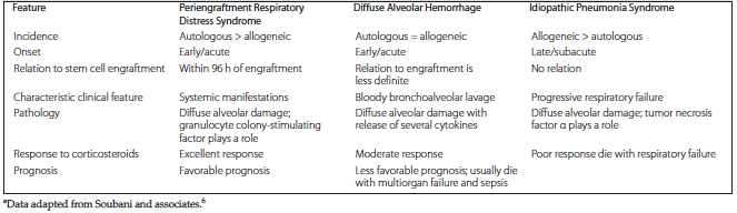
Table 1. Comparison of the Characteristics of the Main Noninfectious Causes of Acute Lung Injury After Hematopoietic Stem Cell Transplanta
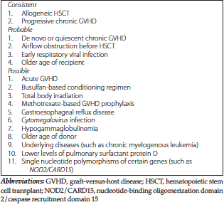
Table 2. Risk Factors for Bronchiolitis Obliterans After Hematopoietic Stem Cell Transplant
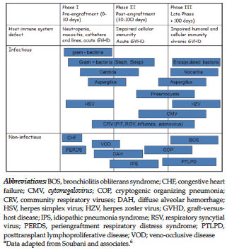
Figure 1. Time Line of the Primary Pulmonary Complications After Hematopoietic Stem Cell Transplant
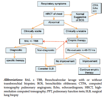
Figure 2. Suggested Algorithm for the Approach to the Diagnosis and Management of Respiratory Complications After Hematopoietic Stem Cell Transplant