Volume: 13 Issue: 6 December 2015
FULL TEXT
Encapsulating peritoneal sclerosis is a rare complication of long-term peritoneal dialysis ranging from moderate inflammation of peritoneal structures to severe sclerosing peritonitis and encapsulating peritoneal sclerosis. Complicated it, ileus may occur during or after peritoneal dialysis treatment or after kidney transplant.
We sought to evaluate 3 posttransplant encapsulating peritoneal sclerosis through clinical presentation, radiologic findings, and outcomes. We analyzed 3 renal transplant patients with symptoms of encapsulating peritoneal sclerosis admitted posttransplant to our hospital with ileus between 2012 and 2013.
Conservative treatment was applied to the patients whenever necessary to avoid surgery. One patient improved with medical therapy. Surgical treatment was delayed and we decided it as a last resort, in 2 cases with no response to conservative treatment for a long time. Finally, patients with peritoneal dialysis history should be searched carefully before renal transplant for intermittent bowel obstruction story.
Key words : Peritoneal fibrosis, Ileus, Peritoneal dialysis
Introduction
Encapsulating peritoneal sclerosis (EPS) is rare but potentially lethal complication of peritoneal dialysis treatment. In recent years, EPS occurring after kidney transplant in patients who were previously being treated with peritoneal dialysis (PD) is increasingly observed. It has been identified as a serious complication after kidney transplant.
Encapsulating peritoneal sclerosis was first reported by Gandhi and colleagues in 1980.1 The cause of EPS seems to be multifactorial. The 2 hypotheses presuppose that prolonged exposure to bioincompatible solutions and superimposition of inflammatory stimuli (eg, infectious peritonitis).2 Other factors are the exposure of the peritoneal membrane to profibrotic immunosuppressants that are used after transplant (ie, calcineurin inhibitors) and genetic predisposition.3
Peritoneal mesothelial cells and fibroblasts play important roles in collagen production through the expression of growth factors and cytokines. This complication may occur during PD therapy or after changing the dialysis type to hemodialysis or renal transplant.
The common symptoms are abdominal pain, nausea, vomiting, dehydration, and abdominal palpable mass and malnutrition, owing to incomplete or complete bowel obstruction.4-6
Clinically, once symptoms occur, it is relatively difficult to diagnose the presence of EPS, the progressive encapsulation, and obstruction of the bowel. In the later stages of the disease, the small intestinal loops stick each other and bind together and become encapsulated by thick adhesions and thus have high morbidity and mortality.1,7
Materials and Methods
We had analyzed 3 renal transplant patients admitted with EPS between 2012 and 2013. Renal insufficiency causes and peritoneal dialysis histories were evaluated. Clinical signs and symptoms, leading to the diagnosis of EPS were recorded. Laboratory data have been reported from the laboratory files. All imaging methods help in diagnosis, including abdominal radiography, abdominal ultrasound, and computed tomography (CT) scans, which are the examinations of choice for diagnosing EPS. It has been found that icodextrin PD solutions had been used in all the patients.
Preoperative care and investigations include hospitalization, examination of general physical condition, decompression of intestine, total parenteral nutrition, continuation of corticosteroids, and immunosuppressive treatment.
If conservative therapy does not improve the symptoms of EPS, surgical therapy is considered. Encapsulating peritoneal sclerosis-related death is defined as death, owing to abdominal sepsis or other EPS-related causes (eg, malnutrition, complicated peritonitis, and infection).
Results
The 3 posttransplant patients were women 28, 34, and 50-years-old at the time of surgery. Two of the patients were lupus nephritis, the other had chronic glomerulonephritis as the underlying diseases. Peritoneal dialysis periods of the patients were 7, 9, and 10 years. Owing to progressive ultrafiltration insufficiency during pretransplant short-term hemodialysis (average, 7 mo) were used until transplant. The reasons behind the transfer to hemodialysis were inadequate ultrafiltration, bacterial, and fungal peritonitis. The number of peritonitis attacks of these patients was on average 3 times in a year. Intestinal obstruction developed 1 month after the transplanting 2 cases and developed within 15 days after transplant in 1 patient. Common presenting symptoms in the posttransplant patients were abdominal pain, anorexia, nausea, vomiting, and weight loss. Blood creatinine levels of the patients were 0.0226, 0.0209, and 0.020 mL/s/m2. Glomerular filtration rate levels were 60, 50, and 72 mL/min. Nonspecific signs of systemic inflammation, such as fever, leukocytosis, elevated C-reactive protein, low serum albumin, and anemia at the time of diagnosis were evident in most patients.
Diagnosis was done by abdominal roentgenography and CT for every patient. Common findings on the CT scan of the abdomen were bowel dilatation, fluid pockets, peritoneal calcification, visceral and parietal peritoneal thickening, and adhesions of bowel loops (Figure 1).
Patients were hospitalized, and conservative treatment was begun as total parenteral nutrition, close monitoring, nasogastric drainage, no oral intake except immunosuppressive drugs which were tacrolimus + mycophenolate mofetil + prednisolone, and bowel resting. Long-term conservative treatment was applied to avoid unnecessary surgery.
One patient improved with medical therapy whereas surgical treatment preferred in 2 cases that had no response to conservative treatment. The surgical procedures were laparotomy and enterolysis if possible. During the operation, a well-known picture of EPS was found characterized by a cocoonlike sclerotic membrane encasing the small bowel (Figure 2). The parietal peritoneum was thickened and visceral peritoneum was solid, as a fibrotic capsule covering the intestines. In 1 of the case of a patient who had deceased-donor transplant, there was no room for safe dissection between the intestines and the peritoneum owing to the severe adhesions. Fortunately, patient improved spontaneously postoperatively. For 1 of the patients who did not respond to conservative treatment, a complete resection of the encapsulating sclerotic membrane and total enterolysis were performed at laparotomy, but the patient died on the postoperative seventh day. After surgery, all patients were managed by total parenteral nutrition until oral food intake becomes possible.
Discussion
Posttransplant encapsulating peritoneal sclerosis causing bowel obstruction has been identified as a serious complication after kidney transplant in patients previously treated with peritoneal dialysis. The prevalence of posttransplant EPS is reported to be 1% to 3% in PD patients undergoing kidney transplant and is associated with significant mortality.8-10 The current prevalence of PD among and-stage renal disease patients is only about 10% worldwide.11 Encapsulating peritoneal sclerosis is regarded primarily as a fatal complication of PD. The incidence of EPS in PD patients has been reported to a range from 0.7% to 7.5%.4,12,13 The EPS mortality rate has been reported to be as high as 43.5% to 69%.4,14 Frequent episodes of peritonitis have been regarded as a significant risk factor for the developing EPS. Peritonitis is the most common pathogenetic risk factor for developing EPS, which is supported by the epidemiologic data but is not a prerequisite for the development of EPS, as EPS occurs even in patients without a previous history of peritonitis.
Also, EPS has been reported in non-PD patients who received β-blockers (ie, atenolol, metoprolol, practolol, propranolol), antiseptics, talc; in patients with malignancies, or inflammatory conditions such as systemic lupus erythematosus and sarcoidosis.15,16
Recently, it has been reported that morphologic changes in the microvasculature supplying the peritoneum were involved in the mechanism of the development of the high transport PD state, and it has been suggested that several humoral factors and angiogenic factors act locally affecting peritoneal permeability in PD.17-19
Encapsulating peritoneal sclerosis was related to PD duration time before transplant. In our presentation, PD duration was around 7 years in the all 3 patients.
The clinical diagnosis of EPS is, in most cases, not difficult to establish. However, EPS develops in a stepwise manner. When PD patients with peritoneal deterioration complain of gastrointestinal symptoms, EPS is to be suspected.2 Encapsulating peritoneal sclerosis, the radiologic findings are signs of ileus and abdominal mass. Detection of thickened edematous changes in the intestinal wall by imaging abdominal CT is a value for diagnosing the inflammatory stage of EPS. The clinical diagnosis of EPS was established based on the guidelines proposed by the International Society for Peritoneal Dialysis: PD patients with bowel obstruction symptoms in whom peritoneal hypertrophy and encapsulation were confirmed by abdominal computed tomography.20
The definitive diagnosis of EPS can be established only by macroscopically confirmed encapsulated intestine at laparotomy or laparoscopy.
Peritoneal biopsy may be useful for demonstrating peritoneal hypertrophy, fibrosis, vascular sclerosis, and neovascularization.21 Although histologic diagnosis of EPS is the only reliable examination, it requires surgery and is invasive. Therefore, the diagnosis is often made postmortem or at late stages of the disease. Peritoneal fibrosis is a common finding in biopsies of uremic patients and patients on PD. Submesothelial thickness was significantly increased in patients undergoing catheter removal for peritoneal membrane failure. Duman and associates reported that sequential peritoneal thickness measurements by using ultrasonography may be useful for early diagnosis of EPS.22
In our study, the reason of not taking peritoneal biopsy is that the presence of EPS was obvious and there was a risk of bowel injury. We suggest that peritoneal biopsy may be taken at the time of catheter removal by laparoscopy for demonstrating early peritoneal fibrosis.
The progression of EPS has been classified into 4 stages by Kawanishi and colleagues: preEPS, inflammatory, encapsulating (with development of ileus), and chronic ileus stages.23
Early diagnosis is difficult before symptoms have developed. At the inflammatory stage, clinical findings related to an inflammatory condition such as fever, general fatigue, appetite and weight loss, ascites, bloody dialysate, and abdominal pain are usual.24,25 Encapsulating peritoneal sclerosis may improve spontaneously, and the patient may stay asymptomatic after removal of the peritoneal catheter and cessation of PD.4,26
If bacterial and fungal peritonitis are ruled out, corticosteroid administration may be considered. Administration of corticosteroids during the inflammatory stages of EPS is useful for minimizing inflammation, thereby reducing the risk of progression and the severity of the bowel obstruction. Certain reports have suggested the use methylprednisolone pulse therapy (500-1000 mg daily) for 2 to 3 days.23 Corticosteroids may be suggested as a routine at the time of cessation of PD, catheter removal, and transfer to hemodialysis.
Recently, Allaria and associates reported the successful use of tamoxifen in 1 case of EPS. They said that tamoxifen probably interferes with transforming growth factor beta-1 and may be useful in the treatment of this peritoneal dialysis complication.27
In the present study, 3 transplanted patients were on usual triple immunosuppression with low-dose steroids during the complication. We tried to do the most possible means for the patients to take immunosuppressive drugs per oral. In one case that had the complication early posttransplant and was vomiting, we gave up oral route and started intravenous antithymocyte globulin at a low dose as the acute rejection prophylaxis. But, we did not give pulse prednisolone therapy to any of our patients.
The key elements in conservative treatment of EPS are early diagnosis, cessation of PD with transfer to hemodialysis, conservative treatment, bowel rest with total parenteral nutrition, and corticosteroids.
If conservative therapy does not improve the symptoms of EPS, surgical intervention must be considered.26,27 In those cases who had successful surgery, bowel obstruction symptoms may improve by the time after enterolysis. Parietal peritoneal biopsy can be performed, but visceral peritoneal biopsy should not be done because of the complications resulting from intestinal perforation. However, poor surgical outcomes also have been reported. In our study, 1 patient was lost after enterolysis, and 2 patients improved with prolonged medical therapy even after the surgery.
In conclusion, there is no relation between kidney transplant and EPS formation. We believe in that EPS is a conclusion of prolonged and complicated peritoneal dialysis. Patients should be searched carefully for intermittent intestinal obstruction symptoms before transplant. Encapsulating peritoneal sclerosis that negatively influences the outcomes and quality of life of kidney recipients.
References:
- Gandhi VC, Humayun HM, Ing TS, et al. Sclerotic thickening of the
peritoneal membrane in maintenance peritoneal dialysis patients. Arch Intern
Med. 1980;140(9):1201-1203.
CrossRef - PubMed - Kawanishi H, Watanabe H, Moriishi M, et al. Successful surgical
management of encapsulating peritoneal sclerosis. Perit Dial Int.
2005;25(suppl 4):S39-S47.
PubMed - Trigka K, Dousdampanis P, Chu M, et al .Encapsulating peritoneal
sclerosis: a single-center experience and review of the literature. Int Urol
Nephrol. 2011;43(2):519-526.
CrossRef - PubMed - Nomoto Y, Kawaguchi Y, Kubo H, et al. Sclerosing encapsulating
peritonitis in patients undergoing continuous ambulatory peritoneal
dialysis: a report of the Japanese Sclerosing Encapsulating Peritonitis
Study Group. Am J Kidney Dis. 1996;28(3):420-427.
CrossRef - PubMed - Nakamoto H, Kawaguchi Y, Suzuki H, et al. Encapsulating peritoneal
sclerosis in patients undergoing continuous ambulatory peritoneal dialysis
in Japan. Adv Perit Dial. 2002;18:119-123.
PubMed - Campbell S, Clarke P, Hawley C, et al. Sclerosing peritonitis:
identification of diagnostic, clinical, and radiological features. Am J
Kidney Dis. 1994;24(5):819-825.
CrossRef - PubMed - Shao JC, Yorioka N, Nishida Y, at al. Effect of pH and glucose on
cultured human peritoneal mesothelial cells. Scand J Urol Nephrol.
1999;33(4):248-256.
CrossRef - PubMed - Fontana I, Bertocchi M, Santori G, et al. Encapsulating peritoneal
sclerosis after kidney transplantation: a single-center experience from 1982
to 2010. Transplant Proc. 2012;44(7):1918-1921.
CrossRef - PubMed - Dębska-Ślizien A, Konopa J, Januszko-Giergielewicz B, et al.
Posttransplant encapsulating peritoneal sclerosis: presentation of cases and
review of the literature. J Nephrol. 2013;26(5):906-911.
CrossRef - PubMed - Korte MR, Habib SM, Lingsma H, et al. Posttransplantation encapsulating
peritoneal sclerosis contributes significantly to mortality after kidney
transplantation. Am J Transplant. 2011;11(3):599-605.
CrossRef - PubMed - Saito A. Peritoneal dialysis in Japan: the issue of encapsulating
peritoneal sclerosis and future challenges. Perit Dial Int. 2005;25(suppl
4):S77-S82.
PubMed - Slingeneyer A. Preliminary report on a cooperative international study
on sclerosing encapsulating peritonitis. Contrib Nephrol. 1987;57:239-247.
PubMed - Rottembourg J, Gahl GM, Poignet JL, et al. Severe abdominal
complications in patients undergoing continuous ambulatory peritoneal
dialysis. Proc Eur Dial Transplant Assoc. 1983;20:236-242.
PubMed - Oulès R, Challah S, Brunner FP. Case-control study to determine the
cause of sclerosing peritoneal disease. Nephrol Dial Transplant.
1988;3(1):66-69.
PubMed - Hall DR, Morrison JB, Edwards FR. Pleural fibrosis after practolol
therapy. Thorax. 1978;33(6):822-824.
CrossRef - PubMed - Krediet RT. Beta-blockers and ultrafiltration failure. Perit Dial Int.
1997;17(6):528-531.
PubMed - Numata M, Nakayama M, Nimura S, et al. Association between an increased
surface area of peritoneal microvessels and a high peritoneal solute
transport rate. Perit Dial Int. 2003;23(2):116-122.
PubMed - Goldman M, Vandenabeele P, Moulart J, et al. Intraperitoneal secretion
of interleukin-6 during continuous ambulatory peritoneal dialysis. Nephron.
1990;56(3):277-280.
CrossRef - PubMed - Zemel D, Krediet RT. Cytokine patterns in the effluent of continuous
ambulatory peritoneal dialysis: relationship to peritoneal permeability.
Blood Purif. 1996;14(2):198-216.
CrossRef - PubMed - Kawaguchi Y, Kawanishi H, Mujais S, et al. Encapsulating peritoneal
sclerosis: definition, etiology, diagnosis, and treatment. International
Society for Peritoneal Dialysis Ad Hoc Committee on Ultrafiltration
Management in Peritoneal Dialysis. Perit Dial Int. 2000;20(suppl 4):S43-S55.
PubMed - Williams JD, Craig KJ, Topley N, et al. Morphologic changes in the
peritoneal membrane of patients with renal disease. J Am Soc Nephrol.
2002;13(2):470-479.
PubMed - Duman S, Ozbek SS, Gunay ES, et al. What does peritoneal thickness in
peritoneal dialysis patients tell us? Adv Perit Dial. 2007;23:28-33.
PubMed - Kawanishi H, Harada Y, Noriyuki T, et al. Treatment options for
encapsulating peritoneal sclerosis based on progressive stage. Adv Perit
Dial. 2001;17:200-204.
PubMed - Nakayama M, Yamamoto H, Ikeda M, et al .Risk factors and preventive
measures for encapsulating peritoneal sclerosis--Jikei experience 2002. Adv
Perit Dial. 2002;18:144-148.
PubMed - Moriishi M, Kawanishi H, Kawai T, et al. Preservation of peritoneal
catheter for prevention of encapsulating peritoneal sclerosis. Adv Perit
Dial. 2002;18:149-153.
PubMed - Nakamoto H, Takane H, Sugahara S, et al. Longitudinal changes of
peritoneal function calculated by personal dialysis capacity in a patient
after long-term continuous ambulatory peritoneal dialysis. Adv Perit Dial.
2003;19:97-102.
PubMed - Allaria PM, Giangrande A, Gandini E, et al. Continuous ambulatory
peritoneal dialysis and sclerosing encapsulating peritonitis: tamoxifen as a
new therapeutic agent? J Nephrol. 1999;12(6):395-397.
PubMed

Volume : 13
Issue : 6
Pages : 588 - 592
DOI : 10.6002/ect.2014.0036
From the 1Department of General Surgery and Transplantation; the
2Department of Molecular Genetics and Typing Tissue; and the 3Department
of Nephrology, Haydarpasa Numune Training and Research Hospital, Istanbul,
Turkey
Acknowledgements: The authors declare that they have no sources of
funding for this study, and they have no conflicts of interest to declare.
Corresponding author: Leyla Özel, Department of General Surgery and
Transplantation, Haydarpasa Numune Training and Research Hospital, Uskudar,
Istanbul, Turkey
Phone: +90 506 541 7209
Fax: +90 216 346 0582
E-mail: drleylaozel@gmail.com
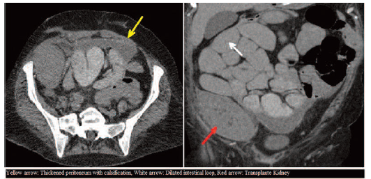
Figure 1. Computed Tomography Scan Findings
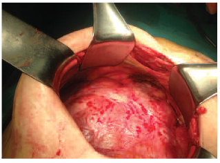
Figure 2. Cocooned Intestine View