Volume: 13 Issue: 6 December 2015
FULL TEXT
Objectives: Cytomegalovirus infection is an important cause of morbidity and mortality among recipients undergoing hematopoietic stem cell and solid-organ transplant. The risk of cytomegalovirus infection is high in cytomegalovirus-seronegative recipients of cytomegalovirus-seropositive organs (donor positive/recipient negative) and recipients with strong immunosuppressive status such as those receiving rituximab induction or antirejection treatment. However, it remains unclear how rituximab affects patients with primary cytomegalovirus infection. We evaluated the effects of low-dose rituximab therapy on clinical and immunologic outcomes in recipients who were donor positive but recipient negative for primary cytomegalovirus infections.
Materials and Methods: We conducted a retrospective review of patients with primary cytomegalovirus infections from January 2005 to March 2014. Patient outcomes were compared between groups administered given rituximab or given no intervention at the time of transplant.
Results: Our study group included 49 recipients with primary cytomegalovirus infection, including 32 who received rituximab therapy (group 1) and 17 who did not (group 2). No significant differences were observed between groups in the duration of cytomegalovirus seroconversion (P = .0570) and initial cytomegalovirus immunoglobulin G titers (P = .8418).
Conclusions: Rituximab induction therapy does not affect clinical or immunologic outcomes of primary cytomegalovirus infection, even in high-risk recipients who are donor positive but recipient negative for primary cytomegalovirus infections.
Key words : Primary cytomegalovirus infection, Rituximab, Renal transplant, Morbidity, Mortality
Introduction
Cytomegalovirus (CMV) is one of the most common causes of viral infection after hematopoietic stem cell and solid-organ transplant. The symptoms of CMV infection vary, from asymptomatic viremia to CMV syndrome and invasive tissue diseases.1 In solid-organ transplant, recipients who are seronegative for CMV but receive allografts from donors who are seropositive for CMV (D+/R-) are at the highest risk of CMV infection.1 In addition to increased morbidity and mortality, CMV infection is a risk factor for graft rejection and can reduce graft function and survival.1 Besides administration of antiviral agents, careful withdrawal of immunosuppressive drugs is sometimes used to treat CMV infections.
Rituximab is a chimeric anti-CD20 monoclonal antibody that depletes B cells through antibody-dependent cellular cytotoxicity, complement-dependent cytotoxicity, and apoptosis.2,3 Rituximab is an effective treatment for malignant B-cell lymphoma4 and various autoimmune disorders.5 Moreover, rituximab has beneficial effects on chronic inflammatory diseases mediated by T cells, such as rheumatoid arthritis6 and multiple sclerosis.7
In patients who are undergoing kidney transplant, rituximab is used to prevent rejection of ABO-incompatible transplants,8 for desensitization,9 and to treat antibody-mediated rejection.10
Because of its strong immunosuppressive effects, rituximab has been associated with increased risk of viral, bacterial, and fungal infections. Several studies have described the effects of rituximab on posttransplant Christ infection.11-13 However, its effects on primary CMV infection at the time of kidney transplant remain unknown. In the present study, we retrospectively evaluated the clinical and immunologic differences between recipients with primary CMV infections who received rituximab and those who did not.
Materials and Methods
The Internal Ethics Review Board of Tokyo Women’s Medical University approved this study. This retrospective analysis followed the principles of the Declaration of Helsinki. Written informed consent was obtained from all patients in our kidney transplant program. Clinical and laboratory results were extracted from an electronic database and patient medical records.
Patients
We retrospectively evaluated 49 patients who received a diagnosis of primary
CMV infection between January 2005 and March 2014 at our department and 2 other
departments (Table 1). All patients were living-donor kidney transplant
recipients with D+/R- CMV serostatus who did not receive any other organ
transplant before or after the kidney transplant. Patients were excluded for
unknown CMV serostatus if splenectomy was performed or if valganciclovir or
ganciclovir prophylaxis was administered. Thirty-two recipients received
rituximab induction therapy within 7 days before transplant (group 1), whereas
17 recipients who served as the control group (group 2) underwent transplant
without rituximab but received immunosuppressive agents, including tacrolimus,
mycophenolate mofetil or mizoribine, methylprednisolone, and basiliximab. Two
patients in group 2 did not receive basiliximab induction therapy because the
immunosuppressive regimen did not include basiliximab for ABO-compatible or
nonsensitized transplant recipients until March 2006. Graft rejection occurred
in 1 patient in group 1 and 4 patients in group 2 before primary CMV infection;
these patients received antirejection treatment (1/32, 3.1% in group 1, 4/17,
23.5% in group 2).
Baseline immunosuppression protocol
Immunosuppression was usually with a triple-drug regimen (tacrolimus,
mycophenolate mofetil or mizoribine, and methylprednisolone) administered 1 week
before transplant (Figure 1). Postoperatively, basiliximab (20 mg) was
administered intravenously on days 0 and 4. Three to five sessions of
plasmapheresis, including double-filtration plasmapheresis or plasma exchange,
were performed before transplant in patients with a history of sensitization.
Some patients also received preoperative double-filtration plasmapheresis or
plasma exchange to prevent recurrence of kidney disease such as focal segmental
glomerular sclerosis.
Rituximab therapy
Rituximab was first administered in our departments in January 2005.
Rituximab induction therapy is indicated for ABO minor mismatch or
ABO-incompatible recipients, patients positive for antibodies against donor
human leukocyte antigen (donor-specific antibodies), and recipients originally
diagnosed with focal segmental glomerular sclerosis as an end-stage kidney
disease. Antibodies against human leukocyte antigen were detected by flow
cytometry or Luminex (One Lambda, Canoga Park, CA, USA). In group one, 28
patients (87.5%) received 200 mg rituximab and 4 patients (12.5%) received 500
mg rituximab (Figure 1).
Detection of donor-specific antibodies using a solid-phase assay
We preoperatively examined the sensitized status of all recipients using a
lymphocyte cytotoxic test/flow cytometric crossmatch test, as previously
reported.14 Solid-phase assay (Luminex) has been used in our departments since
2005. Briefly, 20 μL of sera were added to 5 μL of class I or class II antigen
beads. The beads were incubated for 30 minutes in the dark at room temperature
and washed. The luminescence was read using a LABScreen 100 Luminex system. Data
were analyzed using the LABScreen analysis software, HLA Fusion 2.0 (One
Lambda), and mean fluorescence intensities over 800 were considered positive.
Definition of cytomegalovirus infection and disease
Asymptomatic CMV infection was defined as detection of CMV in blood based
on CMV antigenemia test results. Cytomegalovirus antigen levels were the
total number of virus-positive cells per 150 000 leukocytes, as determined from
blood samples processed and stained with C10/C11 monoclonal antibodies, which
recognize the viral pp65 early antigen. Cytomegalovirus antigen levels
were determined postoperatively at weekly intervals for 3 months, at biweekly
intervals between 3 and 6 months, and at monthly intervals between 6 and 12
months. Cytomegalovirus syndrome was defined as CMV infection associated
with clinical manifestations, including fever > 38°C for at least 2 days,
malaise, leukopenia, or thrombocytopenia. Recurrent CMV infection was defined as
detection of antigenemia more than 1 month after the original antigenemia had
disappeared.
Cytomegalovirus seroconversion and initial cytomegalovirus immunoglobulin
G titers
Seroconversion in CMV-seronegative recipients was defined as the detection of
CMV immunoglobulin G (IgG). Cytomegalovirus IgG and immunoglobulin M were
measured by enzyme-linked immunosorbent assay. Both were measured at least twice
weekly after diagnosis of CMV infection and 2 to 5 times until the transplant
recipients became positive for CMV antibodies. Initial CMV IgG titers evaluated
in this study were defined as the titers measured when seroconversion was
confirmed.
Treatment of primary cytomegalovirus infection
Patients who received any prophylaxis for CMV infection were excluded from
the present study. Patients with CMV antigenemia of more than 4 per 150 000
leukocytes, had fever > 38°C, or who were symptomatic for CMV infection were
intravenously treated with ganciclovir at 10 mg/kg daily until CMV
seroconversion was confirmed or CMV antigenemia disappeared. The ganciclovir
dosage was reduced according to kidney function. Coordinated administration of
antimetabolic drugs included reduction, discontinuation, or conversion to
another metabolic drug.
Rejection diagnosis and treatment
Transplant rejections were confirmed by biopsy. Rejection types were
classified according to the Banff’ 09 Meeting Report,15 with some
modifications. Cases with intimal arteritis v1 and v2, categorized as
T-cell–mediated rejection IIA or IIB in the Banff’ 09 classification scheme,
were diagnosed as antibody-mediated rejection (AMR) based on the Banff’ 13
classification.16 All patients with acute rejection, including
T-cell–mediated rejection and AMR, were first treated intravenously with 500 mg
methylprednisolone for 2 days. Patients with T-cell–mediated rejection who were
resistant to methylprednisolone were administered muromonab-CD3 (OKT3;
Orthoclone: Ortho-Biotech, Tokyo, Japan) at a dosage of 5 mg/day for 10 days or
gusperimus hydrochloride (Spanidin, Tokyo, Japan) at a dosage of 3 mg/kg/day for
7 days (before 2006). Patients with AMR were treated with plasma exchange,
gusperimus hydrochloride at the same dosage as described above, and an
additional single dose of rituximab (200 mg).
Statistical analyses
All analyses were performed using JMP 11 (SAS Institute Inc., Cary, NC, USA).
Quantitative parameters were compared using Mann-Whitney U test; qualitative
parameters were compared using the chi-square and Fisher exact tests. P
values less than .05 were considered statistically significant.
Results
Baseline patient characteristics
The baseline characteristics of patients in both groups were similar (Table 1).
All patients in group 2 underwent ABO-compatible transplant. In group 1, 8
patients underwent ABO-compatible, 7 patients underwent ABO minor mismatch, and
17 patients underwent ABO-incompatible transplant (P < .0001). There were
no significant differences in the original kidney diseases between groups.
Outcomes of primary cytomegalovirus infection
We evaluated clinical and immunologic outcomes to determine the effects of
rituximab on primary CMV infection (Table 2). There were no significant
differences between groups in outcome rates, including duration of onset after
transplant (P = .5776), ratio of patients with CMV syndrome (P =
.2962), peak viral load (P = .9414), hospitalization duration (P =
.8579), seroconversion duration (P = .0570), initial CMV IgG titer (P
= .8418), ratio of immunosuppressive drug coordination (P = .2490), and
recurrence of CMV infection (P = .9093).
Rejection before and after primary cytomegalovirus infection
Figure 2 shows the rejection rates before and after primary CMV infection.
Rejection was observed in 1 of 32 patients (3.1%) in group 1 and in 4 of 17
patients (23.5%) in group 2 before primary CMV infection. After infection,
rejection was shown in 4 of 31 patients (12.9%) in group 1 and 1 of 13 patients
(7.69 %) in group 2. The rejection rate before primary CMV infection was
significantly higher in group 2 (P = .0432); however, there were no
significant differences in the rejection rate after primary CMV infection (P
= .6193) (Table 3). Antibody-mediated rejection persisted in 1 highly sensitized
group 1 patient with donor-specific antibodies who experienced AMR before
infection. We also observed 4 patients with rejection before infection in group
2 (1A, 1B, 1B, 1B) who also demonstrated changes in their condition after
infection, with 1A aggravated to 1B, 1B aggravated to AMR + 1A, 1A disappeared,
and 1B improved to 1A (Table 4).
Risk factors for rejection after primary cytomegalovirus infection
There were 44 patients (89.7%) without rejection before primary CMV infection;
these patients were evaluated to determine risk factors for rejection after
infection (Table 5). Multivariate analysis indicated that administration of
rituximab (P = .8731), existence of donor-specific antibodies (P =
.8963), and ABO incompatibility (P = .4793) were not independent risk
factors for rejection after primary CMV infection. The duration of
seroconversion (P = .6306) and CMV IgG titer (P = .2186) were also
not associated with rejection after infection. We also found that
discontinuation of antimetabolic drugs was not correlated with rejection after
infection (P = .4268). However, although we observed that CMV recurrence
could be an independent risk factor for rejection after infection (odds ratio,
8.554; 95% confidence interval, 0.901-148.866), the P value did not reach
statistical significance (P = .0616).
Survival rates for patients with cancer in the native kidney
In contrast to patients with tumors that developed in the allograft, all 17
cancer-specific deaths were in patients with tumors in the native kidneys. The
5-year and 10-year overall survival rates for patients with cancer in the native
kidney were 59.1% (95% CI, 41.8-72.9) and 37.6% (95% CI, 18.6-56.5) (Figure 2).
The cancer-specific (RCC or TCC) survival rates for patients with cancer in the
native kidney was 68.2% at 5 years (95% CI, 54.1-81.6) and 51.5% at 10 years
(95% CI, 33.9-71.8) (Figure 2).
Of the 17 cancer-specific deaths of patients with cancer in the native kidney, 14 were from RCC and 3 were from TCC (Figure 1). The survival rate for patients who developed RCC in the native kidney was 71.2% at 5 years (95% CI, 57.0-84.0) and 58.5% at 10 years (95% CI, 40.2-77.9). In contrast, the 5- and 10-year survival rates for TCC were 50% (95% CI, 15.5-94.2) and 0%.
Discussion
Our results suggest that rituximab did not significantly affect the clinical or immunologic outcomes of primary CMV infection after living-donor kidney transplant. The influence of rituximab on humoral responses after vaccination has been previously reported. Several studies have shown that antibody response to pneumococcal conjugate17,18 or influenza vaccines19 in patients with rheumatoid arthritis was impaired by rituximab; therefore, it is preferable to administer these vaccinations before rituximab infusion. We hypothesized that rituximab also suppresses CMV seroconversion and secretion of CMV IgG. However, neither the duration of seroconversion nor CMV IgG titer differed significantly between groups in this study. We also observed no significant differences in clinical outcomes, including the ratio of patients with CMV syndrome, peak viral load, hospitalization duration treatment, and the ratio of CMV infection recurrence. Our findings that CMV IgG titer was not dependent on rituximab are discordant from previous studies. One explanation for this difference may be the higher dosage of rituximab administered to patients with rheumatoid arthritis versus those administered in our study. Higher dosage may result in stronger immunosuppression and consequent antibody secretion.
Several studies have investigated the effect of rituximab monotherapy and rituximab combined with other immunosuppressive regimens, such as plasmapheresis or intravenous immunoglobulin, on posttransplant infectious complications in transplant recipients.20 In their studies, Petropoulou and associates11 and Kamar and associates12 suggested that the incidence of bacterial or fungal infections was higher in patients who received rituximab, whereas Lee and associates13 reported that patients with non-Hodgkin lymphoma who were given rituximab before autologous hematologic stem cell transplant tended to have increased risk of CMV infection.
At our centers, a single low dose of RTX is administered within 7 days before transplant. Based on our previous studies,18 we believe that administration of rituximab at low doses may prevent rejection. Toki and associates21 reported that administration of a single low dose of RTX, less than the 375 mg/m2 used in this and previous studies (15, 35, 150, or 300 mg/m2), had a potent effect, depleting B cells in both the spleen and peripheral blood.21 We initially administered rituximab at a dose of 375 mg/m2. However, we have gradually reduced administration to a single dose of 200 mg as a component of the desensitization protocol, with good outcomes. Table 3 also shows the efficacy of low-dose rituximab to prevent rejection during the early posttransplant period, with a significantly lower rejection rate before primary CMV infection in group 2 (P = .0432). Nishida and associates22 previously demonstrated that low-dose rituximab did not affect CMV infection. However, these studies included patients with all donor and recipient CMV serology combinations. Therefore, the relation between rituximab and primary CMV infection in D+/R- patients remains uncertain. Luan and associates23 reported rabbit anti-thymocyte globulin induction to be associated with higher risk of primary CMV infection versus basiliximab in D+/R- kidney and/or pancreas transplant recipients.
There are several explanations regarding why the groups in our study showed no clinical and immunologic differences. First, rituximab does not impair T-cell and natural killer cell responses, which are an important part of the immune system response to viral infection.24,25 Similarly, mature plasma cells in bone marrow, which do not express CD19 or CD20, and immature, transitional B cells and early B-cell progenitors in bone marrow, which do not express CD20, are not affected by rituximab. These unsuppressed cells may play a role in the immune system against CMV infection.22,26 Moreover, Nishida and associates22 showed that CD19-positive, CD5-positive B cells, which produce polyreactive natural antibodies, may be key factors of CMV seroconversion because CMV seroconversion coincided with the appearance of CD19-positive, CD5-positive B cells. They also speculated that CD27-positive memory B cells and plasma cells in the spleen, which are not affected by rituximab,27 may play a role in CMV infection.22 Even in the absence of detectable memory B cells, the majority of plasma cells can survive and secrete antibodies for more than 1 year.28 Previous studies have suggested that CMV IgG titers did not significantly change after rituximab-contained desensitization.22,25 Based on the results of these previous reports and our findings, we believe that, once secreted, CMV IgG titers are not affected by rituximab.
Mycophenolate mofetil is one of the most commonly used and successful antirejection antimetabolic agents. Its strong immunosuppressive effects inhibit both B- and T-cell function, increasing the risk of infectious complications.29 Cautious withdrawal of immunosuppression agents is an option for treatment of moderate to severe CMV infections.1 However, adverse effects such as impaired immunosuppression remain an unsolvable problem and potential cause of graft rejection. Moreover, CMV infection itself is an important contributor to allograft rejection, which is caused by modulation of the immune system.30,31 The findings of the present study indicate that antimetabolic drug coordination had no significant influence on rejection after primary CMV infection, regardless of rituximab administration status. However, 4 patients (3 in group 1 and 1 in group 2) experienced AMR after discontinuation of antimetabolic drugs (Table 6). This result might be caused by immunologic issues attributed to CMV infection. Therefore, discontinuation of antimetabolic drug treatment should be approached cautiously. Moreover, Table 5 suggests that CMV infection recurrence was an independent risk factor for rejection after primary CMV infection, although the P value did not reach statistical significance (P = .0616). Two reasons may explain the association between recurrent CMV infection and rejection: more intense modulation of the immune systems as a result of recurring CMV infection or prolonged immunosuppression during treatment.
The limitations of our study include the small number of patients. In addition, we could not exactly confirm the duration of discontinuation or reduction of antimetabolic drugs. It was difficult to determine whether rejection after CMV infection was caused by the coordination of drugs during immunosuppressive reduction or by the CMV infection itself. Our findings, therefore, should be confirmed in larger and more detailed studies.
In Japan, valganciclovir or ganciclovir prophylaxis is not a common off-label treatment because the Japanese health insurance system has not covered their use even in high-risk D+/R- recipients. Because several studies have previously demonstrated the effectiveness of prophylaxis and preemptive treatment,32-36 further investigations of the relation between low-dose rituximab and primary CMV infection in patients with prophylaxis or preemptive treatment are necessary.
In conclusion, our study suggests that rituximab at low doses does not affect the clinical and immunologic outcomes of patients with primary CMV infection. Therefore, we can safely use low-dose RTX to prevent rejection even in high-risk D+/R- recipients. However, discontinuation of antimetabolic drugs for treatment of CMV infection might trigger rejection such as AMR. Moreover, recurrent CMV infection after primary CMV infection could be associated with rejection.
References:
- Razonable RR, Humar A. Cytomegalovirus in solid organ transplantation.
Am J Transplant. 2013;13 Suppl 4:93-106.
CrossRef - PubMed - Chow KU, Sommerlad WD, Boehrer S, et al. Anti-CD20 antibody (IDEC-C2B8,
rituximab) enhances efficacy of cytotoxic drugs on neoplastic lymphocytes in
vitro: role of cytokines, complement, and caspases. Haematologica.
2002;87(1):33-43.
PubMed - Glennie MJ, French RR, Cragg MS, Taylor RP. Mechanisms of killing by
anti-CD20 monoclonal antibodies. Mol Immunol. 2007;44(16):3823-3837.
CrossRef - PubMed - Coiffier B, Lepage E, Briere J, et al. CHOP chemotherapy plus rituximab
compared with CHOP alone in elderly patients with diffuse large-B-cell
lymphoma. N Engl J Med. 2002;346(4):235-242.
CrossRef - PubMed - Dorner T, Isenberg D, Jayne D, et al. Current status on B-cell depletion
therapy in autoimmune diseases other than rheumatoid arthritis. Autoimmun
Rev. 2009;9(2):82-89.
CrossRef - PubMed - Thurlings RM, Vos K, Wijbrandts CA, Zwinderman AH, Gerlag DM, Tak PP.
Synovial tissue response to rituximab: mechanism of action and
identification of biomarkers of response. Ann Rheum Dis. 2008;67(7):917-925.
CrossRef - PubMed - Bar-Or A, Fawaz L, Fan B, et al. Abnormal B-cell cytokine responses a
trigger of T-cell–mediated disease in MS? Ann Neurol. 2010;67(4):452-461.
CrossRef - PubMed - Fuchinoue S, Ishii Y, Sawada T, et al. The 5-year outcome of
ABO-incompatible kidney transplantation with rituximab induction.
Transplantation. 2011;91(8):853-857.
CrossRef - PubMed - Ishida H, Furusawa M, Shimizu T, Nozaki T, Tanabe K. Influence of
preoperative anti-HLA antibodies on short- and long-term graft survival in
recipients with or without rituximab treatment. Transpl Int.
2014;27(4):371-382.
CrossRef - PubMed - Immenschuh S, Zilian E, Dammrich ME, et al. Indicators of treatment
responsiveness to rituximab and plasmapheresis in antibody-mediated
rejection after kidney transplantation. Transplantation. 2015;99(1):56-62.
CrossRef - PubMed - Petropoulou AD, Porcher R, Peffault de Latour R, et al. Increased
infection rate after preemptive rituximab treatment for Epstein-Barr virus
reactivation after allogeneic hematopoietic stem-cell transplantation.
Transplantation. 2012;94(8):879-883.
CrossRef - PubMed - Kamar N, Milioto O, Puissant-Lubrano B, et al. Incidence and predictive
factors for infectious disease after rituximab therapy in kidney-transplant
patients. Am J Transplant. 2010;10(1):89-98.
CrossRef - PubMed - Lee MY, Chiou TJ, Hsiao LT, et al. Rituximab therapy increased
post-transplant cytomegalovirus complications in Non-Hodgkin's lymphoma
patients receiving autologous hematopoietic stem cell transplantation. Ann
Hematol. 2008;87(4):285-289.
CrossRef - PubMed - Ishida H, Miyamoto N, Shirakawa H, et al. Evaluation of
immunosuppressive regimens in ABO-incompatible living kidney
transplantation--single center analysis. Am J Transplant. 2007;7(4):825-831.
CrossRef - PubMed - Sis B, Mengel M, Haas M, et al. Banff '09 meeting report: antibody
mediated graft deterioration and implementation of Banff working groups. Am
J Transplant. 2010;10(3):464-471.
CrossRef - PubMed - Haas M, Sis B, Racusen LC, et al. Banff 2013 meeting report: inclusion
of c4d-negative antibody-mediated rejection and antibody-associated arterial
lesions. Am J Transplant. 2014;14(2):272-283.
CrossRef - PubMed - Crnkic Kapetanovic M, Saxne T, Jonsson G, Truedsson L, Geborek P.
Rituximab and abatacept but not tocilizumab impair antibody response to
pneumococcal conjugate vaccine in patients with rheumatoid arthritis.
Arthritis Res Ther. 2013;15(5):R171.
CrossRef - PubMed - Bingham CO, 3rd, Looney RJ, Deodhar A, et al. Immunization responses in
rheumatoid arthritis patients treated with rituximab: results from a
controlled clinical trial. Arthritis Rheum. 2010;62(1):64-74.
CrossRef - PubMed - van Assen S, Holvast A, Benne CA, et al. Humoral responses after
influenza vaccination are severely reduced in patients with rheumatoid
arthritis treated with rituximab. Arthritis Rheum. 2010;62(1):75-81.
CrossRef - PubMed - Chung BH, Yun JT, Ha SE, et al. Combined use of rituximab and
plasmapheresis pre-transplant increases post-transplant infections in renal
transplant recipients with basiliximab induction therapy. Transpl Infect
Dis. 2013;15(6):559-568.
CrossRef - PubMed - Toki D, Ishida H, Horita S, Setoguchi K, Yamaguchi Y, Tanabe K. Impact
of low-dose rituximab on splenic B cells in ABO-incompatible renal
transplant recipients. Transpl Int. 2009;22(4):447-454.
CrossRef - PubMed - Nishida H, Ishida H, Tanaka T, et al. Cytomegalovirus infection
following renal transplantation in patients administered low-dose rituximab
induction therapy. Transpl Int. 2009;22(10):961-969.
CrossRef - PubMed - Luan FL, Samaniego M, Kommareddi M, Park JM, Ojo AO. Choice of induction
regimens on the risk of cytomegalovirus infection in donor-positive and
recipient-negative kidney transplant recipients. Transpl Infect Dis.
2010;12(6):473-479.
CrossRef - PubMed - Agarwal A, Vieira CA, Book BK, Sidner RA, Fineberg NS, Pescovitz MD.
Rituximab, anti-CD20, induces in vivo cytokine release but does not impair
ex vivo T-cell responses. Am J Transplant. 2004;4(8):1357-1360.
CrossRef - PubMed - Ge S, Pao A, Vo A, et al. Immunologic parameters and viral infections in
patients desensitized with intravenous immunoglobulin and rituximab. Transpl
Immunol. 2011;24(3):142-148.
CrossRef - PubMed - Zarkhin V, Li L, Sarwal M. "To B or not to B?" B-cells and graft
rejection. Transplantation. 2008;85(12):1705-1714.
CrossRef - PubMed - Ramos EJ, Pollinger HS, Stegall MD, Gloor JM, Dogan A, Grande JP. The
effect of desensitization protocols on human splenic B-cell populations in
vivo. Am J Transplant. 2007;7(2):402-407.
CrossRef - PubMed - Slifka MK, Antia R, Whitmire JK, Ahmed R. Humoral immunity due to
long-lived plasma cells. Immunity. 1998;8(3):363-372.
CrossRef - PubMed - Ritter ML, Pirofski L. Mycophenolate mofetil: effects on cellular immune
subsets, infectious complications, and antimicrobial activity. Transpl
Infect Dis. 2009;11(4):290-297.
CrossRef - PubMed - Sagedal S, Nordal KP, Hartmann A, et al. The impact of cytomegalovirus
infection and disease on rejection episodes in renal allograft recipients.
Am J Transplant. 2002;2(9):850-856.
CrossRef - PubMed - Kranz B, Vester U, Wingen AM, et al. Acute rejection episodes in
pediatric renal transplant recipients with cytomegalovirus infection.
Pediatr Transplant. 2008;12(4):474-478.
CrossRef - PubMed - Manuel O, Kralidis G, Mueller NJ, et al. Impact of antiviral preventive
strategies on the incidence and outcomes of cytomegalovirus disease in solid
organ transplant recipients. Am J Transplant. 2013;13(9):2402-2410.
CrossRef - PubMed - Humar A, Lebranchu Y, Vincenti F, et al. The efficacy and safety of 200
days valganciclovir cytomegalovirus prophylaxis in high-risk kidney
transplant recipients. Am J Transplant. 2010;10(5):1228-1237.
CrossRef - PubMed - Helantera I, Kyllonen L, Lautenschlager I, Salmela K, Koskinen P.
Primary CMV infections are common in kidney transplant recipients after 6
months valganciclovir prophylaxis. Am J Transplant. 2010;10(9):2026-2032.
CrossRef - PubMed - Meije Y, Fortun J, Len O, et al. Prevention strategies for
cytomegalovirus disease and long-term outcomes in the high-risk transplant
patient (D+/R-): experience from the RESITRA-REIPI cohort. Transpl Infect
Dis. 2014;16(3):387-396.
CrossRef - PubMed - Martin-Gandul C, Perez-Romero P, Blanco-Lobo P, et al. Viral load,
CMV-specific T-cell immune response and cytomegalovirus disease in solid
organ transplant recipients at higher risk for cytomegalovirus infection
during preemptive therapy. Transpl Int. 2014;27(10):1060-1068.
CrossRef - PubMed

Volume : 13
Issue : 6
Pages : 573 - 580
DOI : 10.6002/ect.2015.0126
From the 1Department of Urology, Kidney Center, Tokyo Women’s
Medical University, Tokyo; the 2Department of Urology, Okubo Hospital
and Shinjuku, Tokyo; and the 3Department of Urology, Toda-Chuo
General Hospital, Saitama, Japan
Acknowledgements: The authors declare that they have no sources of
funding for this study, and they have no conflicts of interest to declare. We
thank Dr Ishigouka Hidetoshi (Department of Urology, Okubo Hospital), Dr
Tsujimura Kazuma (Department of Urology, Toda-Chuo General Hospital), Ms Makiko
Fujiwara, Research laboratory, Tokyo Women’s Medical University, Shinjuku,
Tokyo, and the STATZ Institute for data collection.
Corresponding author: Hideki Ishida, Department of Urology, Kidney
Center, Tokyo Women’s Medical University, 162-8666, 8-1 Kawada-cho, Shinjuku-ku,
Tokyo, Japan
Phone: +81 3 3353 8111
Fax: +81 3 3356 0293
E-mail: tgphide@gol.com
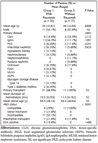
Table 1. Characteristics of Patients With Primary Cytomegalovirus Infection After Kidney Transplant
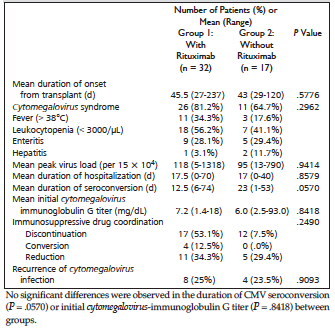
Table 2. Outcome of Primary Cytomegalovirus Infection According to Group
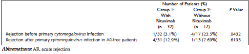
Table 3. Rejection Incidence Before and After Primary Cytomegalovirus Infection
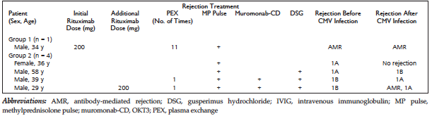
Table 4. Demographics of Rejection Treatments Before Primary Cytomegalovirus Infection and Graft Outcomes After Infection
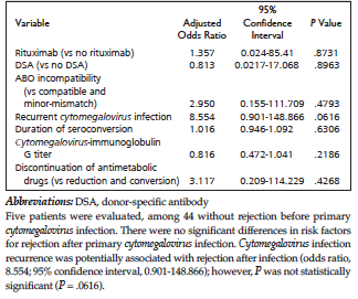
Table 5. Multiple Logistic Regression Analysis of Risk Factors Associated With Graft Rejection After Primary Cytomegalovirus Infection
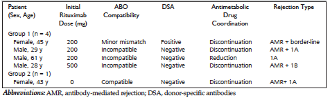
Table 6. Demographics of Patients With Rejection After Primary Cytomegalovirus Infection
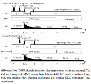
Figure 1. Baseline Immunosuppressive Protocol According to Group
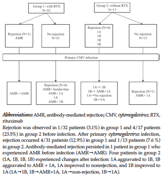
Figure 2. Rejection Time Series Before and After Primary Cytomegalovirus Infection