Volume: 13 Issue: 6 December 2015
FULL TEXT
Objectives: Continuous thrombocytopenia after liver transplant is associated with a less favorable prognosis, but this pathogenesis remains unclear. We focused on the consumption of platelets in the allograft. We assessed platelet consumption in allografts, and evaluated the pathology of platelet aggregation in an allograft tissue and its involve-ment in clinical outcomes.
Materials and Methods: We took biopsy specimens from 20 patients. To examine the localization of platelet aggregation, CD42b was assayed immuno-histochemically, and its level of expression correlated with clinical data and outcomes.
Results: Platelet aggregation in zone 3 was 70%, compared with 30% in zone 1 and 50% in zone 2. Platelets were found mainly as extravasated platelet aggregates in local microenvironments. Patients were stratified according to the extent of extravasated platelet aggregates in zone 3 into extravasated platelet aggregate-negative and -positive groups. Graft weight/recipient body weight ratio with the extravasated platelet aggregate-positive group was significantly lower than that of the extravasated platelet aggregate-negative group. Platelet count after surgery was lower, while total bilirubin and prothrombin time/international normalized ratio were higher in the extravasated platelet aggregate-positive than they were in the extravasated platelet aggregate-negative group.
Conclusions: Extravasated platelet aggregates in the zone 3 of allograft tissue cause the consumption of platelets and continuous thrombocytopenia after transplant, and may be the clinical marker for deterioration of graft function. Platelet activation and degranulation following the release by platelets of some negative regulators may be involved partially in liver damage.
Key words : CD42b, Liver biopsy, Allograft tissue
Introduction
There is increasing evidence that continuous thrombocytopenia soon after liver transplant (LT) is associated with a less favorable prognosis.1,2 Few pathophysiological studies have assessed post-LT thrombocytopenia, because of difficulties in obtaining tissue samples.3 Evaluation of allograft tissue from a living-donor LT (LDLT) recipient with throm-bocytopenia plus sinusoidal obstruction syndrome (SOS) shows platelet aggregation in the space of Disse along the sinusoidal vessels and platelet phagocytosis by hepatocytes, a morphology called extravasated platelet aggregation (EPA).4
In our case, the progression of SOS resulted in graft dysfunction and the patient died. These findings indicate that platelets have the potential to aggregate and be consumed in allograft tissue after transplant. Moreover, platelet aggregation in the allograft may be partly involved in graft dysfunction. We immunohistochemically evaluated the presence and extent of platelet aggregation in allograft tissue obtained after transplant, using antibodies to the platelet marker CD42b (platelet glycoprotein Ib), and assessed the clinical effects of platelet aggregation on graft function.
Materials and Methods
Patients and biopsy material
Of the 45 consecutive patients who underwent LT at our institution from August
2005 to February 2014, we selected those who underwent liver biopsy within 60
days of LDLT (median, 28 d). Liver biopsies were obtained from only LDLT
recipients. Pediatric recipients and recipients of deceased-donor liver
transplants were excluded. Thus, a total of 20 patients were included.
After transplant, all patients received immuno-suppressive therapy, consisting of tacrolimus, prednisolone, and mycophenolate mofetil. All LDLTs were performed after obtaining full informed consent from the patients. The study protocol conformed to the ethical guidelines of the 1975 Declaration of Helsinki, and was approved by the Ethical Review Committee of the Institute.
Evaluation of platelet aggregation in allograft tissues
The avidin-biotin peroxidase complex method was used for immunohistochemistry.
The primary antibody was mouse monoclonal anti-CD42b antibody (1:100, EPR6995;
Abcam, Tokyo, Japan). CD42b (platelet glycoprotein Ib) is a specific marker for
platelets, so a CD42b-positive reaction was taken to indicate the presence of
platelets. We previously reported that normal spleens were positive and normal
livers negative for CD42b.4 The distribution of
CD42b-immunoreactivity in allograft tissues was categorized using the Rappaport
classification.5 The extent of CD42b-immunoreactivity in each zone
was evaluated by light microscopy at ×200 magnification, and was categorized by
2 independent observers as − (no staining); + (< 33% of each zone); ++ (33% to
66%; and +++ (> 66%). The relations between platelet aggregation in allograft
tissues and clinical data, including preoperative status, graft variables,
operative data, and clinical outcomes, were evaluated.
Statistical analyses
Continuous values are presented as means ± standard deviation (SD), and are
compared by the Mann-Whitney U test. Differences in frequency were analyzed by
the chi-square test. All statistical analyses were performed using StatMate IV
software (Release 8.0.1, SAS Institute Japan), with P < .05 considered
statistically significant.
Results
Expression of CD42b in allograft tissues
The results are shown in Table 1. CD42b-immunoreactivity was observed in
liver zone one of 6 of the 20 patients (30.0%), with the intensity of staining
being grade one in all 6. CD42b-immunoreactivity was observed in zone two of 10
patients (50.0%), being grade one in 8 and grade two in 2.
CD42b-immunoreactivity was present in zone three in 14 patients (70.0%), being
grade one in 6, grade two in 5, and grade three in 3. These results indicated
that platelet aggregation was more likely present in liver zone 3 than in zones
1 and 2. Representative results from one of the LDLT recipients with grade 3
CD42b intensity in zone 3 are shown in Figure 1. At low magnification (×100),
CD42b-immunoreactivity was mainly observed in zone 3 (Figure 1A). Higher
magnification (×200) showed that > 66% of zone 3 was immunopositive for CD42b
(Figure 1B).
Histologic localization of CD42b-immunoreactivity in local
microenvironments
High magnification after incubation of tissue samples with antibody to CD42b
showed that platelet aggregation could be classified into 2 patterns: aggregates
attached to hepatocytes along the sinusoids (Figure 2A), and dots in hepatocyte
cytoplasm (Figure 2B). These platelet aggregate morphologies are identical to
those previously reported.4
Relation of platelet aggregation in zone 3 and clinical features during
the perioperative period
To determine the associations between EPA in zone 3 and the clinical features of
LDLT patients, patients were stratified according to the extent of
CD42b-immunoreactivity. Patients with grade 0/1 staining were classified as the
EPA-negative group, and those with grade 2/3 staining as the EPA-positive group.
The groups were comparable in preoperative recipient factors, including age,
sex, underlying diseases, Model for End-Stage Liver Disease score, and
Child-Pugh score; and in donor and graft characteristics including donor age,
donor sex, percentage with ABO incompatibility, and graft type (Table 2). The
graft volume/recipient standard liver volume ratio was lower in the EPA-positive
than it was in the EPA-negative group, although the difference was not
statistically significant. The graft weight/recipient body weight ratio,
however, was significantly lower in the EPA-positive than it was in the
EPA-negative group. There were no significant between-group differences in
intraoperative factors including blood loss, operative time, or the duration of
graft ischemia.
Laboratory data of the recipients after LDLT are shown in the Table 3. Platelet counts on postoperative day (POD) 14 were significantly lower in the EPA-positive than they were in the EPA-negative group. Platelets counts in the EPA-negative group were higher than 15 × 104/mm3 on POD 14, and remained so until POD 56. In contrast, similar platelet counts were not observed in the EPA-positive group until POD 56. Total bilirubin was significantly higher on PODs 14, 28, and 56 in the EPA-positive group, as was prothrombin time/international normalized ratio on POD 14. There were no significant differences in aspartate aminotransferase and alkaline phosphatase concentrations in these 2 groups.
Inasmuch as platelet counts after LDLT are affected by splenic function, we compared postoperative platelet counts only in LDLT recipients without splenic function. Four patients in the EPA-negative group had undergone splenectomy, whereas 5 patients in the EPA-positive group had undergone splenectomy, and 1 had undergone preoperative partial splenic embolization. Platelet counts on PODs 7, 21, and 42 were significantly lower in the EPA-positive patients than they were in the EPA-negative patients without splenic function (Figure 3). Platelet counts in all 4 of these EPA-negative patients were higher than 20 × 104/mm3 by POD 21. By contrast, platelet counts in these 6 EPA-positive patients were not over 20 × 104/mm3 until POD 56.
Postoperative complications and clinical outcomes
Table 4 shows the complications and clinical outcomes for patients in both
groups. Five patients had intra-abdominal hemorrhage, requiring hemostatic
therapy, either interventional radiology or laparotomy. Bacterial infections,
including pneumonia (n = 1), cholangitis (n = 2), sepsis (n = 4), and fungal
infections (n = 1) were diagnosed based on their clinical manifestations and the
isolation of organisms. Ten patients experienced cytomegalovirus infection,
diagnosed as the presence of cytomegalovirus antigenemia. Two patients
experienced acute renal failure, requiring renal replacement therapy. Patients
with biopsy-proven acute (n = 7) and chronic (n = 1) rejection were treated with
augmented immunosuppressive therapy. There were no signi-ficant differences
between the EPA-negative patients and the EPA-positive patients in the rates of
each postoperative complication. In-hospital mortality and 1-year mortality
rates were higher in the EPA-positive group than they were in the EPA-negative
group, although these differences did not reach statistical significance. One
patient in the EPA-negative group died within 12 months of hospital release from
hepatocellular carcinoma recurrence. In contrast, 4 patients in the EPA-positive
group died within 12 months, 1 each from sepsis with overwhelming
postsplenectomy infection, graft dysfunction caused by hepatic artery
thrombosis, small-for-size graft syndrome, and chronic rejection with SOS.
Discussion
Few clinical studies to date have assessed the pathogenesis of continuous thrombocytopenia after LDLT. Platelet consumption because of splenomegaly and hypersplenism was caused by elevated portal venous pressure in a small sized graft,6 and prolonged thrombocytopenia after LDLT was associated with a decrease in ADAMTS13 (a disintegrinlike and metalloproteinase with thrombospondin type-1 motifs 13), which cleaves multimers of von Willebrand factor into smaller sizes and prevents platelet aggregation and/or thrombus formation.2 Pathophysiological examination has shown a relation between platelet aggregation in allograft tissue obtained soon after reperfusion and clinical features.7,8 However, the regional specificity of platelet aggregation in allografts has not been fully assessed. This study showed that platelet aggregation in allografts was mainly present in zone 3 as EPA. Platelet counts were lower in the EPA-positive patients than they were in EPA-negative patients, with similar platelet counts over time in patients with and without splenic function. These findings suggest that the consumption of platelets in allografts, especially in zone 3, can reduce platelet counts after transplant.
We have hypothesized that the pathogenesis of EPA in allograft tissue involves several steps (Figure 4).4 Damage to sinusoidal endothelial cells may be caused by ischemic reperfusion injury,9 the shear stress of high portal venous pressure,10 and/or cytotoxic drugs such as tacrolimus,11 resulting in the denuding of the sinusoidal endothelium or loss of fenestrations, and allowing platelets to enter the space of Disse. This space contains reticulin fibers, most of which contain collagen type three.12 Platelets have been found to bind to, and form aggregates, with collagen type three,13 resulting in platelet aggregation in the space of Disse. Subsequently, platelets may bind to hepatocytes through asialoglycoprotein receptor, resulting in phagocytosis.14,15 We found that graft weight/recipient body weight ratio was significantly lower in the EPA-positive group than it was in the EPA-negative group. Graft weight/recipient body weight ratio is related to portal venous pressure, with recipients of small-sized grafts having significantly higher portal venous pressure recipients of larger grafts.16 Living-donor liver transplant patients implanted with small-sized grafts experience transient portal hypertension, accompanied by sinusoidal endothelial cell damage because of the shear stress of portal hypertension.10 Thus, sinusoidal endothelial cells may be more damaged in EPA-positive than in EPA-negative patients.
Interestingly, EPA appeared mainly in zone 3. Generally, zone 3 hepatocytes contain lower levels of glutathione than those in other zones.17 Glutathione mediates the detoxification of drugs, their metabolites, and reactive oxygen species.18,19 Sinusoidal endothelial cells in LDLT recipients are exposed to the cytotoxic agent tacrolimus,11 as well as to reactive oxygen species induced by liver ischemia/reperfusion injury.20 The relative deficiency of glutathione in zone 3 hepatocytes might result in greater damage to sinusoidal endothelial cells in this zone.21 Additionally, the shear stress of portal hypertension because of small-sized grafts likely exacerbated sinusoidal endothelial cell damage in EPA-positive patients, resulting in a greater likelihood of EPA in zone 3. Sinusoidal obstruction syndrome, previously called veno-occlusive disease, is a liver disease characterized by damage to liver tissue in zone 3 and is clinically diagnosed by the triad of jaundice, painful hepatomegaly, and ascites/weight gain.22 Sinusoidal obstruction syndrome is confirmed histologically, including by fibrous obliteration of small hepatic veins by connective tissue and centrilobular hemorrhagic necrosis.22 Previously, we reported that an allograft parenchyma with EPA in zone 3 progressed from central perivenulitis to centrilobular fibrosis with a finding of SOS.4 Extravasated platelet aggregation in zone 3 soon after transplant may be a sign of SOS.
Postoperative laboratory data showed that total bilirubin and prothrombin time/international normalized ratio were higher in the EPA-positive group than they were in the EPA-negative group. These findings suggested a greater degree of deterioration of graft function, including bile secretion and hepatic synthetic activity in the EPA-positive group. In addition, clinical outcomes, including 1-year mortality rate, tended to be poorer in the EPA-positive group. Similarly, 1-year survival rates were found to be significantly lower in LDLT recipients with platelet counts less than 100 × 103/μL on POD 14, similar to our EPA-positive group, than in patients with higher platelet counts not (58.3% vs 90.3%).2 Increased platelet aggregation in allograft tissue obtained before and 2 hours after reperfusion of deceased-donor liver transplants was associated with significantly higher serum levels of aspartate aminotransferase and lactate, and longer time in the intensive care unit after transplant.8 These results indicate that platelet aggregation or platelet consumption in the allograft may partially contribute to the deterioration in graft function.
Platelet activation and degranulation of EPA, followed by the release from platelets of negative regulators, may be partially involved in liver damage.4 In particular, plasminogen activator inhibitor-1, which is abundant in platelets, suppresses fibrinolysis,and the progression to fibrosis in the tissue micro-environment. Additionally, plasminogen activator inhibitor-1 acts as a negative regulator of hepatocyte proliferation by inhibiting urokinase-type plasminogen activator, which activates hepatocyte growth factor.23,24 Further experimental investigation is necessary to confirm whether negative regulators released by activated platelets contribute to liver damage.
In conclusion, EPA in zone 3 of LDLT allograft tissue causes platelet consumption and continuous thrombocytopenia after transplant, and may be a clinical marker for deterioration in graft function and allograft parenchyma injury with SOS. Measures to reduce damage to the sinusoidal endothelium and platelet aggregation may prevent the occurrence of EPA. Portal venous pressure control because of prophylactic splenic artery modulation, including splenic arterial ligation or splenectomy for small-size graft,25,26 or ischemic preconditioning,8,27 may be effective for endothelial protection. Moreover, prophylactic administration of endothelial protective and antiplatelet agents prior to the development of irreversible damage may be effective. The phosphodiesterase 3 inhibitors cilostazol and milrinone, may be appropriate, owing to their antiplatelet properties, their ability to increase tolerance to ischemia/reperfusion injury,28 and their induction of immune tolerance by enhancing regulatory T-cell responses.29 In rats, phosphodiesterase 3 inhibitors have been found to protect against SOS,30 and to attenuate graft injury in an orthotopic LT model.31
In clinical practice, we have locally infused a phosphodiesterase 3 inhibitor into LDLT recipients and patients with small remnant liver volume after hepatectomy such as trisegmentectomy, with favorable outcomes (unpublished data).
References:
- Pereboom IT, Lisman T, Porte RJ. Platelets in liver transplantation:
friend or foe? Liver Transpl. 2008;14(7):923-931.
CrossRef - PubMed - Nobuoka Y, Wada H, Mizuno S, et al. Prolonged thrombocytopenia after
living-donor liver transplantation is a strong prognostic predictor
irrespective of splenectomy: the significance of ADAMTS13 and graft function
[corrected]. Int J Hematol. 2014;99(4):418-428. Erratum in: Int J Hematol.
2014;100(1):105.
CrossRef - PubMed - Coppell JA, Brown SA, Perry DJ. Veno-occlusive disease: cytokines,
genetics, and haemostasis. Blood Rev. 2003;17(2):63-70.
CrossRef - PubMed - Nakanuma S, Miyashita T, Hayashi H, et al. Extravasated platelet
aggregation in liver zone 3 may correlate with the progression of sinusoidal
obstruction syndrome following living-donor liver transplantation: A case
report. Exp Ther Med. 2015;9(4):1119-1124.
PubMed - Rappaport AM, Borowy ZJ, Lougheed WM, Lotto WN. Subdivision of hexagonal
liver lobules into a structural and functional unit; role in hepatic
physiology and pathology. Anat Rec. 1954;119(1):11-33.
CrossRef - PubMed - Marubashi S, Dono K, Miyamoto A, et al. Impact of graft size on
postoperative thrombocytopenia in living-donor liver transplant. Arch Surg.
2007;142(11):1054-1058.
CrossRef - PubMed - Jassem W, Koo DD, Cerundolo L, Rela M, Heaton ND, Fuggle SV. Cadaveric
versus living-donor livers: differences in inflammatory markers after
transplantation. Transplantation. 2003;76(11):1599-1603
CrossRef - PubMed - Jassem W, Fuggle SV, Cerundolo L, Heaton ND, Rela M. Ischemic
preconditioning of cadaver donor livers protects allografts following
transplantation. Transplantation. 2006;81(2):169-174.
CrossRef - PubMed - Peralta C, Jiménez-Castro MB, Gracia-Sancho J. Hepatic ischemia and
reperfusion injury: effects on the liver sinusoidal milieu. J Hepatol.
2013;59(5):1094-1106.
CrossRef - PubMed - Man K, Fan ST, Lo CM, et al. Graft injury in relation to graft size in
right lobe live donor liver transplantation: a study of hepatic sinusoidal
injury in correlation with portal hemodynamics and intragraft gene
expression. Ann Surg. 2003;237(2):256-264.
CrossRef - PubMed - Ikezoe T, Yang J, Nishioka C, Honda G, Furihata M, Yokoyama A.
Thrombomodulin protects endothelial cells from a calcineurin
inhibitor-induced cytotoxicity by upregulation of extracellular
signal-regulated kinase/myeloid leukemia cell-1 signaling. Arterioscler
Thromb Vasc Biol. 2012;32(9):2259-2270.
CrossRef - PubMed - Yamamoto M, Sumiyoshi H, Nakagami K, Tahara E. Distribution of collagen
types I, III, and V in fibrotic and neoplastic human liver. Acta Pathol Jpn.
1984;34(1):77-86.
CrossRef - PubMed - Jung SM, Takemura Y, Imamura Y, Hayashi T, Adachi E, Moroi M.
Collagen-type specificity of glycoprotein VI as a determinant of platelet
adhesion. Platelets. 2008;19(1):32-42.
CrossRef - PubMed - Brech A, Kjeken R, Synnes M, Berg T, Roos N, Prydz K. Endocytosed ricin
and asialoorosomucoid follow different intracellular pathways in
hepatocytes. Biochim Biophys Acta. 1998;1373(1):195-208.
CrossRef - PubMed - Sørensen AL, Rumjantseva V, Nayeb-Hashemi S, et al. Role of sialic acid
for platelet life span: exposure of beta-galactose results in the rapid
clearance of platelets from the circulation by asialoglycoprotein
receptor-expressing liver macrophages and hepatocytes. Blood. 2009
114(8):1645-54.
CrossRef - PubMed - Ito T, Kiuchi T, Yamamoto H, et al. Changes in portal venous pressure in
the early phase after living-donor liver transplantation: pathogenesis and
clinical implications. Transplantation. 2003;75(8):1313-1317.
CrossRef - PubMed - Kera Y, Penttilä KE, Lindros KO. Glutathione replenishment capacity is
lower in isolated perivenous than in periportal hepatocytes. Biochem J.
1988;254(2):411-417.
PubMed - DeLeve LD. Cellular target of cyclophosphamide toxicity in the murine
liver: role of glutathione and site of metabolic activation. Hepatology.
1996;24(4):830-837.
CrossRef - PubMed - Cacciatore I, Cornacchia C, Pinnen F, Mollica A, Di Stefano A. Prodrug
approach for increasing cellular glutathione levels. Molecules.
2010;15(3):1242-1264..
CrossRef - PubMed - Zhang W, Wang M, Xie HY, et al. Role of reactive oxygen species in
mediating hepatic ischemia-reperfusion injury and its therapeutic
applications in liver transplantation. Transplant Proc.
2007;39(5):1332-1337.
CrossRef - PubMed - Wang X, Kanel GC, DeLeve LD. Support of sinusoidal endothelial cell
glutathione prevents hepatic veno-occlusive disease in the rat. Hepatology.
2000;31(2):428-434.
CrossRef - PubMed - Takamura H, Nakanuma S, Hayashi H, et al. Severe veno-occlusive
disease/sinusoidal obstruction syndrome after deceased-donor and
living-donor liver transplantation. Transplant Proc. 2014;46(10):3523-3535.
CrossRef - PubMed - Mars WM, Zarnegar R, Michalopoulos GK. Activation of hepatocyte growth
factor by the plasminogen activators uPA and tPA. Am J Pathol.
1993;143(3):949-958.
PubMed - Watanabe K, Togo S, Takahashi T, et al. PAI-1 plays an important role in
liver failure after excessive hepatectomy in the rat. J Surg Res.
2007;143(1):13-19.
CrossRef - PubMed - Umeda Y, Yagi T, Sadamori H, et al. Effects of prophylactic splenic
artery modulation on portal overperfusion and liver regeneration in
small-for-size graft. Transplantation. 2008;86(5):673-680.
CrossRef - PubMed - Wang H, Ikegami T, Harada N, et al. Optimal changes in portal
hemodynamics induced by splenectomy during living-donor liver
transplantation. Surg Today. 2014 Aug 2.
CrossRef - PubMed - Arai M, Thurman RG, Lemasters JJ. Contribution of adenosine A(2)
receptors and cyclic adenosine monophosphate to protective ischemic
preconditioning of sinusoidal endothelial cells against Storage/Reperfusion
injury in rat livers. Hepatology. 2000;32(2):297-302.
CrossRef - PubMed - Iba T, Kidokoro A, Fukunaga M, Takuhiro K, Ouchi M, Ito Y. Comparison of
the protective effects of type III phosphodiesterase (PDE3) inhibitor
(cilostazol) and acetylsalicylic acid on intestinal microcirculation after
ischemia reperfusion injury in mice. Shock. 20Table06;26(5):522-526.
CrossRef - PubMed - Wang S, Yan C, Xu H, Zhao X, Han Y. Suppression of encephalitogenic
T-cell responses by cilostazol is associated with upregulation of regulatory
T cells. Neuroreport. 2010;21(9):629-635.
CrossRef - PubMed - Narita M, Hatano E, Ikai I, et al. A phosphodiesterase III inhibitor
protects rat liver from sinusoidal obstruction syndrome through heme
oxygenase-1 induction. Ann Surg. 2009;249(5):806-813.
CrossRef - PubMed - Ikegami T, Nishizaki T, Hiroshige S, Ohta R, Yanaga K, Sugimachi K.
Experimental study of a type 3 phosphodiesterase inhibitor on liver graft
function. Br J Surg. 2001;88(1):59-64.
CrossRef - PubMed

Volume : 13
Issue : 6
Pages : 556 - 562
DOI : 10.6002/ect.2015.0063
From the Gastroenterologic Surgery, Division of Cancer Medicine, Graduate
School of Medical Science, Kanazawa University, Kanazawa, Japan
Acknowledgements: The authors have no conflicts of interest to disclose,
and there was no funding for this study.
Corresponding author: Shinichi Nakanuma, MD, PhD, Department of
Gastroenterologic Surgery, Division of Cancer Medicine, Graduate School of
Medical Science, Kanazawa University, 13-1 Takara-machi, Kanazawa, Ishikawa
920-8641, Japan
Phone: +81 76 265 2362
Fax: +81 76 234 4260
E-mail: n_shin@gj8.so-net.ne.jp
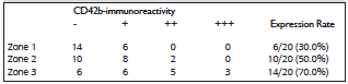
Table 1. Evaluation of Platelet Aggregation in Allograft Tissues
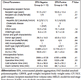
Table 2. Evaluation of Platelet Aggregation in Allograft Tissues
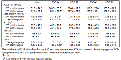
Table 3. Perioperative Laboratory Data of the Living-Donor Liver Transplant Recipients
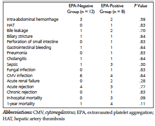
Table 4. Postoperative Complications and Clinical Outcomes
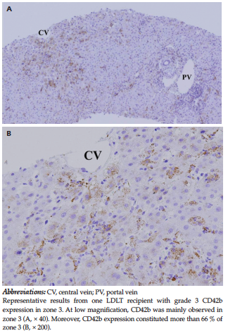
Figure 1. Immunohistochemical Staining of Liver Allograft Tissues With Antibody to CD42b
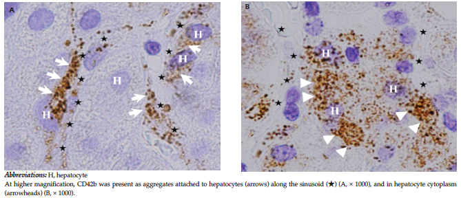
Figure 2. Histologic Localization of CD42b-Immunoreactivity in Local Liver Microenvironments
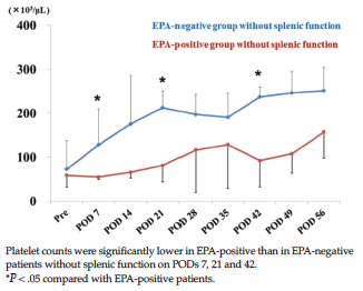
Figure 3. Perioperative Platelet Counts of the Living Donor Liver Transplant Recipients Without Splenic Function
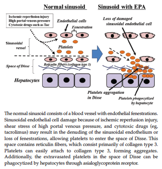
Figure 4. Schematic Model of the Pathogenic Mechanism of EPA-Induced Sinusoid Damage