Volume: 13 Issue: 6 December 2015
FULL TEXT
Objectives: We evaluated a continuous, immediate, localized ischemic preconditioning regimen in a rat model of ischemia-reperfusion injury and assessed whether it attenuated injury at the histologic and molecular levels.
Materials and Methods: Fifteen adult male Lewis rats received sham operation, left unilateral warm ischemia (45 minutes of cross-clamping of the renal pedicle; ischemia-reperfusion injury group), or 15 minutes of ischemia followed by a 20-minute reperfusion period, 45 minutes of ischemia-reperfusion injury, and subsequent reperfusion (ischemic preconditioning/ischemia-reperfusion injury group). Kidney tissue was retrieved 48 hours later, sectioned, stained with hematoxylin and eosin, and examined. We used RNA extraction and real-time quantitative polymerase chain reaction analysis to assess acute kidney injury markers, cytokines, and microRNA-21.
Results: Forty-five minutes of unilateral ischemia-reperfusion injury caused marked changes in histology at 48 hours, characterized by endothelial loss, tubulointerstitial damage (inflammation, cast formation), tubular cell necrosis, and glomerular capsule thickening. The ischemia-reperfusion injury and ischemic preconditioning/ischemia-reperfusion injury groups showed no measurable differences in histology. Expression of the acute kidney injury markers was significantly increased in the ischemia-reperfusion injury versus Sham group; however, no difference was found between the ischemia-reperfusion injury and ischemic preconditioning/ischemia-reperfusion injury groups. Similarly, expression of interleukin 17, interleukin 18, and tumor necrosis factor α was significantly increased in the ischemia-reperfusion injury versus Sham group. No significant difference was found between the ischemia-reperfusion injury and ischemic preconditioning/ischemia-reperfusion injury groups for interleukin 17 and interleukin 18; however, tumor necrosis factor α expression was significantly increased in the ischemic preconditioning/ischemia-reperfusion injury versus ischemia-reperfusion injury group.
Conclusions: In our ischemic preconditioning model, tumor necrosis factor α expression was increased without altering the sequelae of ischemia-reperfusion injury. The long-term consequences of this augmented early inflammatory response and whether these consequences are altered by variations in ischemic preconditioning or a subsequent injury require further study.
Key words : Ischaemia reperfusion injury, Ischemic preconditioning, Tumor necrosis factor alpha, MicroRNA-21
Introduction
One strategy proposed to improve ischemia-reperfusion injury (IRI) is ischemic preconditioning (IPC). In IPC, a brief period of ischemia is followed by reperfusion, inducing mechanisms that allow the organ to develop a degree of protection to a subsequent prolonged episode of ischemia. Several IPC methods have been described, including having the stimulus localized at the target organ or remotely from it, having 1 continuous episode of ischemia or several cycles of brief ischemia and reperfusion, and varying the interval between the IPC stimulus and the index ischemia.1-3
A recent systematic review and meta-analysis2 concluded that, overall, IPC reduces histologic damage and serum creatinine levels; however, some studies reported that IPC does not result in any protection at all.4,5 Most IPC investigations in kidneys have used an adult male rat as the animal model and a 40- or 45-minute IPC and IRI period. A 45-minute period has been shown to be robust and reliable in studies that have evaluated the effects of treatment on IRI.6,7
For an IPC regimen to be a practical and clinically transferrable practice in kidney transplant, a relatively short period of ischemia and reperfusion localized to the organ is the most desirable option. Indeed, a recent meta-analysis showed that most regimens have used localized IPC.2 Of particular note, a continuous regimen of 15 minutes of IPC before injury has been shown to result in histologic and functional protection.1
MicroRNAs (miRNAs) are a large family of endogenous, small non-coding RNAs that are thought to regulate the expression of the majority of human protein-coding genes.8-11 They have been implicated in diverse physiological and pathological cellular processes, and accumulating evidence has suggested that miRNAs play key roles in chronic kidney disease and acute kidney injury from various causes, including IRI.12-14 One study identified a unique miRNA expression profile in a mouse model of IRI and described a significant role for microRNA-21 (miR-21) in preventing tubular epithelial cell death.15 Another study identified the importance of miR-21 in a mouse model of IRI and IPC.16 In that study, 15 minutes of localized IPC significantly increased miR-21 expression, resulting in attenuation of IRI 4 days later, whereas knockdown of miR-21 significantly increased tubular cell apoptosis.16
The purpose of this study was to determine whether a 15-minute localized IPC regimen attenuated injury in a rat model of unilateral IRI. To this end, we evaluated histologic damage and analyzed the expression of selected acute kidney injury markers (kidney injury molecule 1 [KIM-1] and neutrophil gelatinase-associated lipocalin [NGAL]),17-19 cytokines (interleukin 17 [IL-17]), interleukin 18 [IL-18], and tumor necrosis factor α [TNF-α]),20-23 and miR-21.
Materials and Methods
General reagents
All general reagents were purchased from Sigma-Aldrich Corp. (Poole, UK), Life
Technologies, Inc. (Paisley, UK), or New England Biolabs (Ipswich, MA, USA),
unless otherwise stated. Oligonucleotides were purchased from Life Technologies.
Animal experiments
Fifteen adult (8- to 12-week-old) male Lewis rats weighing 180 to 220 g were
used (Harlan Laboratories, Ltd., Derby, UK). The rats acclimated to their
surroundings for 7 days, with housing, handling, and experimental procedures in
accordance with the local institutional policies and procedures licensed by the
UK Home Office under the Animals (Scientific Procedures) Act (1986).
Rats (n = 5 in each treatment group) were provided with analgesics (200 μg of buprenorphine dissolved in 500 mL of drinking water) from 24 hours before surgery until kidney retrieval. Animals were anesthetized with isoflurane, a midline laparotomy incision made, and the left renal pedicle was identified and clamped for 45 minutes using a vascular clip (IRI group). The kidney was visually assessed for both ischemia upon clamping and reperfusion upon release of the clamp. Rats in the Sham group underwent the same operation without renal pedicle clamping. Animals in the IPC/IRI group underwent 15 minutes of left renal pedicle clamping followed by 20 minutes of reperfusion before the IRI. During the procedure, the abdominal cavity was covered with saline-soaked gauze. Kidneys were retrieved 48 hours after terminal anesthesia.
Histology
Kidney tissue was embedded in paraffin, sectioned, and stained with hematoxylin
and eosin. All slides were scored under blinded assessment by one
histopathologist according to the system detailed in Table 1. This comprehensive
scoring system has been adapted specifically for animal research on kidney
tissue in the context of injury and provides a quantitative measurement of the
histologic damage of the kidney.
Real-time quantitative polymerase chain reaction analyses
All tissue was placed in RNA later solution (Life Technologies) immediately
after retrieval and stored at −80°C. Total RNA was extracted using TRIzol
reagent (Life Technologies) according to the manufacturer’s instructions. RNA
quality was assessed using the Agilent Technologies 2100 Bioanalyzer with RNA
6000 Nano chips (Palo Alto, CA, USA) and quantified before real-time
quantitative polymerase chain reaction (RT-qPCR). cDNA was generated using a
High Capacity Reverse Transcription Kit (Life Technologies) with random primers
or specific stem loop primers for the TaqMan miRNA assays. Real-time
quantitative polymerase chain reaction was performed on a 7900-HT Fast Real-Time
PCR System (Life Technologies). We quantified NGAL, KIM-1, IL-17, IL-18, and
TNF-α using POWER SYBR Green PCR Master Mix (Life Technologies) with
gene-specific primers. The amplification of a single PCR product was confirmed
by melting curve analysis. Expression of NGAL, KIM-1, IL-17, IL-18, and TNF-α
was normalized to glyceraldehyde 3-phosphate dehydrogenase. MicroRNA-21 was
quantified by TaqMan miRNA assay according to the manufacturer’s instructions,
and its expression was normalized to microRNA-16. The relative changes in gene
expression were analyzed by the 2 to the power of minus delta delta cycle
threshold (2-ΔΔCT) method.24 TaqMan miRNA gene expression assay
identification numbers were hsa-miR-16 (000391) and hsa-miR-21 (000397).
Primers were designed to mRNA sequences as follows: glyceraldehyde 3-phosphate dehydrogenase (forward: 5ʹ-CCTCTGACTTCAACAGCGACAC-3ʹ; reverse: 5ʹ-TGTCATACCAGGAAATGAGCTTGA3ʹ), NGAL (forward: 5ʹ-GGGCTGTCCGATGAACTGA-A-3ʹ; reverse: 5ʹ-CATTGGTCGGTGGGAACAGA-3ʹ), KIM-1 (forward:5ʹCGGCTAACCAGAGTGACTTGT-3ʹ; reverse: 5ʹ-TACAGAGCCTGGAAGAAGCAG-3ʹ), IL-17 (forward: 5ʹ-CCATCCATGTGCCTGATGCT-3ʹ; reverse: 5ʹ-GTTATTGGCCTCGGCGTTTG-3ʹ), IL-18 (forward: 5ʹ-GACCGAACAGCCAACGAATC-3ʹ, reverse: 5ʹ-ATAGGGTCACAGCCAGTCCT-3ʹ), and TNF-α (forward: 5ʹ-ATGGGCTCCCTCTCATCAGT-3ʹ; reverse: 5ʹ-GCTTGGTGGTTTGCTACGAC-3ʹ).
Statistical analyses
Statistical analyses were performed using GraphPad Prism Version 6 software (La
Jolla, CA, USA). Data are expressed as either median (and range) or mean (± SEM)
and assessed for statistical significance by unpaired t test or Mann-Whitney U
test. Differences with P < .05 were considered statistically significant.
Results
Effects of ischemia-reperfusion injury
Histologic architecture
Figure 1 shows that 45 minutes of unilateral IRI in the rat caused marked
histologic damage at 48 hours when compared with sham controls as characterized
by endothelial loss, tubulointerstitial damage (inflammation, cast formation,
and necrosis), and glomerular capsule thickening. The median (and range)
histology score was 10 (8-11) in the IRI group and 0 (0-1) in the Sham group (P
= .0079). No mortality was associated with IRI
Acute kidney injury markers
We selected KIM-1 and NGAL for analysis because reports have suggested that they
are accurate and robust biomarkers of acute kidney injury.17-19 As
shown in Figure 2, KIM-1 mRNA synthesis increased by 300-fold and NGAL mRNA
synthesis increased by 30-fold in the IRI group (P < .0001). The
histologic and acute kidney injury marker results shown in Figures 1 and 2
suggest that our IRI model is reliable, producing significant nonfatal kidney
injury.
Cytokine profile
We selected IL-17, IL-18, and TNF-α for analysis because reports have shown that
all play an important role in the pathophysiology of IRI, including kidney
IRI.20-23 As shown in Figure 3, mRNA expression levels of IL-17 (P =
.4614), IL-18 (P = .3807), and TNF-α (P = .0757) were increased in
IRI but not significantly.
MicroRNA changes
Several reports have suggested that miRNA expression changes are important in
IRI, with particular emphasis on the role of miR-21. Figure 4 shows that there
was a 2-fold statistically significant increase in the expression of miR-21 in
the IRI group compared with the sham group (P = .0347).
Effects of ischemic preconditioning on ischemia-reperfusion injury
Histologic architecture
Marked histologic damage was observed at 48 hours in both the IRI and IPC/IRI
groups. However, results between the IRI and IPC/IRI groups were not
significantly different, with median (and range) histology scores of 10 (8-11)
in the IRI group and 9
(8-12) in the IPC/IRI group (P = .8095) (Figure 5). No mortality was
associated with IPC/IRI.
Acute kidney injury markers
As shown in Figure 6, no difference was found in the mRNA expression of KIM-1
and NGAL between the IRI group (P = .0610) and the IPC/IRI group (P
= .7903).
Cytokine profile
There was an overall increase in the mRNA expression of IL-17, IL-18, and TNF-α
in the IPC/IRI group compared with the IRI group. For IL-17 (P = .2401)
and IL-18 (P = .1683), this increase was not statistically significant.
However, expression of TNF-α was increased 2-fold in the IPC/IRI group compared
with IRI group (P = .0328) (Figure 7).
MicroRNA changes
As shown in Figure 8, the increased miR-21 expression shown in the IPC/IRI group
was not significantly different compared with the IRI group (P = .4519).
Discussion
The major finding of this study is that the immediate, localized IPC regimen did not protect the rat kidney against 45 minutes of IRI. Significant up-regulation of TNF-α mRNA synthesis was observed in the IRI and IPC/IRI groups, suggesting that this inflammatory cytokine plays a role in the early mechanisms of IPC. Expression of miR-21 also was significantly increased with IRI but not following IPC.
The rat model used in the present study is a well-established in vivo model of acute kidney injury18,25 and is supported by a recent systematic review and meta-analysis.2 Cross-clamping of the renal pedicle for 45 minutes produces significant but not fatal ischemic injury.6,7 In the present study, the 48-hour reperfusion period led to clear histologic and molecular alterations, which are in line with previous studies.1,4,26
Our finding that IPC does not alter histologic or molecular markers of renal injury in this model should be placed in the context of the current controversy as to whether IPC protects from subsequent renal IRI. Although several investigators have found IPC to be effective in protecting against renal IRI in various animal species,3,27-30 other studies have found no evidence that IPC is protective against renal injury.4,5,31 A recent meta-analysis by Wever and associates2 analyzed 58 animal studies and concluded that IPC was associated with significantly improved serum creatinine, blood urea nitrogen, and histologic injury score after IRI.5
The mechanisms underlying IPC remain undefined, with the majority of insight from studies on the heart, in which IPC has become the criterion standard to which other cardiac interventions are compared.32 One of the main pathways conferring IPC benefit in the heart is the myocardial reperfusion injury salvage kinase pathway. Some of the components within this pathway appear to be involved in renal IPC protection, such as protein kinase C and heme oxygenase-1.33,34 Another mechanism, apparently independent of the reperfusion injury salvage kinase pathway, is the survivor activating factor enhancement pathway, components of which include TNF-α.35 In the present study, although the IPC regimen conferred no benefit to the renal injury, TNF-α was significantly increased, suggesting a role in early IPC mechanisms.
We selected miR-21 for our study because of its significant role in kidney IRI in previous reports.15,36,37 Tubular cell apoptosis contributes significantly to renal IRI, and antiapoptotic functions have been attributed to miR-21.15,38 Although the role of miR-21 remains to be fully elucidated, some evidence supports a protective as well as pathological role for miR-21 in this context.39 In the present study, miR-21 expression increased significantly in IRI, supporting its potential as a biomarker of IRI-mediated injury. However, results observed between IRI and IPC/IRI groups were not significantly different. This is in contrast to a previous study in which a 15-minute IPC regimen attenuated IRI induced 4 days later, associated with up-regulated miR-21 and hypoxia-inducible factor 1α expression.16 Similar to our study, this previous study used an IPC regimen of 15 minutes of continuous ischemia, but the extended period of 4 days between IPC and IRI may be key in terms of understanding the different outcomes. A beneficial effect after this extended recovery may support improvements in tissue robustness to subsequent injury that take hours rather than minutes to set in place. MicroRNAs act predominantly by posttranscriptional repression of their targets, leading to diminished synthesis. This diminished synthesis may lead to a delayed alteration in tissue phenotype or response, requiring first a change in miRNA expression or activity and then the time required for expression of key protein targets to “decay” to the necessary threshold level for the effect, as determined by the degree of posttranscriptional repression elicited and the stability of the protein target. Other miRNAs involved in cardiac, hepatic, and brain IRI and IPC also have been identified, including microRNA-200, microRNA-24, microRNA-1, and microRNA-192.40-43 Further evaluation of miRNAs in renal IRI and IPC/IRI is warranted.
One of the major limitations of this model, and consequently this study, is that unilateral IRI does not lead to changes in overall kidney function that are easy to measure. Because the contralateral (right) kidney was not subjected to nephrectomy or ischemia, it was not possible to provide a meaningful measurement of functional markers such as serum creatinine. However, despite this shortcoming, the histologic damage correlated well with 2 molecular markers of acute kidney injury (NGAL and KIM-1). Further investigations that assess different IPC regimens in a model, such as one with bilateral IRI, and that also allow for functional assessment of renal response to IRI are warranted.
The significant increase of TNF-α that we measured in our model (Figure 7) suggests that IPC had an additive inflammatory and potentially harmful effect. Although not demonstrated in our study, one may speculate that this effect may be part of the IPC hypothetical series of events that would eventually confer protection, as seen in other studies that used a longer interval between IPC and IRI. This hypothesis requires further investigation.
References:
- Torras J, Herrero-Fresneda I, Lloberas N, et al. Promising effects of
ischemic preconditioning in renal transplantation. Kidney Int.
2002;61(6):2218-2227.
CrossRef - PubMed - Wever KE, Menting TP, Rovers M, et al. Ischemic preconditioning in the
animal kidney, a systematic review and meta-analysis. PLoS One.
2012;7(2):e32296.
CrossRef - PubMed - Joo JD, Kim M, D'Agati VD, Lee HT. Ischemic preconditioning provides
both acute and delayed protection against renal ischemia and reperfusion
injury in mice. J Am Soc Nephrol. 2006;17(11):3115-3123.
CrossRef - PubMed - Obal D, Dettwiler S, Favoccia C, et al. Effect of sevoflurane
preconditioning on ischaemia/reperfusion injury in the rat kidney in vivo.
Eur J Anaesthesiol. 2006;23(4):319-326.
CrossRef - PubMed - Kadkhodaee M, Aryamanesh S, Faghihi M, Zahmatkesh M. Protection of rat
renal vitamin E levels by ischemic-preconditioning. BMC Nephrol. 2004;56.
CrossRef - PubMed - Delbridge MS, Shrestha BM, Raftery AT, El Nahas AM, Haylor JL. The
effect of body temperature in a rat model of renal ischemia-reperfusion
injury. Transplant Proc. 2007;39(10):2983-2985.
CrossRef - PubMed - Wystrychowski W, Wystrychowski G, Zukowska-Szczechowska E, et al.
Nephroprotective effect of pentoxifylline in renal ischemia-reperfusion in
rat depends on the timing of its administration. Transplant Proc.
2014;46(8):2555-2557.
CrossRef - PubMed - He L, Hannon GJ. MicroRNAs: small RNAs with a big role in gene
regulation. Nat Rev Genet. 2004;5(7):522-531.
CrossRef - PubMed - Krol J, Loedige I, Filipowicz W. The widespread regulation of microRNA
biogenesis, function and decay. Nat Rev Genet. 2010;11(9):597-610.
CrossRef - PubMed - Pritchard CC, Cheng HH, Tewari M. MicroRNA profiling: approaches and
considerations. Nat Rev Genet. 2012;13(5):358-369.
CrossRef - PubMed - Chen K, Rajewsky N. The evolution of gene regulation by transcription
factors and microRNAs. Nat Rev Genet. 2007;8(2):93-103.
CrossRef - PubMed - Chandrasekaran K, Karolina DS, Sepramaniam S, et al. Role of microRNAs
in kidney homeostasis and disease. Kidney Int. 2012;81(7):617-627.
CrossRef - PubMed - Li JY, Yong TY, Michael MZ, Gleadle JM. Review: The role of microRNAs in
kidney disease. Nephrology (Carlton). 2010;15(6):599-608.
CrossRef - PubMed - Mas VR, Dumur CI, Scian MJ, Gehrau RC, Maluf DG. MicroRNAs as biomarkers
in solid organ transplantation. Am J Transplant. 2013;13(1):11-19.
CrossRef - PubMed - Godwin JG, Ge X, Stephan K, et al. Identification of a microRNA
signature of renal ischemia reperfusion injury. Proc Natl Acad Sci U S A.
2010;107(32):14339-14344.
CrossRef - PubMed - Xu X, Kriegel AJ, Liu Y, et al. Delayed ischemic preconditioning
contributes to renal protection by upregulation of miR-21. Kidney Int.
2012;82(11):1167-1175.
CrossRef - PubMed - Vaidya VS, Ozer JS, Dieterle F, et al. Kidney injury molecule-1
outperforms traditional biomarkers of kidney injury in preclinical biomarker
qualification studies. Nat Biotechnol. 2010;28(5):478-485.
CrossRef - PubMed - Mishra J, Ma Q, Prada A, et al. Identification of neutrophil
gelatinase-associated lipocalin as a novel early urinary biomarker for
ischemic renal injury. J Am Soc Nephrol. 2003;14(10):2534-2543.
CrossRef - PubMed - Mishra J, Dent C, Tarabishi R, et al. Neutrophil gelatinase-associated
lipocalin (NGAL) as a biomarker for acute renal injury after cardiac
surgery. Lancet. 2005;365(9466):1231-1238.
CrossRef - PubMed - Li L, Huang L, Vergis AL, et al. IL-17 produced by neutrophils regulates
IFN-gamma-mediated neutrophil migration in mouse kidney ischemia-reperfusion
injury. J Clin Invest. 2010;120(1):331-342.
CrossRef - PubMed - Parikh CR, Jani A, Melnikov VY, Faubel S, Edelstein CL. Urinary
interleukin-18 is a marker of human acute tubular necrosis. Am J Kidney Dis.
2004;43(3):405-414.
CrossRef - PubMed - Donnahoo KK, Shames BD, Harken AH, Meldrum DR. Review article: the role
of tumor necrosis factor in renal ischemia-reperfusion injury. J Urol.
1999;162(1):196-203.
CrossRef - PubMed - Perry BC, Soltys D, Toledo AH, Toledo-Pereyra LH. Tumor necrosis
factor-alpha in liver ischemia/reperfusion injury. J Invest Surg.
2011;24(4):178-188.
CrossRef - PubMed - Livak KJ, Schmittgen TD. Analysis of relative gene expression data using
real-time quantitative PCR and the 2(-Delta Delta C(T)) Method. Methods.
2001;25(4):402-408.
CrossRef - PubMed - Kato J, Nakayama M, Zhu WJ, Yokoo T, Ito S. Ischemia/reperfusion of
unilateral kidney exaggerates aging-induced damage to the heart and
contralateral kidney. Nephron Exp Nephrol. 2014;126(4):183-190.
CrossRef - PubMed - Toosy N, McMorris EL, Grace PA, Mathie RT. Ischemic preconditioning
protects the rat kidney from reperfusion injury. BJU Int.
1999;84(4):489-494.
CrossRef - PubMed - Jefayri MK, Grace PA, Mathie RT. Attenuation of reperfusion injury by
renal ischemic preconditioning: the role of nitric oxide. BJU Int.
2000;85(9):1007-1013.
CrossRef - PubMed - Salehipour M, Khezri A, Monabbati A, et al. Ischemic preconditioning
protects the dog kidney from ischemia-reperfusion injury. Urol Int.
2007;79(4):328-331.
CrossRef - PubMed - Guye ML, Mc Gregor B, Weil G, Arnal F, Piriou V. [Ischemic and
pharmacologic preconditioning: desflurane reduces renal reperfusion injury
in rabbits]. Ann Fr Anesth Reanim. 2010;29(7-8):518-523.
CrossRef - PubMed - Hernandez DJ, Roberts WB, Miles-Thomas J, et al. Can ischemic
preconditioning ameliorate renal ischemia-reperfusion injury in a
single-kidney porcine model? J Endourol. 2008;22(11):2531-2536.
CrossRef - PubMed - Herrero F, Morales D, Baamonde C, et al. Ischemic preconditioning and
kidney transplantation: in vivo nitric oxide monitoring in a rat
ischemia-reperfusion experimental model. Transplant Proc.
2006;38(8):2600-2602.
CrossRef - PubMed - Gumina RJ, Gross GJ. If ischemic preconditioning is the gold standard,
has a platinum standard of cardioprotection arrived? Comparison with NHE
inhibition. J Thromb Thrombolysis. 1999;8(1):39-44.
CrossRef - PubMed - Lee HT, Emala CW. Protein kinase C and G(i/o) proteins are involved in
adenosine- and ischemic preconditioning-mediated renal protection. J Am Soc
Nephrol. 2001;12(2):233-240.
PubMed - He X, Xu X, Fan M, et al. Preconditioning with hyperbaric oxygen induces
tolerance against renal ischemia-reperfusion injury via increased expression
of heme oxygenase-1. J Surg Res. 2011;170(2):e271-277.
CrossRef - PubMed - Lecour S. Activation of the protective Survivor Activating Factor
Enhancement (SAFE) pathway against reperfusion injury: Does it go beyond the
RISK pathway? J Mol Cell Cardiol. 2009;47(1):32-40.
CrossRef - PubMed - Li YF, Jing Y, Hao J, et al. MicroRNA-21 in the pathogenesis of acute
kidney injury. Protein Cell. 2013;4(11):813-819.
CrossRef - PubMed - Shapiro MD, Bagley J, Latz J, et al. MicroRNA expression data reveals a
signature of kidney damage following ischemia reperfusion injury. PLoS One.
2011;6(8):e23011.
CrossRef - PubMed - Lieberthal W, Levine JS. Mechanisms of apoptosis and its potential role
in renal tubular epithelial cell injury. Am J Physiol. 1996;271(3 Pt
2):F477-488.
PubMed - Xu X, Kriegel AJ, Jiao X, et al. miR-21 in ischemia/reperfusion injury:
a double-edged sword? Physiol Genomics. 2014;46(21):789-797.
CrossRef - PubMed - Xu CF, Yu CH, Li YM. Regulation of hepatic microRNA expression in
response to ischemic preconditioning following ischemia/reperfusion injury
in mice. OMICS. 2009;13(6):513-520.
CrossRef - PubMed - Yin C, Salloum FN, Kukreja RC. A novel role of microRNA in late
preconditioning: upregulation of endothelial nitric oxide synthase and heat
shock protein 70. Circ Res. 2009;104(5):572-575.
CrossRef - PubMed - Lee ST, Chu K, Jung KH, et al. MicroRNAs induced during ischemic
preconditioning. Stroke. 2010;41(8):1646-1651.
CrossRef - PubMed - Salloum FN, Yin C, Kukreja RC. Role of microRNAs in cardiac
preconditioning. J Cardiovasc Pharmacol. 2010;56(6):581-588.
CrossRef - PubMed

Volume : 13
Issue : 6
Pages : 535 - 542
DOI : 10.6002/ect.2015.0039
From the 1Institute of Nephrology, Cardiff University, Cardiff,
United Kingdom; and the 2Cardiff Transplant Unit, Department of
Nephrology & Transplant Surgery, University Hospital of Wales, Cardiff, United
Kingdom
Acknowledgements: This work was funded by the Kidney Wales Foundation.
The authors have no conflicts of interest to declare.
Corresponding author: Usman Khalid, Cardiff Transplant Unit, Department
of Nephrology & Transplant Surgery, University Hospital of Wales, Heath Park,
Cardiff, CF14 4XW, UK
Phone: +44 2920 746 647
Fax: +44 2920 746 661
E-mail:
usman.khalid@doctors.org.uk
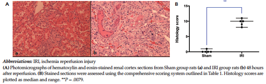
Figure 1. Characterization of the Rat Model of Unilateral Ischemia-Reperfusion Injury
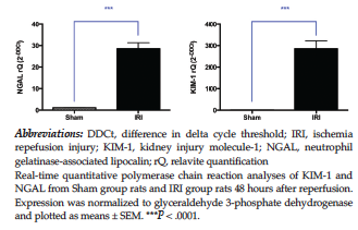
Figure 2. Expression of Acute Kidney Injury Markers in Ischemia-Reperfusion Injury
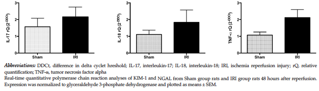
Figure 3. Expression of IL-17, IL-18, and TNF-α in Ischemia-Reperfusion Injury
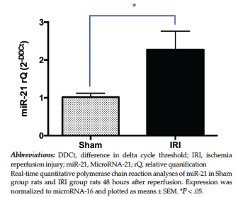
Figure 4. Expression of miR-21 in Ischemia-Reperfusion Injury
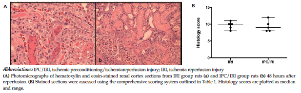
Figure 5. Characterization of Unilateral Ischemia-Reperfusion Injury and Ischemic Preconditioning Followed by Ischemia-Reperfusion Injury
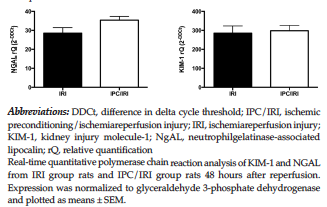
Figure 6. Expression of Acute Kidney Injury Markers in the IRI and IPC/IRI Groups
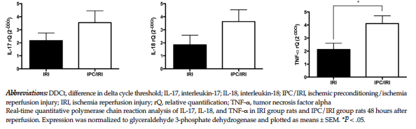
Figure 7. Expression of IL-17, IL-18, and TNF-α in the IRI and IPC/IRI Groups
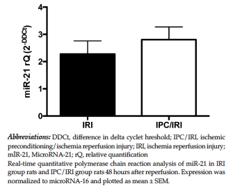
Figure 8. Expression of miR-21 in the IRI and IPC/IRI Groups
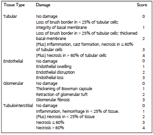
Table 1. The Histology Scoring System