Volume: 13 Issue: 6 December 2015
FULL TEXT
During the past decades, dual kidney transplant has enabled greater use of marginal kidneys and reduced waiting time. Since the first description of dual transplant in 1996, the techniques and outcomes have improved. No clear allocation criteria for donors and suitable candidates have been outlined; however, in general, an older for older approach is followed by many centers. Many centers are hampered by the lack of a clear allocation policy and the fact that decisions for dual kidney transplant are solely clinician based. Unilateral placement of both kidneys is the technique of choice in many centers. En block pediatric dual transplant and several vascular reconstruction methods for dual kidneys have been adopted by surgeons to enable single arterial and venous anastomosis and to reduce complications. Although there is a higher prevalence of vascular complications, mainly in the form of graft thrombosis, the overall complication rate with dual kidney transplant is comparable to single kidney transplant. Kidney survival and function are encouraging and close to results with standard criteria single kidney transplant. Although the technique is well established in many centers, standardized guidelines are lacking. Here, we review the current experience with dual kidney transplant.
Key words : Donation after cardiac death, En block kidney transplant, End-stage renal disease, Extended criteria donors, Marginal donors
Introduction
During the past decades, there have been major improvements in kidney transplant. Better perioperative care and immunosuppressive agents have improved patient outcomes. Renal transplant is the treatment of choice for patients with end-stage renal disease. In the United States, patients waiting for kidney transplant now number 50 000 with an annual death rate of 6.3%.1 To increase the number of organs available for transplant, the use of kidneys from donors from age extremes (pediatric or elderly) has been suggested. In late 2003, the United Network for Organ Sharing (UNOS) implemented the use of kidneys from extended criteria donors (ECDs). Extended criteria donors include donors older than 60 years or those who are older than 50 with a history of hypertension, had cause of death due to cerebrovascular events, or have serum creatinine level at retrieval of more than 1.97 mg/dL.2 Donation after cardiac death also has been accepted worldwide as source of organs despite organs regarded as marginal due to association with warm ischemic injury.3
The transplant of a single marginal kidney (from ECDs, donors with cardiac death, and pediatric donors) may result in a suboptimal number of functional nephrons to allow recipients to become dialysis independent. Episodes of acute rejection, medication toxicity (particularly from calcineurin inhibitors), and effect of the recipient’s comorbidities on the transplanted kidney will adversely affect these limited functional nephrons.4 The concept of transplanting both donor kidneys into 1 recipient as dual renal transplant has been adopted to increase available “nephron mass.” The hypothesis is, if 2 marginal organs are given to the same recipient, more functioning nephrons should be available versus with a single suboptimal organ or as many functioning nephrons versus with a single ideal kidney.5 Nephron mass as a determinant of chronic allograft failure has been experimentally tested in animals, with results showing that increasing the size of viable nephron mass by transplanting 2 kidneys to the same recipient effectively prevents progressive deterioration in renal function compared with single transplant controls.
The first adult dual kidney transplant (DKT) was in the United States in 1996 by Johnson and assoiates.6 Two decades earlier, dual kidneys from pediatric donors had been transplanted into adults.7 Currently, centers perform DKTs using different organ selection criteria and techniques. Although no randomized prospective studies comparing the results of DKT to single transplant have been published, several authors have reported acceptable results even with kidneys considered unacceptable by others. Dual kidney transplant carries a potentially higher risk of surgical complications because of the longer surgical procedure and the 2-fold risk associated with double vascular and ureteric anastomoses. As a result, many centers remain reluctant and lack experience with DKT.8 Differences in practice among different centers, absence of clear guidelines and allocation policies, and the belief that these grafts are suboptimal can contribute to this reluctance. In a review of UNOS practices by Gill and associates, between 2000 and 2005, DKTs from donors > 50 years old accounted for only 4% of transplants.9 Moreover, 54% of the kidneys from donors > 65 years old in the United States and 12% in Europe are discarded.10 These limitations clearly indicate the need to expand DKT practice. Here, we provide a literature review of different DKT aspects, techniques, and results and address points for further research.
Which kidney is suitable for dual transplant?
Dual kidney transplant is a waste of resources if a single kidney will keep
the recipient dialysis independent. Equally, having a DKT with insufficient
function is extremely unwelcomed. Thus, the decision of which kidney is suitable
for dual transplant is crucial. There is so far no global consensus as to which
donor kidney is best for DKT, with one reason being that DKTs are not routinely
performed in many transplant centers. In addition, the variations in practice
and protocols between different surgeons and centers have led to differences in
kidney selection criteria. In the first DKT report, Johnson and associates6 used
kidneys from donors older than 60 years and/or long history of hypertension or
diabetes with cold ischemia time less than 30 hours. They picked donors who had
creatinine clearance levels between 80 and 40 mL/min and with kidneys that
showed less than 40% glomerulosclerosis without severe interstitial fibrosis or
arteriosclerosis on biopsy. Kidneys being rejected for a single kidney
transplant (SKT) by other units were criteria. The group reported 100% patient
and graft survival at 6 months.6
In 1999, Remuzzi and associates4 suggested a scoring system based on pretransplant biopsy for selection of a kidney for DKT. Brain dead donors older than 60 years, donors who were diabetic, or donors with presence of proteinuria of less than three grams/24 hours were considered. Kidneys with macroscopic major vascular abnormality or evidence of focal scarring (ie, chronic pyelonephritis) were excluded. Both kidneys were biopsied, and those with less than 25 glomeruli were excluded. Dual kidney transplants were performed if a priori score was between 4 and 6 (Table 1). In the 24 DKTs reported in the study, patient and graft survival was 100% at 6 months.4
In a 2000 study by a Spanish group, kidneys from brain dead donors with normal serum creatinine levels were considered for DKT if donors older than 75 years old. In addition, donors between 60 and 74 years old with glomerulosclerosis of 15% to 50% at biopsy also were included. Both kidneys were biopsied, and the higher glomerulosclerosis percentage was considered. Kidneys with less than 15% glomerulosclerosis were transplanted separately, and kidneys with greater than 50% glomerulosclerosis were discarded. Mean donor age in the study was 75 ± 7 years. Graft survival at 6 months was 95% in 21 recipients. This series reported the oldest donor for DKT.11
In a review of UNOS database published on 2008, five hundred twenty-five DKTs were performed from 2000 to 2005. Donors were considered for DKT if any 2 of the following criteria present: age greater than 60 years, creatinine clearance greater than 65 mL/min, rising serum creatinine greater than 2.5 mg/dL at retrieval, chronic hypertension or type 2 diabetes mellitus, and glomerulosclerosis on biopsy between 15% and 50%. Three-year graft survival was 79.8%.9
In another large Italian series of 100 unilateral DKTs performed between 2003 and 2009, allocation was based on donor age and clinical and histologic findings (Figure 1). Mean donor age was 72 ± 5 years old. Interestingly, a Tru-Cut biopsy needle (Medline Industries, Inc., One Medline Place, Mundelein, IL, USA) was used rather than the conventional wedge open biopsy. Three-year graft survival reached 90.9%.8
Severe histologic findings are associated with impaired allograft function and are the rationale of using biopsy to determine suitability of marginal kidneys. Five-year survival was 80% when protocol biopsy showed no glomerulosclerosis and dropped to 35% when sclerosis was > 20% in donated kidneys.5 Some authors also have reported higher delayed graft function (80%) with sclerosis > 20%.
One of the limitations is that most surgeons obtain an elliptical biopsy from kidney surface. This could be misleading as glomeruli sclerose from outside in. As a result, these biopsies could overestimate glomerulosclerosis. Needle biopsy is usually avoided by surgeons because of risk of bleeding or arteriovenous fistula formation. Moreover, there might be an element of individual interpretation variations in biopsy scoring, despite a structured scoring system.12 This may have led some centers to allocate some kidneys inappropriately to DKT. In an analysis of the UNOS database from 2000 to 2005, Gill and associates9 reported that 12% of DKTs were performed using grafts from standard criteria donors. In the Spanish series, 4 of the 21 DKTs recipients had single kidney thrombosis. Renal function was maintained in 3 of recipients by the single nonthrombosed kidney raising the question whether the patients should have received SKT initially.11 Ironically, greater use of kidneys for DKT that are more suitable for SKT could result in a reduced organ pool for transplant.
To avoid biopsy-based decisions, the estimated glomerular filtration rate (eGFR) in the donor as a criterion was used instead. Donors older than 65 years old with at least 1 of the following risk factors were considered: hypertension, type 2 diabetes mellitus, atherosclerotic disease, or death from a cardiovascular event. Depending on maximal eGFR calculated with use of the Cockcroft and Gault formula, donors with eGFR > 60 mL/min were considered for SKT. Kidneys were discarded when eGFR was < 30 mL/min, and eGFR between these results was an indicator for DKT. Snanoudj and associates performed 81 DKTs using this protocol, with 2-year graft survival of 90%.10
Similarly, United Kingdom Kidney Advisory Group suggested that kidneys from donors who are age 70 years or older could be used for DKTs if 1 or more of the following clinical risk factors were present: history of hypertension, myocardial infarction, type 2 diabetes mellitus, cerebrovascular event as cause of death, serum creatinine level of > 1.97 mg/dL at retrieval, or presence of any anatomic anomaly (renal artery stenosis, polycystic, small kidneys).2 Our center also uses this approach. Kidneys with prolonged warm ischemia time, small kidneys, eGFR < 60 mL/min, kidneys with multiple cysts, and kidneys from elderly donors (> 70 y) are considered for DKTs, especially in association with donors who have a history of hypertension or type 2 diabetes mellitus.
Kidney donations after cardiac death are generally considered marginal because of warm ischemic injury. In 2008, a Newcastle team published their experience using kidney donations after cardiac death as a source of DKT. Dual kidney transplants from donors with Maastricht category two (15 donors) and category three (8 donors) were reported. All kidneys had hypothermic machine perfusion. Pressure flow index, defined as flow per 100 grams renal mass divided by systolic blood pressure, and concentration of glutathione transferase, an enzyme marker of ischemic injury, in the perfusate were measured. Kidneys were used for SKT if pressure flow index was 0.4 mL/min per 100 g/mm Hg and glutathione transferase was less than 100 IU/L/100 grams renal mass. If pressure flow index was less than 0.4, kidneys were discarded. Kidneys were considered for DKT if pressure flow index was satisfactory but GST was higher than cutoff value for SKT or if other risk factors were present (eg, comorbidities or cold ischemia). Mean donor age in the study was 49 ± 11 years, with delayed graft function rate of 81% but 3-month GFR of 46.2 ± 13.7 mL/min.3
Pediatric donors were first considered for pediatric recipients. Because of increased surgical complications, inadequate nephron mass, relative sensitivity regarding rejection, and risk of hyperfiltration injury, pediatric SKT remains controversial. Hobart and associates7 suggested using en block kidney transplant (EBK) when donor age is less than 2 years. The study reported results of 33 EBKs, with 3-year graft survival reaching 87.3%. In UNOS registry review of EBK published in 2005, EBK was recommended when brain dead donors were less than 5 years old. The study reported graft survival rates of 85% at 1 year, 76% at 3 years, and 71% at 5 years.13
Who is a suitable recipient?
Similarly, there is debate about the best candidate for DKTs. Many authors
suggested matching recipients with donors by age and size. Because most DKTs
involve donors at age extremes, matching an older donor with an older recipient
is suggested. One reason is that the limited functioning nephron mass would be
enough for an elderly recipient with limited metabolic demands and the elderly
would not require graft survival of greater than 20 years based on their
expected lifespan. In addition, lower acute rejection rates in elderly
recipients as a result of antiproliferative agents with lower dose levels of
nephrotoxic calcineurin inhibitors would reduce long-term nephron insult.14
To reduce possible injury to limited nephron mass, most authors have suggested
DKTs to recipients with lower immunologic risk (ie, recipients without previous
transplant and panel reactive antibody titer < 50%).4
In contrast, the encouraging results of DKTs with excellent creatinine levels and lesser acute rejections allow for greater consideration in younger recipients. Younger candidates can more easily recover from the longer operative time necessary for DKTs. To support this approach, Andres and associates performed DKTs in recipients with mean age of 60 ± 5 years old from donors with mean age 75 ± 7 years.11
Hobart and associates failed to show a difference in recipient or graft survival to support using age or weight as variables to guide selection in his 33 pediatric EBK into adult recipients,7 although it has been suggested that candidates of pediatric EBK should weigh less than 80 kg.15
In general, DKT is offered to patients 60 years or older. Our group prefers offering DKTs to patients with low immunological risk, who are less than 60 years old, and who have minimal comorbidities and body mass index < 30 kg/m2.
Surgical technique
Johnson and associates6 described the first DKT; the original
technique included bilateral Gibson incision and transplanting 1 kidney to each
side. This method required more tissue dissection and a longer operative time.
Because most recipients are 55 years or older, reduction of total anesthetic, a
shorter operative time, and less vascular anastomoses are desirable.
A midline extraperitoneal approach was described to minimize dissection and operative time. Through midline infraumbilical incision, blunt dissection of extraperitoneal space bilaterally to expose iliac vessels. Because the left iliac vein is in a deeper anatomic position, the left kidney with its longer renal vein was placed on the left side. Drains were placed bilaterally. Haider and associates described this technique in 2007.16 The single midline incision has the advantage of a shorter operative time and dissection and fewer hernia complications in comparison to a bilateral Gibson incision. In addition, a potential wound infection would be far away from the graft. This method can be converted easily to an intraperitoneal approach, although the latter has higher ileus and bowel complications, mobile grafts, and is more difficult to biopsy.6
Unilateral placement of both kidneys was described by Mason and Hefty in 1998.17 The right kidney was placed superiorly with renal artery anastomosed into the common iliac artery and renal vein into the inferior vena cava. Clamps were released, allowing perfusion of transplanted kidney before both the external iliac vein and external iliac artery were clamped distally to allow the anastomosis of left kidney vascular pedicle to external iliac vessels. With this method, both ureters were spatulated and joined to each other. The conjoint ureters were anastomosed to the bladder with ureteric double J stent. This technique reduced trauma from the surgical procedure and operative time. Moreover, the contralateral side remained untouched for possible future transplant.17 This technique results in graft survival at 1 year similar to that shown with the bilateral technique (96% vs 93%) and shorter operative time (261 ± 31 vs 351 ± 76 min; P = .0001).18
A modification was described in Ekser and associates in a report of 29 unilateral extraperitoneal DKTs.18 The right kidney was placed superiorly, but the renal vein was extended using the donor’s inferior vena cava patch. It was anastomosed to the external iliac vein instead of the inferior vena cava. The group also anastomosed the transplant ureters through 2 separate extravesical ureteroneocystostomies on ureteric stents. The upper kidney ureter was placed lateral to the lower kidney ureter. Less dissection as the inferior vena cava is not used for anastomosis is an advantage. In the report, there were 2 incidents of renal vein thrombosis in the unilateral group. This technique has shorter operative time and length of hospital stay and a lower delayed graft function rate versus bilateral placement of DKT done by same team.18 In a larger series, unilateral DKT was performed in 100 patients by the same team. Shorter operative time was observed (260 ± 35 min), with 95% of placements on the right side. Outcomes and surgical complications were similar to that shown in an SKT control group operated by the same team.8 Many surgeons now prefer this technique for DKT. At our center, we have used a similar technique in 22 patients. We prefer to extend the short right renal vein using the donor inferior vena cava anastomosed to the recipient’s inferior vena cava. We use the common iliac artery as an inflow artery (Figures 2 and 3).
Pediatric kidneys have higher risk of surgical complications, especially vascular as a result of small vessel size. En block kidney transplant using pediatric donor aorta and the inferior vena cava can theoretically reduce that risk. En block kidney transplant was described 40 years ago. Kidneys were retrieved en block and prepared. The proximal aorta and inferior vena cava were closed with running sutures and ligated lumbar vessels. Through a unilateral modified Gibson incision, the donor’s distal inferior vena cava was anastomosed to the recipient’s external iliac vein. Distal aorta was anastomosed to external iliac artery. Others have used use the ovarian or internal iliac artery for arterial inflow. In addition, interrupted knot anastomosis to allow the graft to grow and to avoid anastomotic stenosis also has been suggested. Conjoint ureters were anastomosed to the bladder using extravesical techniques.7
Newcastle team described a modification for EBK. One of the main concerns of the original technique was torsion of the whole block or 1 kidney at risk for thrombosis due to a long vascular pedicle. Moreover, at the end of the transplant, kidneys usually lay high in the pelvis, placing the short pediatric ureters under tension to reach the bladder. This might explain the higher rate of ureteric complications with pediatric versus adult transplanted kidneys. The infrarenal aortic and inferior vena cava segment of the donor was transposed to the top, and the new inferior stump was sewn. The new vascular pedicle was anastomosed to iliac vessels, with kidneys able to drop to the extraperitoneal pelvic space.19 In a 2003 report of 15 patients, 1-year graft survival was 93%, with 100% patient survival. The only graft failure was secondary to recurrence of the original disease. In addition, no vascular complications were reported.20
Theoretically, performing a single composite anastomosis for both renal arteries and venous drainage could reduce vascular complications, duration of warm ischemia, and length of the surgical procedure. Therefore, EBK with 2 adult kidneys performed in the same manner as with pediatric donors was also described. An Iranian center transplanted adult kidneys en block. The difference was closing the aorta below the origin of renal arteries, with the donor inferior vena cava closed above the origin of renal veins and the lower end used for anastomosis into iliac vessels.21 Joining both renal arterial patches into 1 and using an inferior vena cava patch to elongate the right renal vein and then attaching both veins into a common ostium to allow single arterial and venous anastomosis was also described.22 Even multiple renal arteries were anastomosed to a donor’s inferior vena cava patch to allow quicker single arterial anastomosis. One possible complication is aneurysmal degeneration of the thinner venous graft.23
Graft and patient survival
The earliest report of DKT documented 100% graft and patient survival in 9 DKTs.
Although the mean follow-up was short (6 months; range, 2-14 months), recipients
of DKT had better graft function than both control groups (recipients of SKT
from donors > 60 y or from donors < 50 y).6
Lee and associates14 suggested an older donor for older recipient strategy. They reported similar patient and graft survival at 1 year (98% and 89% in DKT vs 97% and 90% in SKT) and 2 years (86% and 77% in DKT vs 95% and 86% in SKT; P = not significant). The group reported 2-year follow-up results from 41 recipients of bilateral DKT. There were significant differences in age between DKT donors (59 ± 12 y) and SKT donors (42 ± 17 y; P = .0001).14
The 2007 review of the UNOS database from 2000 until 2005 followed 625 recipients of DKT for 48 months. Recipients of DKTs from ECDs had similar death-censored graft survival, which reached 70%. During the same follow-up, recipients of kidneys from standard criteria donors had better survival of 80%.9
European reports have revealed encouraging results. A French team with older donors (age, 75 ± 5.8 y) recorded DKT patient and graft survival at 3 years that reached around 50%.10 In a study of ECD donors, D’Arcy and associates24 from Ireland reported superior 3-month patient (92% vs 100%; P = .05) and graft survival (88% vs 93%; P = .02) in recipients of SKT vs DKT. At 3 years, patient and graft survival results were comparable between both groups.24 A Spanish report that compared survival results of 21 recipients of kidneys from donors up to 75 years old for DKT with recipients of SKT with 74-year-old donors or recipients who received kidneys from 60- to 74-year-old donors (follow-up of 15 ± 5 mo) found no significant differences in patient (100%) or graft survival (95%) at 1 year.11
Similarly, reports on pediatric kidneys transplanted into adults as EBK have shown encouraging results. A review of the UNOS database from 1987 to 2003 reported results of 2160 recipients of EBK. The report showed that 77% of EBKs were from donors < 5 years old; however, graft survival at 1, 3, and 5 years was superior (85%, 76%, 71%) versus SKT (81%, 68%, 63%; P = .001).13 In an early study of 33 adult recipients of pediatric EBK, no differences in 3-year graft or patient survival rates were shown versus rates shown in recipients of SKTs. A delayed graft function was reported in 15% of recipients of EBK, which was not statistically different from control group results.7 A study with of pediatric EBK 22-year follow-up from Austria found creatinine levels of 1.08 mg/dL at 2 years after transplant.15 Tables 2 and 3 summarizes the major dual kidney transplant studies and the graft survival graft function.
The quality of DKT function is as important as how long it will keep functioning since the aim is to keep the patient dialysis independent for as long as possible. Many factors affect graft function, including donor factors, donation circumstances, cold ischemia time, and perioperative events. In the initial reports of DKT, delayed graft function was 11%. In the same study, delayed graft function reached 50% in SKT from older donors (> 60 y) and 20% in SKT from younger donors (< 50 y).6
Two large analyses of the UNOS database have been published. The 2008 report documented 625 DKTs performed between January 2000 and December 2005. Despite a longer cold ischemia time in the DKT group versus that shown for groups who underwent SKT with ECD and standard criteria donor kidneys (22.2 ± 9.7, 19.5 ± 8.8, 18.7 ± 8.1 h; P < .001), delayed graft function incidence (29.3%) was lower than shown in recipients of SKT from
ECD (33.6%; P = .03) and similar to recipients of SKT from standard criteria donors (28.3%; P = .62). Primary nonfunction was as low as 1.8%.9 A recent study by Tanriover and associates25 reported on transplant procedures conducted between 2002 and 2012, comparing 1160 recipients of DKT versus 13 543 recipients of a single first transplant from ECD. In their multivariate analysis, DKT had a protective effect against delayed graft function (odds ratio = 0.76; 95% confidence interval, 0.6-0.97). Interestingly, the incidence of primary nonfunction was the same for both groups. Kidney discard rate was 3 times lower when the high-risk ECD kidneys were offered as DKT. The Newcastle team reported similar eGFR results at 3 and 12 months posttransplant in a comparison of DKT with donations after cardiac death versus SKT.3 Table 3 summarizes the outcomes of the major DKT reports. Outcomes of EBK procedures from the UNOS database were also analyzed. En block kidney transplant was associated with slightly increased risk of graft loss (adjusted hazard ratio, 1.18; P = .004) when compared to SKTs. This analysis included 2160 recipients of EBK performed between 1987 and 2003. Delayed graft function was reported in 17.9% of EBK candidates (300/1677), with 23.44% shown in the SKT arm (537/2291; P = .0001).13
Concerns related to pediatric EBK include more difficult surgical anastomosis, raising the risk of complications, inadequate nephron mass, relative sensitivity of pediatric kidneys to rejection, and hyperfiltration injury. Hyperfiltration is considered a functional and structural adaptation of the kidney. Increased glomerular filtration leads to compensatory glomerular hypertrophy and sequentially focal segmental glomerulosclerosis, which ends by sclerosis. No differences in proteinuria, the timing of first diagnosis, or average serum creatinine level were shown in a study comparing 33 EBKs and SKTs.7
In the longest follow-up of EBK (22 y), serum creatinine level was 1.3 ± 0.6 mg/dL at 5 years and 1.37 ± 0.7 mg/dL at 10 years in 11 patients. At 20 years, the average serum creatinine level was 1.08 mg/dL in 2 patients. Proteinuria was slightly increased during follow-up. Of note, the pediatric kidneys grew to approach adult kidney size in the first months.15
Complications
Complications with DKT are perceived to be higher due to technical difficulties,
with longer operative time and lower kidney quality. Lee and associates14
reported a local wound dehiscence rate of 5% in their series of 41 DKTs. Their
approach was midline extraperitoneal bilateral placement of both kidneys.
Early renal graft thrombosis is a cause of concern in DKT. In a report, 5 kidneys (12%) of 42 transplanted kidneys showed thrombosis. One patient had bilateral thrombosis, and 3 recipients had single kidney thrombosis but with renal function preserved by the other kidney.11
Urinary tract complications also have been reported to be slightly higher, especially urinary tract fistulas requiring surgery (17%; P = .06), although no significant difference in overall surgical complications was shown.4 In a French report, 11% of recipients had ureteric stenosis and a similar rate of urinary fistula. The group also reported higher incidence of vascular thrombosis in the DKT group (13% vs 4.3%; P = .049). The kidneys for DKT were from older donors; these more atherosclerotic vessels might suggest this higher risk. Postoperatively, recipients of DKT have a higher risk of developing myocardial infarctions (12% vs 0%; P = .002) when compared to SKT candidates. Longer operative procedure and anesthetic time in addition to older recipients might provide an explanation.10 Higher postoperative admission rates to an intensive care unit also have been reported by D'Arcy and associates.24
Monolateral placement of both kidneys reduces length of the surgical procedure and hospital stay. Ekser and associates18 reported 2% renal vein thrombosis in both the bilateral and ipsilateral DKT groups (total of 58 DKTs), and no difference in the complication rates between both techniques. A larger study also showed no differences in complication rates between ipsilateral DKT and SKT. The ipsilateral placement reduced dissection time and made lymphocele risk similar to that for SKT procedures.8
The goal of techniques that permit single arterial and venous anastomosis is to reduce vascular complications. Nghiem and associates26 described such a technique in their series of 12 patients, with a single venous thrombosis observed due to twist of 1 of the dual kidneys.26 Other authors have reported no surgical complications with vascular reconstruction of both kidneys.22,23
Hobart and associates7 reported a higher EBK complication rate (39.4%) versus SKT (P < .001). As expected, the most serious and common vascular complications were due to smaller vessels, affecting 5 of 33 EBKs in their study. Thrombosis of renal vein, artery, or even the donor’s aorta was observed. Renal artery stenosis occurred as a late complication in 2 recipients of EBK. In another series, rates of 10% arterial thrombosis and 6% renal vein thrombosis were shown with EBK.7 The UNOS database study reported a graft thrombosis rate of 4.11% (69/2291).13 This high rate was not reported with the modified surgical technique described by the Newcastle group,20 who reported rates of ureteral complication or urine leak of 9.25% and rates of wound infection of 4.6% with ipsilateral DKT.3 Finally, an interesting radiologic finding rather than a true complication is a “wandering kidney,” as described by Anavy and associates27 in 2012. The group reported the results of a recent recipient of DKT who presented with increasing abdominal pain and elevated levels of serum creatinine. A renogram showed only a single kidney on posteroanterior imaging despite 2 kidneys shown on earlier scans, giving a false impression of single kidney thrombosis. Subsequent computed tomography and ultrasonography confirmed normal perfusion of both grafts, with the upper graft wandering to lay posterior to the lower graft.27 Figure 4 shows the close projection of both kidneys on renogram that may develop wandering kidney.
Conclusions
Dual kidney transplant has increased the pool of potential organs by increasing use of marginal kidneys. Ipsilateral placement of both grafts is widely accepted and performed. Results of graft and patient survival, complications, and quality of renal function provided by DKTs are comparable to SKTs. Moreover, the use of pediatric kidneys by an en block technique into adult recipients has resulted in similar encouraging outcomes despite the higher surgical complication rate. The main question is which kidney is more suitable as DKT or SKT. Selection criteria for DKTs are still variable and center dependent. There is reluctance of many centers to perform DKTs because of lack of clear guidelines regarding allocation, which is usually left to the transplant clinicians to decide. More research should help to develop standardized criteria for DKT kidney allocation.
References:
- Bunnapradist S, Gritsch HA, Peng A, Jordan SC, Cho YW. Dual kidneys from
marginal adult donors as a source for cadaveric renal transplantation in the
United States. J Am Soc Nephrol. 2003;14(4):1031-1036.
CrossRef - PubMed - Allen J, Hudson A. Dual kidney transplantation. Kidney Advisory Group. 2011;16(11):1-9.
- Navarro AP, Sohrabi S, Reddy M, Carter N, Ahmed A, Talbot D. Dual
transplantation of marginal kidneys from nonheart beating donors selected
using machine perfusion viability criteria. J Urol. 2008;179(6):2305-2309;
discussion 2309.
CrossRef - PubMed - Remuzzi G, Grinyo J, Ruggenenti P, et al. Early experience with dual
kidney transplantation in adults using expanded donor criteria. Double
Kidney Transplant Group (DKG). J Am Soc Nephrol. 1999;10(12):2591-2598.
PubMed - Codreanu I, Cravedi P, Remuzzi G, Ruggenenti P. Dual kidney transplantation. Tx Med. 2004;16:13-18.
- Johnson LB, Kno PC, Dafoe DC, et al. Double adult renal allografts: a
technique for expansion of the cadaveric kidney donor pool. Surgery.
1996;120(4):580-583; discussion 583-584.
CrossRef - PubMed - Hobart MG, Modlin CS, Kapoor A, et al. Transplantation of pediatric en
bloc cadaver kidneys into adult recipients. Transplantation.
1998;66(12):1689-1694.
CrossRef - PubMed - Ekser B, Furian L, Broggiato A, et al. Technical aspects of unilateral
dual kidney transplantation from expanded criteria donors: experience of 100
patients. Am J Transplant. 2010;10(9):2000-2007.
CrossRef - PubMed - Gill J, Cho YW, Danovitch GM, et al. Outcomes of dual adult kidney
transplants in the United States: an analysis of the OPTN/UNOS database.
Transplantation. 2008;85(1):62-68.
CrossRef - PubMed - Snanoudj R, Rabant M, Timsit MO, et al. Donor-estimated GFR as an
appropriate criterion for allocation of ECD kidneys into single or dual
kidney transplantation. Am J Transplant. 2009;9(11):2542-2551.
CrossRef - PubMed - Andres A, Morales JM, Herrero JC, et al. Double versus single renal
allografts from aged donors. Transplantation. 2000;69(10):2060-2066.
CrossRef - PubMed - Lu AD, Carter JT, Weinstein RJ, et al. Excellent outcome in recipients
of dual kidney transplants: a report of the first 50 dual kidney transplants
at Stanford University. Arch Surg. 1999;134(9):971-975; discussion 975-976.
CrossRef - PubMed - Dharnidharka VR, Stevens G, Howard RJ. En-bloc kidney transplantation in
the United states: an analysis of United Network of Organ Sharing (UNOS)
data from 1987 to 2003. Am J Transplant. 2005;5(6):1513-1517.
CrossRef - PubMed - Lee CM, Carter JT, Weinstein RJ, et al. Dual kidney transplantation: older donors for older recipients. J Am Coll Surg. 1999;189(1):82-91; discussion 91-82.
- Hafner-Giessauf H, Mauric A, Muller H, et al. Long-term outcome of en
bloc pediatric kidney transplantation in adult recipients - up to 22 years
of center experience. Ann Transplant. 2013;18:101-107.
CrossRef - PubMed - Haider HH, Illanes HG, Ciancio G, Miller J, Burke GW. Dual kidney
transplantation using midline extraperitoneal approach: description of a
technique. Transplant Proc. 2007;39(4):1118-1119.
CrossRef - PubMed - Masson D, Hefty T. A technique for the transplantation of 2 adult
cadaver kidney grafts into 1 recipient. J Urol. 1998;160(5):1779-1780.
CrossRef - PubMed - Ekser B, Baldan N, Margani G, et al. Monolateral placement of both
kidneys in dual kidney transplantation: low surgical complication rate and
short operating time. Transpl Int. 2006;19(6):485-491.
CrossRef - PubMed - Talbot D, Balupuri S, Gerstenkorn C, Rix D, Abusin K, Manas D. "En bloc"
paediatric renal donors into adult recipients -- the Newcastle technique.
Transpl Int. 1999;12(2):152-155.
CrossRef - PubMed - El-Sheikh MF, Gok MA, Buckley PE, et al. En bloc pediatric into adult
recipients: the Newcastle experience. Transplant Proc. 2003;35(2):786-788.
CrossRef - PubMed - Salehipour M, Bahador A, Nikeghbalian S, et al. En-bloc Transplantation:
an Eligible Technique for Unilateral Dual Kidney Transplantation. Int J
Organ Transplant Med. 2012;3(3):111-114.
PubMed - Veroux P, Giuffrida G, Cappellani A, et al. Two-as-one monolateral dual
kidney transplantation. Urology. 2011;77(1):227-230.
CrossRef - PubMed - Veroux M, Giuffrida G, Ekser B, et al. Vascular reconstruction in
monolateral dual kidney transplantation with multiple arteries. Clin
Transplant. 2012;26(3):443-446.
CrossRef - PubMed - D'Arcy FT, O'Connor KM, Shields W, et al. Dual kidney transplantation
with organs from extended criteria cadaveric donors. J Urol.
2009;182(4):1477-1481.
CrossRef - PubMed - Tanriover B, Mohan S, Cohen DJ, et al. Kidneys at higher risk of
discard: expanding the role of dual kidney transplantation. Am J Transplant.
2014;14(2):404-415.
CrossRef - PubMed - Nghiem DD. Simultaneous double adult kidney transplantation using single
arterial and venous anastomoses. Urology. 2006;67(5):1076-1078.
CrossRef - PubMed - Anavy ND, Desai CS, Avery R, Kuo PH. Wandering kidney in dual-kidney
transplant recipient presents a pitfall in the interpretation of renal
scintigraphy. J Nucl Med Technol. 2012;40(4):275-277.
CrossRef - PubMed - Lu AD, Carter JT, Weinstein RJ, et al. Outcome in recipients of dual
kidney transplants: an analysis of the dual registry patients.
Transplantation. 2000;69(2):281-285.
CrossRef - PubMed

Volume : 13
Issue : 6
Pages : 500 - 509
DOI : 10.6002/ect.2015.0130
From the Sheffield Kidney Institute, Sheffield, United Kingdom S5 7AU
Acknowledgements: The authors declare that they have no sources of
funding for this study, and they have no conflicts of interest to declare.
Corresponding author: Ahmed Halawa, Northern General Hospital, Sheffield
Teaching Hospitals NHS Foundation Trust, Herries Road, Sheffield, UK S5 7AU
Phone: +44 114 271 4847
Phone: +44 114 256 0472
E-mail: Ahmed.Halawa@sth.nhs.uk
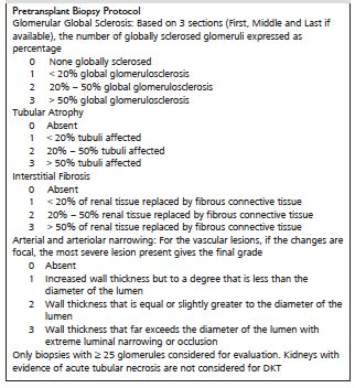
Table 1. Pretransplant Biopsy Scoring System4
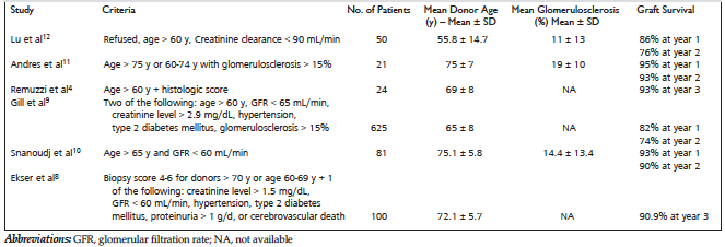
Table 2. Major Dual Kidney Transplant Studies and Allocation Criteria
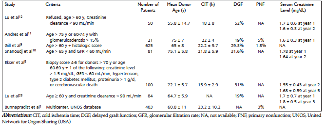
Table 3. Outcomes of Major Dual Kidney Transplant Studies – Values described as Mean ± SD
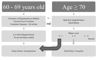 Figure 1. Allocation Flow Chart for Dual Kidney Transplants as
Proposed by Ekser and Associates8" style="margin-bottom: 10px; cursor: pointer">
Figure 1. Allocation Flow Chart for Dual Kidney Transplants as
Proposed by Ekser and Associates8" style="margin-bottom: 10px; cursor: pointer">
Figure 1. Allocation Flow Chart for Dual Kidney Transplants as Proposed by Ekser and Associates8
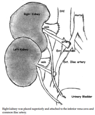
Figure 2. Unilateral Extraperitoneal Dual Kidney Transplant
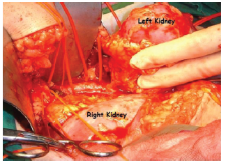
Figure 3. Unilateral Placement of Dual Kidney Transplant
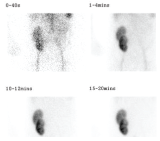
Figure 4. Dual Kidney Transplant on Isotope Mercaptoacetyltriglycine Scanning