Volume: 13 Issue: 6 December 2015
FULL TEXT
After the first clinical application of ex vivo lung perfusion in 2001, the technique has been used in many lung transplant centers worldwide. In addition, many modifications have been tested, leading to the development of various ex vivo lung perfusion systems and application protocols. Currently, the Lund protocol, the Toronto protocol, and Organ Care System Lung protocol are the clinically applied ex vivo lung perfusion protocols, based on the favorable results of the safety studies. Accordingly, the comparison among these EVLP systems and protocols should be an important research target, in order to provide the evidence based medical data that would recommend one protocol over the others. In this manuscript, the current experience with EVLP is reviewed and some molecular and clinical targets, that could be used to compare the various protocols of the technique, are introduced.
Key words : Lung preservation, Lung reconditioning Lung transplant
Introduction
The increased incidence of end-stage pulmonary diseases, such as chronic obstructive pulmonary disease, idiopathic pulmonary fibrosis, pulmonary hypertension, cystic fibrosis, sarcoidosis, and bronchiectasis, has raised the frequency of lung transplants.1 However, there is a significant unmet gap between the demands and the availability of lung grafts, which is worsened by lung transplant having the lowest success rate among all other solid-organ transplants.2,3 About 40% of lung grafts declined for transplant were found to have no significant histologic, microbiologic, or physiologic bases for rejection.4 This shows the importance of objective rather than subjective graft assessment.
Recently, the ex vivo lung perfusion (EVLP) system was introduced as a method to keep the lung graft perfused under normothermic conditions in the interval between donor retrieval and transplant. The technique involves the placement of the graft in a hard shell, connecting the tracheal stump to a ventilator, connecting the pulmonary artery stump to a perfusate circuit, and reconstructing the left atrium or leaving it open for drainage of the perfusate.5,6 The principle of the ex vivo perfusion technique is based on the graft perfusion using a hyperoncotic perfusate, which washes out the inflammatory cells in the vasculature, blood clots, and waste products and, in turn, potentially reduces ischemia-reperfusion injury (IRI). In addition, EVLP allows the objective assessment of graft performance over time, by monitoring the airway and pulmonary vascular pressures and the oxygenation capacity.5,6 Since the first clinical application of EVLP by Steen and associates in 2001, the technique has been used in many lung transplant centers worldwide. In addition, many modifications have been tested, leading to development of various EVLP systems and application protocols. At present, the Lund protocol, the Toronto protocol, and the Organ Care System (OCS) Lung protocol are the clinically applied EVLP protocols, based on the favorable results of the safety studies.7 Accordingly, comparing various EVLP systems and protocols should be an important research target. This would provide the evidence-based medical data needed to recommend a protocol over the others.
Conducting clinical trials to compare the various protocols is not practically applicable, considering the costs, intricate and precious nature of the procedure, and many other factors, including the variations among centers in the preoperative, perioperative, and postoperative care of patients. In this manuscript, the present experience with EVLP is reviewed, and some molecular and clinical targets that could be used to compare the various EVLP lung transplant protocols are introduced.
State of the art of ex vivo lung perfusion
Lung transplant involves the surgical retrieval of the donor graft, retrograde
and antegrade perfusion of cold preservation solution (4°C), and transport of
the graft to the recipient’s hospital while statically preserved in cold
preservation solution. The only known exception of this stage is the application
of EVLP according to the OCS lung protocol, where normothermic ex vivo perfusion
starts immediately after graft retrieval.7,8
Previously, high K+ preservation solutions were used, which had been associated with pulmonary vascular spasms and increased production of reactive oxygen species. Low potassium (dextran) solutions are now widely used, which are associated with better clinical outcomes.9 Although 2 studies have confirmed Celsior solution to produce similar clinical outcomes to those of Perfadex solution, with additional trends toward better survival and less incidence of chronic lung allograft dysfunction, especially with prolonged periods of ischemia, Perfadex remains the most widely used preservation solution in most lung transplant centers.10,11 According to the present practice, lung grafts that meet the standard criteria are kept cold and statically preserved until transplant, whereas grafts that are found to be marginal are declined, otherwise EVLP (Figure 1). Up to 80% of donor lung grafts could be potentially injured and cannot be considered for transplant. During cold static lung preservation, cell metabolism is slowed down and requirements for oxygen and essential nutrients are reduced, which slows down organ deterioration.12 Normothermic EVLP under physiologic conditions allows pulmonary cells and tissues to remain metabolically active and viable for several hours. During this period, injured donor lungs could be assessed and perhaps improved.7,12 These improvements could be achieved through several mechanisms: (1) resolving pulmonary edema through the high oncotic pressure of the perfusate; (2) removal of harmful and toxic waste products (blood clots, leukocytes, inflammatory cytokines) with filters and membranes in the circuit; (3) recruitment of atelectatic areas for better ventilation-perfusion matching; and (4) application of delivered therapy and or preventive measures against immunologic reactions. Although the technique can be performed using various institutionally built systems, 4 commercial devices for clinical EVLP are available in the market.7 These are (1) OCS Lung (TransMedics, Andover, MA USA); (2) Vivoline LS1 (Vivoline Medical, Lund, Sweden); (3) Lung Assist (Organ Assist, Groningen, The Netherlands); and (4) XPS (XVIVO Perfusion AB). The technical differences among these devices are listed in Table 1.7
The application of EVLP in the practice of lung transplant has widened the scope of lung donation. Ex vivo lung perfusion has allowed the use of grafts from brain dead donors, which were previously declined being unable to meet the standard acceptance criteria. In addition, EVLP has allowed the use of lung donations after cardiac death, with results similar to those shown with donations after brain death.3,12
Many centers consider EVLP before transplant. In Lund, using their own protocol, EVLP improved 66.6 % of the previously declined grafts, with PaO2 / FiO2 Ratio improved from 21.1 kPa to 68.7 kPa. During Lund experience with six EVLP transplants, the 3 month -survival was 100%, one patient died due to sepsis after 95 days, and one due to rejection after 9 months. Four recipients were alive and well 24 months after transplant, without any sign of bronchiolitis obliterans syndrome12
Another Swedish team conducted a nonrandomized clinical study to compare standard versus EVLP lung transplant procedures, applying the Lund protocol and using the Vivoline LS1 system. From 172 lung grafts, 47 were directly accepted for transplant, whereas 11 of 125 lungs (8.8%) that were rejected for transplant underwent EVLP, through which PaO2/FiO2 improved from 27.9 to 59.6 kPa. However, there was a significantly longer time for extubation (12 vs 6 h; P = .05) and longer duration in the intensive care unit (152 vs 48 h; P = .01) for the EVLP group versus the conventionally treated group, respectively. At 72 hours post-operative, primary graft dysfunction grade 2 or more occurred in 2 patients in the EVLP group and 6 patients in the control group. Length of hospital stay did not differ between groups (P = .21). However, three patients died in the control group (due to multi-organ failure, anoxic brain injury and pneumonia combined with kidney failure) before discharge from hospital compared to none in the EVLP group. Consequently, all patients in the EVLP group were discharged from the hospital. At the 3-month follow-up visit, 11/11 patients (100%) were still alive in the EVLP and 44/47 patients (94%) in the standard treatment groups. Spirometry tests performed at that time point showed a median forced expiratory volume in the first second of expiration of 79% for patients who had double lung transplant and 40% for patients who had single lung transplant in the EVLP group, compared to 85% for double-lung transplant patients and 55% for single-lung transplant patients in the standard treatment group.13
The Danish experience published in 2014, using Vivoline LS1 system and applying Lund protocol, showed a conversion rate of 87.5% for the previously non-acceptable grafts (conversion from non-acceptable to acceptable), with PaO2 / FiO2 Ratio improved from 23.1kPa to 58.8 kPa.14
Based on the success achieved by Lund team, Toronto team has considered the application of EVLP in the practice of lung transplant, modifying the technique to develop their own protocol, for which the XVIVO lung perfusion system was designed. The success achieved by Toronto team reported for the first time the ability of keeping the lung graft perfused under normothermic conditions for up to 12 hours, and presented very encouraging results, which inspired many other transplant teams worldwide to consider EVLP in their practice according to the Toronto protocol.15,16
Vienna group, following the Toronto technique, considered the initial decision to either transplant or continue EVLP after two hours of perfusion. In addition, they recruited the lung alveoli with peak airway pressure of 20 cm H2O 10 minutes before the hourly assessment. Furthermore, they ventilated the lungs for 15 minutes at 1.0 FiO2 for each assessment instead of 5 minutes.17 Between March 2010 and June 2011, 13 lung grafts from brain dead donors were subjected to EVLP reconditioning, where 9 grafts were accepted for transplant (69% conversion rate). This EVLP experience was compared to 119 standard lung transplants in a nonrandomized study, where the clinical outcomes of both groups showed no significant differences. Survival after 30 days was 100% in the EVLP group; however, 1 patient died 54 days posttransplant due to sudden cardiac death and another patient died 280 days posttransplant due to sepsis.17 The French and Italian experiences, published in 2014, applying the Toronto protocol, documented the valuable results of EVLP, where 32 grafts declined by all French centers were converted into acceptable grafts through EVLP, at a conversion rate of 96% versus the 87% conversion rate achieved by the Toronto team.18 This difference in the conversion rate resulted from the difference in the strategy for the selection of the graft to be subjected to EVLP, where the French team adopted a selective trend excluding injured grafts, while the Toronto team adopts an aggressive EVLP recruitment strategy. In addition, the French team ensured rapid harvest of the donor grafts to minimize the exposure to the pro-inflammatory signals and the neurogenic edema associated with brain death. Moreover, they performed broncho-alveolar lavage early before reaching the normothermic temperature to remove stagnant secretions.18 According to the Italian experience, the incidence rates of primary graft dysfunction (PGD) and other short term post-transplant complications did not show a statistically significant difference between the EVLP reconditioned grafts and the standard grafts19. All those studies confirmed the similarity between the EVLP transplant and the standard transplant for extubation period, ICU stay, hospital stay, the incidence of primary graft dysfunction, 30-day survival and long term post-transplant complications (chronic lung allograft dysfunction, CLAD). In addition, a large, multi-center, non-randomized ongoing clinical study, the NOVEL trial, is currently being conducted according to the Toronto protocol, with its initial results showing no significant differences between the EVLP and the traditionally transplanted lungs15. Interestingly, the initial results showed a tendency of better long term outcome for EVLP transplants of brain death donors.20 In a retrospective study from Harefield Hospital in the United Kingdom that documented experience with the Toronto technique, 13 lung grafts from brain dead donors were subjected to EVLP, leading to 6 transplants, where the average PaO2/FiO2 was significantly elevated from 42.36 ± 14.13 kPa (pre-retrieval) to 57.32 ± 9.1 kPa (at the end of EVLP). The 3-month survival rate for allografts subjected to EVLP was 100%.21
Another group from Newcastle University investigated the effects of high-dose, empiric antimicrobial agents in the perfusate of EVLP. The study documented the presence of positive microbial cultures in the bronchoalveolar lavage fluid from 13 of 18 lung grafts. Those microbial burdens were significantly reduced by antibiotics and amphotericin B after 3 to 6 hours of EVLP that followed the Toronto technique. Results showed that 6 grafts were accepted for transplant (33% conversion rate), with 100% survival at the time of hospital discharge; however, 1 patient died after 11 months.22
A third EVLP strategy was also developed: the OCS Lung protocol. This system was developed to minimize injury from ischemia-reperfusion as much as possible through the immediate application of normothermic lung perfusion after retrieval of the organ from the donor. This is a mobile system used to keep the graft perfused during transport from the donor hospital to the recipient hospital. The first human application of this system was in Hanover, Germany and Madrid, Spain, where 12 lung grafts were perfused with Perfadex followed by Steen solution containing 2 to 3 red blood cell concentrates. This succeeded to maintain similar PaO2 / FiO2 ratios preharvst and at the end of perfusion (P = .72). Further research on this system is being conducted through a prospective, randomized multicenter (INSPIRE) trial, which aims to compare preservation with OCS Lung versus standard cold storage.8 Nevertheless, a case has been recently presented to document the successful use of OCS system for the perfusion of a graft, after being cold statically transported with low potassium dextran (Perfadex).23 The technical differences among the three clinically applied EVLP protocols are listed in Table 2.7 However, to compare among the effectiveness of those protocols, general markers that reflect the ability of the technique to attenuate the IRI should be assessed.
Molecular markers
Ischemia-reperfusion injury has been shown to activate toll-like receptor 4,
leading to graft edema and increased production of versican.24 During ischemia,
the absence of shear stress mechanical signaling (mechano-signaling) results in
membrane depolarization, inhibition of potassium channels, and increased
production of reactive oxygen species, which activates inflammasomes and caspase
1, resulting in the activation and the release of interleukin (IL)-1β and IL-18,
both cytokines that induce IL-6. In addition, the enhanced production of IL-1β
and tumor necrosis factor α up-regulates toll-like receptor 2 on the surface of
pulmonary cells. Versican binds toll-like receptor 2, leading to a marked
increase in the production of IL-8, which starts to increase 2 hours after the
onset of perfusion and persists for at least 6 to 7 hours.25,26
Thus, IL-6, IL-8, and versican were found to correlate with the clinical prognosis after lung transplant and the incidence of acute and chronic graft dysfunction.27,28 Together, the assessment of inflammasome activation, level of reactive oxygen species production, activity of potassium channels, level of versican, and levels of various inflammatory cytokines (IL-1ß, IL-18, IL-6, IL-8) could be reliable molecular targets for the assessment of the degree of IRI and hence the efficacy of various EVLP protocols or other protective measures to attenuate its hazards.
Clinical markers
Almost all of the clinical studies on lung transplant and EVLP have concentrated
on graft parameters, periods of extubation, length of intensive care unit stay,
requirement of extracorporeal membrane oxygenation, hospital stay, incidence of
primary graft dysfunction, and survival rates (30 d, 1 y, 5 y). However, lung
transplant has been indicated for end-stage lung diseases such as chronic
obstructive pulmonary disease, idiopathic pulmonary fibrosis, cystic fibrosis,
pulmonary hypertension, and sarcoidosis, which significantly affects the
pulmonary vasculature and may affect the function of the right ventricle and
cardiac output.
Accordingly, in addition to the aforementioned clinical parameters, a special light should be shed on the cardiac parameters after lung transplant. Thus, right ventricular and correspondingly the left ventricular parameters and functions have been reported to improve following lung transplant.29 However, improved cardiac index, echocardiography, and other cardiovascular parameters are not the only indicators of good prognosis. Renal functions and liver functions may, in addition, provide a reliable clinical prognostic evaluation. When the cardiac output is improved, following lung transplant, renal perfusion and the urine output would correspondingly improve. However, if renal injury develops, urine output would not be able to reflect the improvements of cardiac functions, and the cardiovascular system may instead be affected secondary to the renal injury.
High rates of incidence of acute and chronic kidney injuries have been reported following lung transplant, with complete recovery from the acute kidney injury did not decrease the risk for the development of chronic kidney disease or long-term mortality.30,31 Though renal injury following lung transplant depends on many risk factors, including the original status of the patient's kidneys and the effects of the immunosuppression, especially calcineurin inhibitor therapy, the increased production of inflammatory cytokines due to the IRI and the donor-recipient contact can be propagated to significant levels that lead to renal and other organs injury, dysfunction and or failure.32 In addition, reducing the pro-inflammatory stimuli associated with lung transplant, may affect the long term immunosuppression regime. Hence, effective EVLP might, to some degree, affect the risk of acute and or chronic kidney injuries following lung transplant. Taking these concepts into consideration, a nonrandomized retrospective study has been recently reported by the Toronto team to compare 52 standard lung transplants to 13 EVLP transplants regarding the incidence of acute kidney injury following transplant. The results showed no significant differences.33 Thus, the incidence of renal injuries could be added to the comparing factors of the efficiency of various EVLP protocols.
On the other hand, the increased levels of the circulating TNFα have been found to increase the expression of monocyte chemotactic protein-1 in the liver, which is the major monocytes recruiter across the endothelial cells. In addition, the expression of the inflammatory cytokines was found to increase in the liver following lung transplant34. This might initiate an adverse inflammatory reaction in the liver secondary to lung transplant, leading to impaired liver functions and or increased serum liver enzymes. As effective EVLP would lead to the attenuation of the circulating levels of the inflammatory cytokines post-transplant, the consideration of cardiac parameters, renal and liver functions, beside the other assessment parameters, has the potential to provide reliable clinical prognostic markers to indicate the EVLP of choice.34
A view to the future
Recently, a new trend of EVLP practice was theoretically described. This
included a new EVLP protocol and a modified system (Shehata EVLP protocol and
device).35 In addition to adopting modified running parameters, the major
modifications introduced in this protocol are the use of Steen solution
supplemented with K+ channel agonists, antioxidants, insulin and TNF-alpha
antagonists for both graft transportation (cold static preservation) and ex vivo
perfusion, and the inclusion of the bronchial arteries in the procedure of ex
vivo perfusion, in addition to the reconstruction after transplant.
The modifications introduced in the Shehata protocol and system were based on basic medical knowledge, and constituted a translational medical view, where the bases of the development of the graft edema, as well as the immunologic bases of acute and chronic graft dysfunction were looked up. Hence, Shehata technique tried to treat the reasons rather than the symptoms to prevent and oppose graft dysfunction.30,35,36
Till moment, Shehata protocol and system are not used in the clinical application, being subjected to the preclinical and the clinical assessment. However, accumulative and synergistic data are found to support the new suggestions from various studies that have been published simultaneously or shortly after the introduction of Shehata theories. For instance, Carnevale and associates have discovered new properties of Steen solution, which stated the solution can inhibit nicotinamide adenine dinucleotide phosphate oxidase and decrease the production of reactive oxygen species.37 In addition, Chatterjee and associates confirmed that the major hazards associated with IRI in the lung result from sustained cell membrane depolarization, inhibited potassium ATP channels, and enhanced activity of nicotinamide adenine dinucleotide phosphate oxidase and xanthine oxidase enzymes, resulting in increased production of reactive oxygen species. Thus, these studies have confirmed an expected significant effect of antioxidants and potassium ATP channel agonists to oppose the hazards of IRI.38
Nevertheless, the trend toward better survival and less incidence of chronic lung allograft dysfunction, observed with Celsior over Perfadex, concurs with the Shehata recommendations as Celsior contains glutathione, mannitol, glutamic acid, and histidine, which act as potent antioxidants and can provide the protective functions of Steen solution. In addition, 2 separate studies have confirmed a significant reduction in graft cytokine production, a significant improvement of graft physiologic and functional parameters, and a better clinical outcome after transplant, in response to 2% hydrogen supplementation of the ventilated air during EVLP.39,40 Hydrogen is a potent antioxidant and up-regulates heme oxygenase 1, which catalyzes the production of carbon monoxide, activating ATP-sensitive potassium channels and large conductance calcium-sensitive potassium channels.41
Moreover, a recent study has investigated a dual EVLP system in a rat model and reported that the inclusion of the bronchial arteries in the ELVP procedure had a significant effect, associated with improved graft histology and physiology, and attenuated production of various inflammatory cytokines.42 These findings show the importance of the experimental investigations of the Shehata technique.
Conclusion
This manuscript discussed the current progress regarding EVLP, showing its significant impact to widen the donor pool, to recruit previously declined grafts, and to improve the clinical outcome after lung transplant. Moreover, innovative molecular and clinical targets were discussed, which could be used in future studies to compare among various EVLP and lung transplant protocols.
References:
- Kotloff RM, Thabut G. Lung transplantation. Am J Respir Crit Care Med.
2011;184(2):159-171.
CrossRef - PubMed - Pomfret EA, Sung RS, Allan J, Kinkhabwala M, Melancon JK, Roberts JP. Solving
the organ shortage crisis: the 7th annual American Society of Transplant
Surgeons' State-of-the-Art Winter Symposium. Am J Transplant. 2008;8(4):745-752.
CrossRef - PubMed - Cypel M, Keshavjee S. Strategies for safe donor expansion: donor management,
donations after cardiac death, ex-vivo lung perfusion. Curr Opin Organ
Transplant. 2013;18(5):513-517.
CrossRef - PubMed - Ware LB, Wang Y, Fang X, et al. Assessment of lungs rejected for
transplantation and implications for donor selection. Lancet.
2002;360(9333):619-620.
CrossRef - PubMed - Steen S, Sjoberg T, Pierre L, Liao Q, Eriksson L, Algotsson L.
Transplantation of lungs from a non-heart-beating donor. Lancet.
2001;357(9259):825-829.
CrossRef - PubMed - Cypel M, Yeung JC, Hirayama S, et al. Technique for prolonged normothermic ex
vivo lung perfusion. J Heart Lung Transplant. 2008;27(12):1319-1325.
CrossRef - PubMed - Van Raemdonck D, Neyrinck A, Cypel M, Keshavjee S. Ex-vivo lung perfusion.
Transpl Int. 2015;28(6):643-656.
CrossRef - PubMed - Warnecke G, Moradiellos J, Tudorache I, et al. Normothermic perfusion of
donor lungs for preservation and assessment with the Organ Care System Lung
before bilateral transplantation: a pilot study of 12 patients. Lancet.
2012;380(9856):1851-1858.
CrossRef - PubMed - Kelly RF, Murar J, Hong Z, et al. Low potassium dextran lung preservation
solution reduces reactive oxygen species production. Ann Thorac Surg.
2003;75(6):1705-1710.
CrossRef - PubMed - Menezes AQ, Pego-Fernandes PM, Cardoso PF, et al. Comparison of Celsior and
Perfadex lung preservation solutions in rat lungs subjected to 6 and 12 hours of
ischemia using an ex-vivo lung perfusion system. Clinics (Sao Paulo).
2012;67(11):1309-1314.
CrossRef - PubMed - Gohrbandt B, Simon AR, Warnecke G, et al. Lung Preservation With Perfadex or
Celsior in Clinical Transplantation: A Retrospective Single-Center Analysis of
Outcomes. Transplantation. 2015;99(9):1933-1939.
CrossRef - PubMed - Ingemansson R, Eyjolfsson A, Mared L, et al. Clinical transplantation of
initially rejected donor lungs after reconditioning ex vivo. Ann Thorac Surg.
2009;87(1):255-260.
CrossRef - PubMed - Wallinder A, Ricksten SE, Silverborn M, et al. Early results in
transplantation of initially rejected donor lungs after ex vivo lung perfusion:
a case-control study. Eur J Cardiothorac Surg. 2014;45(1):40-44; discussion
44-45.
CrossRef - PubMed - Henriksen IS, Moller-Sorensen H, Moller CH, et al. First Danish experience
with ex vivo lung perfusion of donor lungs before transplantation. Dan Med J.
2014;61(3):A4809.
PubMed - Cypel M, Yeung JC, Machuca T, et al. Experience with the first 50 ex vivo
lung perfusions in clinical transplantation. J Thorac Cardiovasc Surg.
2012;144(5):1200-1206.
CrossRef - PubMed - Cypel M, Yeung JC, Liu M, et al. Normothermic ex vivo lung perfusion in
clinical lung transplantation. N Engl J Med. 2011;364(15):1431-1440.
CrossRef - PubMed - Aigner C, Slama A, Hotzenecker K, et al. Clinical ex vivo lung
perfusion--pushing the limits. Am J Transplant. 2012;12(7):1839-1847.
CrossRef - PubMed - Sage E, Mussot S, Trebbia G, et al. Lung transplantation from initially
rejected donors after ex vivo lung reconditioning: the French experience.
Eur J Cardiothorac Surg. 2014;46(5):794-799.
CrossRef - PubMed - Boffini M, Ricci D, Bonato R, et al. Incidence and severity of primary graft
dysfunction after lung transplantation using rejected grafts reconditioned with
ex vivo lung perfusion. Eur J Cardiothorac Surg. 2014;46(5):789-793.
CrossRef - PubMed - Tikkanen JM, Cypel M, Machuca TN, et al. Functional outcomes and quality of
life after normothermic ex vivo lung perfusion lung transplantation. J Heart
Lung Transplant. 2015;34(4):547-556.
CrossRef - PubMed - Zych B, Popov AF, Stavri G, et al. Early outcomes of bilateral sequential
single lung transplantation after ex-vivo lung evaluation and reconditioning. J
Heart Lung Transplant. 2012;31(3):274-281.
CrossRef - PubMed - Andreasson A, Karamanou DM, Perry JD, et al. The effect of ex vivo lung
perfusion on microbial load in human donor lungs. J Heart Lung Transplant.
2014;33(9):910-916.
CrossRef - PubMed - Mohite P, Maunz O, Popov AF, Zych B, Patil N, Simon A. Utilization of the
organ care system as ex-vivo lung perfusion after cold storage transportation.
Perfusion. 2015.
CrossRef - PubMed - Mohamed MS. Could Ex Vivo Lung Perfusion Be a Platform to Decrease the
Incidence of Chronic Lung Allograft Dysfunction? Arch Med Res.
2015;46(3):240-243.
CrossRef - PubMed - Kakishita T, Oto T, Hori S, et al. Suppression of inflammatory cytokines
during ex vivo lung perfusion with an adsorbent membrane. Ann Thorac Surg.
2010;89(6):1773-1779.
CrossRef - PubMed - Sadaria MR, Smith PD, Fullerton DA, et al. Cytokine expression profile in
human lungs undergoing normothermic ex-vivo lung perfusion. Ann Thorac Surg.
2011;92(2):478-484.
CrossRef - PubMed - Christie JD, Kotloff RM, Pochettino A, et al. Clinical risk factors for
primary graft failure following lung transplantation. Chest.
2003;124(4):1232-1241.
CrossRef - PubMed - Trulock EP, Edwards LB, Taylor DO, Boucek MM, Keck BM, Hertz MI. Registry of
the International Society for Heart and Lung Transplantation: twenty-second
official adult lung and heart-lung transplant report--2005. J Heart Lung
Transplant. 2005;24(8):956-967.
CrossRef - PubMed - Kusano KF, Date H, Fujio H, et al. Recovery of cardiac function after
living-donor lung transplantation in a patient with primary pulmonary
hypertension. Circ J. 2002;66(3):294-296.
CrossRef - PubMed - George TJ, Arnaoutakis GJ, Beaty CA, et al. Acute kidney injury increases
mortality after lung transplantation. Ann Thorac Surg. 2012;94(1):185-192.
CrossRef - PubMed - Wehbe E, Duncan AE, Dar G, Budev M, Stephany B. Recovery from AKI and short-
and long-term outcomes after lung transplantation. Clin J Am Soc Nephrol.
2013;8(1):19-25.
CrossRef - PubMed - Tisoncik JR, Korth MJ, Simmons CP, Farrar J, Martin TR, Katze MG. Into the
eye of the cytokine storm. Microbiol Mol Biol Rev. 2012;76(1):16-32.
CrossRef - PubMed - Hauck J, Osho A, Castleberry A, et al. Acute kidney injury after ex vivo
lung perfusion (EVLP). Transplant Proc. 2014;46(10):3598-3602..
CrossRef - PubMed - Rancan L, Huerta L, Cusati G, et al. Sevoflurane prevents liver inflammatory
response induced by lung ischemia-reperfusion. Transplantation.
2014;98(11):1151-1157.
CrossRef - PubMed - Mohamed MS. New protocol for ex vivo lung perfusion prior to transplantation. Global Adv Res J Med Med Sci. 2014;3:335-341.
- Mohamed MS. A modified model for ex vivo lung perfusion device. Global Adv Res J Med Med Sci. October 2014;3(10):342-346.
- Carnevale R, Biondi-Zoccai G, Peruzzi M, et al. New insights into the Steen
solution properties: breakthrough in antioxidant effects via NOX2
downregulation. Oxid Med Cell Longev. 2014;2014:242180.
CrossRef - PubMed - Chatterjee S, Nieman GF, Christie JD, Fisher AB. Shear stress-related
mechanosignaling with lung ischemia: lessons from basic research can inform lung
transplantation. Am J Physiol Lung Cell Mol Physiol. 2014;307(9):L668-680.
CrossRef - PubMed - Haam S, Lee S, Paik HC, et al. The effects of hydrogen gas inhalation during
ex vivo lung perfusion on donor lungs obtained after cardiac death. Eur J
Cardiothorac Surg. 2015;48(4):542-547.
CrossRef - PubMed - Noda K, Shigemura N, Tanaka Y, et al. Hydrogen preconditioning during ex
vivo lung perfusion improves the quality of lung grafts in rats.
Transplantation. 2014;98(5):499-506.
CrossRef - PubMed - Barbagallo I, Marrazzo G, Frigiola A, Zappala A, Li Volti G. Role of carbon
monoxide in vascular diseases. Curr Pharm Biotechnol. 2012;13(6):787-796.
CrossRef - PubMed - Tanaka Y, Noda K, Isse K, et al. A novel dual ex vivo lung perfusion
technique improves immediate outcomes in an experimental model of lung
transplantation. Am J Transplant. 2015;15(5):1219-1230.
CrossRef - PubMed

Volume : 13
Issue : 6
Pages : 493 - 499
DOI : 10.6002/ect.2015.0128
From the Thoracic Transplantation Department, University Clinic Essen, Essen,
Germany
D-45147
Acknowledgements: The author of this manuscript has no conflicts of interest to
disclose, and no funding was provided for the development of this work. The
intellectual activities included in this manuscript belong solely to the author.
The author is welcoming cooperation for molecular and clinical studies.
Corresponding author: Mohamed S A Mohamed, Thoracic Transplantation Department,
University Clinic Essen, Hufeland Straße 55, Essen, Germany D-45147
Phone: +49 201/723-3779
Phone: +49 201/723-5471
E-mail: Mohammed.Shehatta1@gmail.com
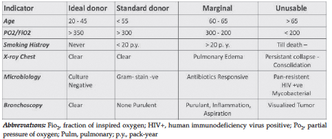
Figure 1. Criteria of Lung Graft Classification
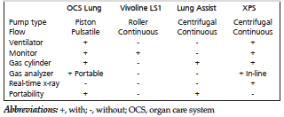
Table 1. Technical Comparison Among Commercially Available EVLP Devices7
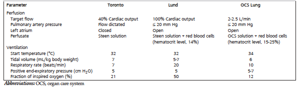
Table 2. EVLP Protocols Vary in Composition of the Perfusate, and in Perfusion and Ventilation Settings