Volume: 13 Issue: 2 April 2015
FULL TEXT
Objectives: Beta thalassemia major is a genetic hemoglobin disorder that affects bone density. The disease leads to deteriorating bone structure but can be treated with hematopoietic stem cell transplant. We aimed to assess bone mineral density changes in pediatric beta thalassemia major patients who had undergone a hematopoietic stem cell transplant compared with similarly affected patients who had not undergone a hematopoietic stem cell transplant.
Materials and Methods: Forty beta thalassemia major patients, 20 transplant and 20 nontransplant, younger than 16 years of age were enrolled. The mean age of transplant patients was 8.15 years and nontransplant patients was 9.5 years (P = .242). The female:male ratio was 1:1 in both groups. None of the patients reached puberty during this study. Bone mineral density was evaluated in transplant patients before and 1 year after hematopoietic stem cell transplant. Bone mineral density of nontransplant patients also was evaluated 1 year after their initial bone mineral density test. A Norland XR-46 densitometer was used to make all bone mineral density measurements. None of the patients had a z score < -2.
Results: Mean bone mineral density changes in the femur and spine during this study were 0.008 ± 0.075 g/cm2 and 0.048 ± 0.045 g/cm2 in transplant patients and 0.045 ± 0.072 g/cm2 and 0.036 ± 0.058 g/cm2 in nontransplant patients. No significant differences between bone mineral density changes in transplant and nontransplant patients were detected during the study.
Conclusions: No significant effects on bone mineral density were detected in hematopoietic stem cell transplant pediatric beta thalassemia major patients compared with similarly affected nontransplant patients. Studies of longer duration may be required to identify significant changes in bone mineral density in hematopoietic stem cell transplant patients.
Key words : Bone marrow transplantation, Children, Skeletal changes, Osteoporosis risk factors
Introduction
Beta thalassemia is a hereditary red blood cell disorder caused by reduced or absent synthesis of hemoglobin beta globin chains. When synthesis is absent in 2 beta globin chain genes, beta thalassemia major occurs. Affected patients develop anemia, impaired growth, and skeletal anomalies during infancy.1
Ineffective erythropoiesis, bone expansion, and extramedullary hematopoiesis are accompanied by different types of bone disorders including bone pain, bone and spinal deformities, bone age delay, short stature, and low bone mineral density (BMD).2 The common finding of low BMD has been reported, based on different bone densitometry methods,3 some of which are not generally in good agreement.4 Several factors are addressed in the reduction of bone mass in thalassemic patients, including endocrinopathies, growth delay,5-6 and liver disease.7 On a molecular basis, an increase in the ratio of sRANKL/OPG is reported in thalassemic patients,8 indicating enhanced osteoclastic bone resorption and bone loss.9 A negative correlation has been found between 17-beta oestradiol in female thalassemics and free testosterone in male thalassemics and the sRANKL/OPG ratio indicates the role of the RANKL/OPG system as a modulator for sex steroid actions on bone remodeling.10 It appears that vitamin D deficiency is another contributing factor to low bone mass associated with thalassemia; however, the prevalence of this problem as reported in different studies varies.5 Gradual liver iron overloading (and deficiency in liver hydroxylation of vitamin D) and a defect in vitamin D absorption in older thalassemic patients, have been reported as disease mechanisms.6
As suggested previously, thalassemic patients need frequent blood transfusions during their life.1 This regimen causes progressive iron overload. Osteoid maturation and mineralization, are interfered by iron. Iron binds into calcium hydroxyapatite crystals; thus, growth of hydroxyapatite crystals is affected and osteoid tissue increases in bone tissue as a result.8 In addition, hemochromatosis-induced iron overload, is commonly associated with diabetes, hypogonadism, hypothyroidism, and hypoparathy-roidism.11 These problems also are risk factors for low BMD. Even the intensive use of chelating agents used to remove the excess iron12 may cause growth failure5 and low BMD.
Correction of the genetic defects with hemato-poietic stem cell transplant (HSCT) is the only definitive cure for thalassemia.13 However, many studies report vulnerability to bone loss and osteoporosis after any type of transplant.14-16
Several factors including gonadal failure, prolonged immobility, decreased osteoprogenitor cells, conditioning regimens, vitamin D deficiency, secondary hyperparathyroidism, cyclosporine, high corticosteroid use, and graft-versus-host disease (GVHD) have been reportedly involved in bone loss and osteoporosis after bone marrow transplants.17-18 However, all studies did not identify bone density deteriorating effects for all of the factors mentioned.19
Because after HSCT, the background disease with deleterious effects on bone is completely cured, the hypothesis emerges that bone density also improves. This hypothesis is strengthened by studies that report positive effects of HSCT on BMD.20 However, some studies found no positive or negative effects on BMD,19 and Petryk and associates found the incidence of preexisting osteopenia and osteoporosis increased 1 year after bone marrow transplants in patients with different background diseases.21
Because Iran has one of the largest populations of major thalassemic patients (nontransplanted and transplanted),22 and these patients have various bone complications, we attempted to evaluate bone density changes in major thalassemic patients after HSCT compared with similarly afflicted patients who did not undergo HSCT.
Materials and Methods
Subjects
The original study population from which the participants of the present study
were drawn, included 26 patients (age < 16 y) diagnosed with beta thalassemia
major who were candidates for HSCT. Deferoxamine, an iron-chelating agent, was
used by all patients. Of the 26 enrolled, only 20 completed all phases of the
study. During the study, 2 patients died and 4 (1 from abroad) withdrew because
the distance from their residences to the study site was too far. All patients
underwent a standard monitoring program recommended for patients after HSCT.
Because 20 transplanted patients ultimately completed the study, we chose 20
nontransplanted age- and sex-matched patients to compare results. Ultimately, 40
pediatric beta thalassemia major patients (20 transplant and 20 nontransplant),
< 16 years of age were enrolled in this prospective study. The female:male ratio
was 1:1 in both groups. None of the patients was pubertal. This study was
approved by the local institutional review board and the parents of the enrolled
patients signed the consent forms. The protocol conforms to the ethical
guidelines of the 1975 Helsinki Declaration.
Osteoporosis risk factors in the medical records of patients were considered study exclusion criteria. Hematopoietic stem cell transplant procedures for thalassemia major patients were performed in the pediatric unit of the Hematology, Oncology, and Stem Cell Transplantation Research Center.
All transplant patients were assessed before and after HSCT. Nontransplant patients also were assessed initially and 1 year later. The mean time intervals for bone mineral densitometry in transplant patients and nontransplant patients were 13 and 17.8 months. We expect nontransplant patients to not have any significant changes in BMD in less than 1 year10; therefore, we think statistical significant difference between time intervals of 2 BMDs in 2 group of pateints was not clinically significant. Bone marrow density in all patients was determined with a Norland XR-46 densitometer (CooperSurgical, Inc. USA). No patients had a z score < -2.
Patient characteristics for hematopoietic stem cell transplant
Histocompatibility was determined by low-resolution molecular typing for
HLA-A and -B antigens and by allelic typing for -DRB1. All hematopoietic stem
cells were provided by related donors.
Patients were categorized according to the criteria of Lucarelli.23 Class 1 and class 2 patients were prepared for transplant with a combination of oral busulfan 3.5 mg/kg daily, in divided doses for 4 days (-8 to -5), cyclophosphamide 50 mg/kg once daily IV for 4 days (-4 to -1) and horse antithymocyte globulin 5 mg/kg daily IV for 2 days (-2 to -1); class 3 patients were conditioned using oral busulfan 3.5 mg/kg in divided doses daily for 4 days (-8 to -5) and cyclophosphamide 40 mg/kg once daily IV for 4 days (-4 to -1). No patients in this study received total body irradiation. Prophylaxis for GVHD included cyclosporine (1.5 mg/kg IV from day -2 until day +7 and 3 mg/kg IV from day +7) and methotrexate (10 mg/m² on day +1 and 6 mg/m² on days +3 and +6). All 20 patients who completed 3 phases of this study were transfusion-free up to 1 year after HSCT. During the first month after hospital discharge patients were followed-up in our post-HSCT clinic weekly, then every 2 weeks to day +100, and thereafter according to the condition of each patient, until 1 year. Anthropometric factors (eg, age, sex, weight, and height) also were recorded. Acute GVHD (aGvHD) and chronic GVHD (cGvHD) were graded according to standard criteria.24-25
Bone mineral density measurement
Bone mineral density at the lumbar spine (L2-L4; anteroposterior view) and
femoral neck of patients were measured by dual-energy X-ray absorptiometry with
a Norland XR-46 densitometer. All measurements were made on the same machine at
Tehran Special Medical Center, the medical branch of Charity Foundation for
Special Diseases). Results were analyzed using specific pediatric software.
World Health Organization osteoporosis and osteopenia criteria are mainly applicable to adults; therefore, we used the term low bone mineral density for age according to the “2007 ISCD Pediatric Position Development Conference, criterion” for a child with a z score < -2.0.26
Statistical analyses
Continuous parameters are reported as means, standard deviations, and ranges.
Categoric parameters are reported as simple percentages. For test of normality
of distribution of our parameters (because our sample size was small (20
cases)), we used normality plots with tests (Shapiro-Wilk statistic is
calculated when the weighted sample size lies between 3 and 50). The independent
samples t test was used to compare the means of the 2 groups. A bivariate
correlation procedure was used to measure correlations, that is how variables or
rank orders were related. Pearson’s correlation coefficient was used for
normally distributed parameters and Spearman’s rho was used for abnormally
distributed parameters. Significance was determined as α (or P value) =
.05, and statistical analyses were performed with SPSS software (SPSS: An IBM
Company, version 16.0, IBM Corporation, Armonk, NY, USA).
Results
Patient characteristics are shown in Table 1. No participants had z scores < -2.0 in the beginning of study and none reached the level of “low density for age” during the progress of this study. The mean femur and spine BMD in the beginning of study in transplant patients was 0.623 ± 0.107 g/cm2 and 0.472 ± 0.082 g/cm2. After HSCT, mean femur and spine BMD was 0.632 ± 0.108 g/cm2 and 0.521 ± 0.078 g/cm2. Bone mineral density of femur and spine in the beginning of study in nontransplant patients was 0.661 ± 0.155 g/cm2 and 0.520 ± 0.123 g/cm2. In the second phase of the study, in nontransplant patients, mean femur and spine BMD was 0.706 ± 0.143 g/cm2 and 0.556 ± 0.101 g/cm2.
The relation between corticosteroid use and BMD changes was not significant. In contrast, chronic and acute GVHD after transplant, especially stage IV, showed a significant effect on BMD changes in femur (P = .011 and .004) and spine (P = .006 and 0.021).
During the study, mean BMD changes in the femur and spine of transplant patients was 0.008 ± 0.075 g/cm2 and 0.048 ± 0.045 g/cm2. Mean BMD changes in the femur and spine in nontransplant patients were 0.045 ± 0.072 g/cm2 and 0.036 ± 0.058 g/cm2. There were no significant differences in BMD changes between nontransplant and transplant patients for 1 year. Figures 1 and 2 show the patient results.
Discussion
Beta thalassemia major is an inherited hemoglobin disorder for which HSCT has opened a new window of hope for a cure. When transplant is not a choice (especially in patients without a compatible donor), patients must be treated by regular monthly blood transfusions and chelation therapy. Both HSCT and hypertransfusion may lead to bone problems; thus, further research is warranted to compare the effects of treatments on bone density. To the best of our knowledge, to date there has been no other study focused on this area, particularly in children. Although we did not find any significant differences between BMD in transplanted and nontransplanted patients, these results are not surprising because we found no significant relation between corticosteroid use and BMD changes in HSCT cases.
These findings are in contrast with those of many investigators such as D'Souza and associates17 and Schulte and associates,27 who reported a significant lowering effect of corticosteroids on the BMD of transplant patients. Their studies did not specifically concern pediatric thalassemic patients. Studies such as the one by Daniels and associates28 did not find a statistically significant correlation between corticosteroid use and BMD in children that received transplants. The nonsignificant effect of concomitant glucocorticoid and cyclosporine use on BMD in some animal studies can be explained by the interference of glucocorticoids and cyclosporine suppresses the individual effect of each agent on bone turnover. Thus, in the first posttransplant phase, when doses of corticosteroid are high, the steroid effect may predominate, leading to a low bone turnover state. Later, lowering the dosage of maintenance corticosteroids may “unmask” the high turnover state induced by cyclosporine.19
We also identified the deleterious effect of GVHD on BMD changes in HSCT patients. In some studies, GVHD was identified as a risk factor for low BMD after HSCT27,29; however, in another study, GVHD was not associated with a significantly lower BMD.30 In our study, chronic GVHD showed a significant relation to bone changes, and GVHD grade IV patients after 1 year had a lower BMD than did all the other acute GVHD patients in all regions.
It may appear conflicting that we did not find a negative effect for corticosteroid use and its duration on BMD changes, while GVHD (associated with high dose corticosteroids use) had negative effects. However, it must not be forgotten that GVHD is a major inflammatory process in which other factors such as inflammation, activity, and nutrition also may affect BMD in these patients. Also, the low prevalence of these complications (4 cases of chronic GVHD and 2 cases of stage IV acute graft-versus-host disease) may have neutralized the adverse effects on mean BMD in the transplant patient group.
If the duration of our study was longer, we might have observed more significant changes, as Schulte and associates showed in their long-term study. They observed patients for 24 months after bone marrow transplants and found that femoral neck BMD was decreased.27
At the beginning of this study, our patients did not have severe osteopenia and no cases of “low bone mineral density for age” were identified, which could be due to the low mean age of our participants compared with other studies. Generally the mean age of participants in other studies was higher and the majority of patients had low BMD.11,20,31,33 Vogiatzi and associates found that adolescence is a critical period for the augmentation of loss of bone mass in thalassemia and suggested that bone turnover plays an important role in this process.33 These findings may better explain why our patients, who were all < 16 years of age, showed no significant changes in BMD. In our previous study of transplant patients we found that patients with a mean age of 7.4 years did not have z scores < -2 initially and 12 months after HSCT.34 The positive bone mass results of our patients may be due to the developing health network that has formed resulting from the large population of thalassemic patients in our country. However, with a larger sample size, we could identify more significant BMD changes in patients in different phases of disease.
In summary, we found no negative or positive effects of HSCT on bone density in transplant pediatric beta thalassemic patients with to nontransplant patients. We believe that using healthy controls and the results of patients’ gonadal, hormonal, and bone marker changes, can aid in identifying the pathogenesis of the disease.
References:
- Muncie HL Jr, Campbell J. Alpha and beta thalassemia. Am Fam Physician.
2009;80(4):339-344.
PubMed - Karimi M, Ghiam AF, Hashemi A, Alinejad S, Soweid M, Kashef S. Bone
mineral density in beta-thalassemia major and intermedia. Indian Pediatr.
2007;44(1):29-32.
PubMed - Baldini M, Forti S, Marcon A, et al. Endocrine and bone disease in
appropriately treated adult patients with beta-thalassemia major. Ann
Hematol. 2010;89(4):1207-1213.
CrossRef -
PubMed - Voskaridou E, Terpos E. New insights into the pathophysiology and
management of osteoporosis in patients with β thalassaemia. Br J Haematol.
2004;127(2):127-139.
CrossRef - PubMed - Hamidi Z. What We Learn from Bone Complications in Congenital Diseases? Thalassemia, an Example. www.intechopen.com Web site. http://www.intechopen.com/books/osteoporosis/what-we-learn-from-bone-complications-in-congenital-diseases-thalassemia-an-example. Accessed September 27, 2014.
- Saffari F, Mahyar A, Jalilolgadr S. Endocrine and metabolic disorders in
β-thalassemiamajor patients. Caspian J Intern Med. 2012;3(3):466-472.
PubMed - La Rosa C, De Sanctis V, Mangiagli A, et al. Growth hormone secretion in
adult patients with thalassaemia. Clin Endocrinol (Oxf). 2005;62(6):667-671.
CrossRef - PubMed - Toumba M, Skordis N. Osteoporosis syndrome in thalassaemia major: an
overview. J Osteoporos. 2010;2010:537673.
CrossRef - Morabito N, Gaudio A, Lasco A, et al. Osteoprotegerin and RANKL in the
pathogenesis of thalassemia-induced osteoporosis: new pieces of the puzzle.
J Bone Miner Res. 2004;19(5):722-727.
CrossRef - PubMed - Haidar R, Musallam KM, Taher AT. Bone disease and skeletal complications
in patients with β thalassemia major. Bone. 2011;48(3):425-432.
CrossRef - PubMed - Shamshirsaz AA, Bekheirnia MR, Kamgar M, et al. Metabolic and
endocrinologic complications in beta-thalassemia major: a multicenter study
in Tehran. BMC Endocr Disord. 2003;3(1):4.
CrossRef - PubMed - Cao A, Galanello R. Beta-thalassemia. Genet Med. 2010;12(2):61-76.
CrossRef - PubMed - Lucarelli G, Andreani M, Angelucci E. The cure of thalassemia by bone
marrow transplantation. Blood Rev. 2002;16(2):81-85.
CrossRef - PubMed - Kulak CA, Borba VZ, Kulak Júnior J, Campos DJ, Shane E.
Post-transplantation osteoporosis. Arq Bras Endocrinol Metabol.
2010;54(2):143-149.
CrossRef - PubMed - Yu TM, Lin CL, Chang SN, Sung FC, Huang ST, Kao CH. Osteoporosis and
fractures after solid organ transplantation: a nationwide population-based
cohort study. Mayo Clin Proc. 2014;89(7):888-895.
CrossRef - PubMed - Helenius I, Remes V, Salminen S, et al. Incidence and predictors of
fractures in children after solid organ transplantation: a 5-year
prospective, population-based study. J Bone Miner Res. 2006;21(3):380-387.
CrossRef - PubMed - D'Souza AB, Grigg AP, Szer J, Ebeling PR. Zoledronic acid prevents bone
loss after allogeneic haemopoietic stem cell transplantation. Intern Med J.
2006;36(9):600-603.
CrossRef - PubMed - Angelopoulos NG, Katounda E, Rombopoulos G, et al. Evaluation of bone
mineral density of the lumbar spine in patients with beta-thalassemia major
with dual-energy x-ray absorptiometry and quantitative computed tomography:
a comparison study. J Pediatr Hematol Oncol. 2006;28(2):73-78.
CrossRef - PubMed - Hamidieh AA, Hamidi Z, Nedaeifard L, et al. Changes of bone density in
pediatric patients with β-thalassemia major after allogenic hematopoietic
stem cell transplantation. Arch Iran Med. 2013;16(2):88-92.
PubMed - Leung TF, Hung EC, Lam CW, et al. Bone mineral density in children with
thalassaemia major: determining factors and effects of bone marrow
transplantation. Bone Marrow Transplant. 2005;36(4):331-336.
CrossRef - PubMed - Klopfenstein KJ, Clayton J, Rosselet R, Kerlin B, Termuhlen A, Gross T.
Prevalence of abnormal bone density of pediatric patients prior to blood or
marrow transplant. Pediatr Blood Cancer. 2009;53(4):675-677.
CrossRef - PubMed - Ghavamzadeh A, Alimoghaddam K, Hamidieh AA, et al. Comparisons between allogeneic peripheral blood stem cell transplantation versus bone marrow stem cell transplantation in Thalassaemia major. Bone Marrow Transplant. 2010;45(suppl 2):S23.
- Lucarelli G, Galimberti M, Giardini C, et al. Bone marrow
transplantation in thalassemia. The experience of Pesaro. Ann N Y Acad Sci.
1998;850:270-275.
CrossRef - PubMed - Glucksberg H, Storb R, Fefer A, et al. Clinical manifestations of
graft-versus-host disease in human recipients of marrow from HL-A-matched
sibling donors. Transplantation. 1974;18(4):295-304.
CrossRef - PubMed - Lee SJ, Vogelsang G, Flowers ME. Chronic graft-versus-host disease. Biol
Blood Marrow Transplant. 2003;9(4):215-233.
CrossRef - PubMed - Baim S, Leonard MB, Bianchi ML, et al. Official Positions of the
International Society for Clinical Densitometry and executive summary of the
2007 ISCD Pediatric Position Development Conference. J Clin Densitom. 2008
r;11(1):6-21.
CrossRef - PubMed - Schulte CM, Beelen DW. Bone loss following hematopoietic stem cell
transplantation: a long-term follow-up. Blood. 2004;103(10):3635-3643.
CrossRef - PubMed - Daniels MW, Wilson DM, Paguntalan HG, Hoffman AR, Bachrach LK. Bone
mineral density in pediatric transplant recipients. Transplantation.
2003;76(4):673-678.
CrossRef - PubMed - Kerschan-Schindl K, Mitterbauer M, Füreder W, et al. Bone metabolism in
patients more than five years after bone marrow transplantation. Bone Marrow
Transplant. 2004;34(6):491-496.
CrossRef - PubMed - Kaste SC, Shidler TJ, Tong X, et al. Bone mineral density and
osteonecrosis in survivors of childhood allogeneic bone marrow
transplantation. Bone Marrow Transplant. 2004;33(4):435-441.
CrossRef - PubMed - Di Stefano M, Chiabotto P, Roggia C, et al. Bone mass and metabolism in
thalassemic children and adolescents treated with different iron-chelating
drugs. J Bone Miner Metab. 2004;22(1):53-57.
CrossRef - PubMed - Vogiatzi MG, Autio KA, Mait JE, Schneider R, Lesser M, Giardina PJ. Low
bone mineral density in adolescents with beta-thalassemia. Ann N Y Acad Sci.
2005;1054:462-466.
CrossRef - PubMed - Vogiatzi MG, Macklin EA, Fung EB, et al. Bone disease in thalassemia: A
frequent and still unresolved problem. J Bone Min Res. 2009;24(3):543-557.
CrossRef - PubMed - Hamidieh AA, Hamidi Z, Nedaeifard L, et al. Bone mineral density in
ex-thalassemic pediatric patients. Hematol Oncol Stem Cell Ther.
2012;5(1):70-71.
CrossRef - PubMed
Volume : 13
Issue : 2
Pages : 173 - 178
DOI : 10.6002/ect.2014.0178
From the 1Hematology-Oncology and Stem Cell Transplantation
Research Center/Tehran University of Medical Sciences; and the 2Endocrinology
and Metabolism Research Center, Endocrinology and Metabolism Clinical Sciences
Institute, Tehran University of Medical Sciences, Tehran, Iran
Acknowledgements: The authors declare that they have no sources of
funding for this study, and they have no conflicts of interest to declare. The
authors thank the Special Medical Center of Charity Foundation for Special
Disease of Iran, and Mrs. Asieh Oojaghi and Miss Fatemeh Ghorbanali for their
valuable assistance in this study. We extend our gratitude to Miss Hamideh
Hesabgar, Dr. Sheema Rezaian, and Mr. Mohammad Rava for their help in editing
the manuscript.
Corresponding author: Zohreh Hamidi MD, MPH, Endocrinology and Metabolism
Research Center, Endocrinology and Metabolism Clinical Sciences Institute,
Tehran University of Medical Sciences, Shariati Hospital, Karegar Ave. P.C.
14114-13137, Tehran, Iran
Phone: +98 21 8822 0037
Fax: +98 21 8822 0052
E-mail: zohreh.hamidi@gmail.com
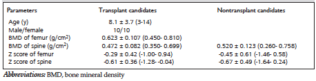
Table 1. Baseline Patient Characteristics *
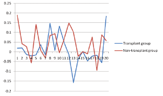
Figure 1. Changes in Bone Mineral Density of the Femurs
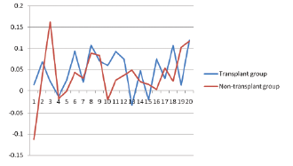
Figure 2. Changes in Spinal Bone Mineral Density