Volume: 13 Issue: 2 April 2015
FULL TEXT
Objectives: Cardiac allograft rejection and infection are leading causes of morbidity and mortality after transplant. The lack of an animal model has hindered related research. We have developed a rat model of pulmonary infection after cardiac transplant to address this issue.
Materials and Methods: Lewis rats received Wistar rat heart allografts, cardiac rejection was induced by cessation of cyclosporine injection, and pulmonary infection was induced by Pseudomonas aeruginosa intrabronchial inoculation. Development of pul-monary infection and/or heart rejection was assessed by histopathology.
Results: Histopathologic findings showed that a dose of 2 × 10⁸ colony forming units of Pseudo-monas aeruginosa intrabronchial inoculation is sufficient to cause severe pneumonia without being lethal to transplanted animals. Daily administration of 10 mg/kg of cyclosporine reliably suppressed rejection, and withdrawal for 7 days can obtain a consistent International Society for Heart and Lung Transplantation 3R rejection.
Conclusions: The current study represents a simple and effective rat model of pulmonary infection along, or in combination, with rejection after heart transplant, which can be used for research of infection-rejection in cardiac transplant settings.
Key words : Heart transplant, Rejection, Lung infection, Animal model
Introduction
Cardiac allograft rejection and infection remain significant sources of morbidity and mortality after heart transplant, accounting for nearly 40% of reported deaths during the first posttransplant year.1,2 In the first 3 years after a transplant, 36% of patients require hospitalization for rejection treatment.1,2 More than 40% of patients experience at least 1 episode of clinically significant infection. Infections occur within the first month after a transplant secondary to nosocomial bacterial pathogens; the lung is the most common site of these infections in heart transplant recipients.3-5
Rejection and infection are inflammatory processes with similar nonspecific symptoms such as fatigue, dyspnea, and low-grade fever. Immuno-suppressive medications used to prevent or treat allograft rejection can result in progression of an existing infection and/or increased risk of a new infection. Conversely, tapering of an immuno-suppressant as part of treating an infection may exacerbate acute rejection.6
To improve the clinical outcome of a cardiac transplant, the pathophysiology of rejection and infection must be understood. However, the lack of an infection/transplant animal model has hindered further investigation. To address this issue, we have developed a rat model of pulmonary infection after a heterotopic cardiac transplant.
Materials and Methods
Animals
Inbred male rats, weighing 200-250 g, were purchased from Slac Laboratory
(Shanghai, China) and were maintained in pathogen-free, laminar flow cages at a
temperature of 24°C and 10-hour light–14-hour dark cycle throughout the
experimental period, and were allowed to drink water and eat rodent chow ad
libitum. Lewis rats served as the recipients, Wistar rats as either donors
(allogeneic transplant) or recipients (syngeneic transplant). The protocol
described in the current study was approved by the Animal Care and Use Committee
of our institution.
Heterotopic cardiac transplant
Cardiac transplants were performed using a modified version of the heterotopic
cardiac transplant model reported by Yokoyama and associates.7
Animals were anesthetized with 40 mg/kg of intraperitoneal pentobarbital. After
heparinization with 1000 units of unfractionated heparin, donor hearts were
procured and placed in an iced lactated Ringer’s solution. The coronary
circulation was immediately flushed with 10 mL cold lactated Ringer’s solution.
Preparation of the donor heart for transplant entails ligation of the main pulmonary artery, superior vena cava, and inferior vena cava, creation of a right atriotomy and an atrial septal defect, disruption of the tricuspid valve leaflets, and en bloc ligation of the pulmonary vessels. The procured hearts are replaced in a cold lactated Ringer’s solution while recipient animals are prepared. The inferior vena cava and abdominal aorta of the recipient animals are exposed through a longitudinal laparotomy with a left subcostal takeoff. The donor ascending aorta is anastomosed to the recipient abdominal aorta with a running 8-0 Prolene using microsurgical techniques. Similarly, the donor right atrium is anastomosed to the recipient inferior vena cava. Upon re-establishment of blood flow, all transplanted hearts resumed spontaneous contractions, were in sinus rhythm, and were free of gross surgical injury at the time of closure.
Daily bodyweight was monitored after surgery. Graft viability was assessed daily by palpation of the donor heart. Rejection was defined as cessation of a palpable heart beat and was confirmed by direct inspection at laparotomy upon organ recovery.
Induction of pulmonary infection
Bacterial lung infection was induced using a modified version of Pseudomonas
aeruginosa pneumonia rat model.8 Through a tracheostomy, 0.2 mL
Pseudomonas aeruginosa ATCC 27853 (1 × 10⁹ CFUs/mL, American
Type Culture Collection, Manassas, VA, USA) was injected into the stem bronchus
of recipient animals assigned to the infection group under direct vision, while
0.2 mL normal saline was injected into the stem bronchus of recipient animals
assigned to the noninfection control group. The bacteria type and inoculums were
determined to be effective from the pilot experiments.
Experimental design
Animals that survived the transplant procedures and were without major
comorbidities were entered into the study. Allogeneic animals (Wistar donor rats
to Lewis recipient rats) underwent a heart transplant. All recipient animals
were started on daily cyclosporine injection (10 mg/kg/d subcutaneously) to
suppress rejection. On postoperative day 6, the allogeneic recipient animals
were randomized to either have their cyclosporine continued (nonrejection group)
or their cyclosporine discontinued, and were begun on a normal saline placebo
injection (10 mg/kg/d subcutaneously, rejection group). On postoperative day 13,
all allogeneic recipient animals were further randomized to receive either
intratracheal inoculation with Pseudomonas aeruginosa (infection group)
or to receive intratracheal inoculation with the same amounts of normal saline
(noninfection group). A syngeneic heart transplant (Wistar donor rats to Wistar
recipient rats, begun daily with 10 mg/kg/d normal saline subcutaneous injection
until postoperative day 14) and saline intratracheal inoculation group served as
controls, so that each animal had a study group randomized to 1 of following:
group 1 (nonrejection and infected), group 2 (nonrejection and noninfection),
group 3 (rejected and infected), group 4 (rejected and noninfection), and the
syngeneic control group 5. Each group contained 5 animals.
Histopathology assessment
Lungs and transplanted hearts were procured after blood withdrawals on
postoperative day 14. Cross sections of heart and lung were preserved in 10%
buffered formalin for hematoxylin and eosin stain. Heart and lung tissue were
confirmed for histologic rejection and infection, blindly, by a pathologist.
Each transplanted heart tissue underwent histologic grading based on the
International Society of Heart and Lung Transplantation (ISHLT) system for
rejection.9 Each lung was evaluated to determine an infection score
based on the extent and severity of pneumonia.10
Statistical analyses
Statistical analyses were performed with SPSS software (SPSS: An IBM Company,
version 13.0, IBM Corporation, Chicago, IL, USA). The t test was used for group
comparison. A 1-way analysis of variance was used when multiple groups were
compared. Two-sided P values less than .05 were considered statistically
significant.
Results
Overall mortality of heart transplants was approximately 10%. The presented modalities of rejection induction and bacterial inoculation did not cause any death over the entire experiment. The mean total transplant operation time was 55 minutes (range, 48-65 min). The mean ischemic time of donor hearts was 35 minutes (range, 31-47 min). Major morbidities such as myocardial infarctions, hemorrhage, and thrombosis were ruled out by direct observation and histopathology.
Development of pulmonary infection
Animals assigned to the noninfection groups recovered their activity and
respiratory rate within several hours after inoculation. In the infected group
animals, decreases in activity in response to audible stimuli and increased
respiratory rate were observed. Histologic examination revealed minimal changes
in the noninfection groups compared with the syngeneic controls (Figures 1A,
1B), whereas all animals of the infected groups had severe pneumonia with
alveolar septal congestion, edema, focal alveolar hemorrhage, and infiltration
of neutrophils and macrophages (Figures 1C, 1D). Pneumonia scores of the
infected groups (group 1 [10.6 ± 0.5], group 3 [10.8 ± 0.5]) were significantly
higher than those of the noninfection groups (group 4 [1.0 ± 0.4], group 4 [1.4
± 0.2]) and syngeneic controls (group 5 [0.8 ± 0.4]; [P < .0001]; [Figure
2]). No statistically significant differences were found among the noninfection
groups and the control group or between the infection groups.
Development of rejection
Daily pulsatility check found that the transplanted heart beat of the
nonrejection groups remained energetic during the study, while in the rejected
groups, the transplanted heart gradually lost its contractions over time after
cyclosporine cessation. Upon recovery, nonrejection hearts were red in color,
soft, normally sized, and beating vigorously; whereas rejected hearts were pale
and firm, and mostly enlarged and not beating. Histology of the cardiac grafts
showed normal staining patterns, with no evidence of interstitial edema or
inflammation (ISHLT grade 0 R) in nonrejection groups (Figures 3A and 3B)
compared with syngeneic controls, whereas all grafts of the rejected groups
(Figures 3C and 3D) showed a diffuse inflammatory process and necrosis (ISHLT
grade 3 R). International Society Of Heart and Lung Transplantation rejection
scores of the rejected groups (group 3 [2.8 ± 0.2], group 4 [3.0 ± 0.0]) were
significantly higher than those of the nonrejection groups (group 1 [0.4 ± 0.2],
group 2 [0.6 ± 0.2]) and syngeneic controls (group 5 [0.2 ± 0.2]; [P <
.0001]; [Figure 4). No statistically significant differences were found among
the nonrejection groups and the control group, or between rejection groups.
Discussion
To date, cardiac transplant is the only established treatment for selected patients with end-stage heart disease. Both immune rejection and infection are frequent, serious complications after heart transplant that cause significant morbidities and mortalities. Although animal models of lung infection and heart transplant have been well developed, to the best of our knowledge, there is no such an animal model of coexisting lung infection and heart transplant, which can be used to explore the pathophysiology and shed light onto the underlying mechanisms of infection/rejection after a heart transplant.
The present model incorporates 2 well-established animal models of heterotopic heart transplant and lung bacterial inoculation. The fundamental heterotopic abdominal heart transplant model in rats was originally reported by Ono and Lindsey,11 of which the major limitation results from lack of hemodynamic loading. Therefore, we used a modified version of the abdominal cardiac transplant model.7,12 By removing the tricuspid valve and interatrial septum, the left atrium and ventricle of the graft are loaded with blood from the right atrium through the interatrial communication. This working left heart model is easily reproducible, thus practical for routine studies.
In our early pilot study, we determined that daily subcutaneous administration of 10 mg/kg of cyclosporine in this model could reliably suppress rejection, but will permit the emergence of rejection upon its discontinuation. This was verified in the current study that cyclosporine withdrawn for 7 days obtained a fairly consistent level of ISHLT grade 3 R rejection. Our results also demonstrate that a dose of 2 × 10⁸ colony forming units of intrabronchial Pseudomonas aeruginosa is sufficient to cause severe pulmonary infection and systemic inflammatory response (ie, increased TNF-alpha and IL-6 levels, and white blood cell counts) along, or in combination, with heart rejection, without being immediately lethal in transplanted rats. Pseudomonas aeruginosa was selected in this study because it is one of the most common pathogens causing pulmonary infection during the early postoperative period in heart transplanted recipients who receive immuno-suppression agents.12
When compared with a rat abdominal infection/heart transplant model reported by Kobayashi and associates,13 the present model might be more favorable if: (1) the lung is the most common site of infections after a heart transplant, whereas the abdomen is less common; (2) the current model yielded a zero mortality rate, all animals survived over the study period, whereas the intraperitoneal infection induced by cecal ligation caused high mortalities; (3) the present model has the advantage of feasibility; the severity of infection and/or rejection could be modulated to a desired degree by adjusting the bacterial type and inoculating dosage, immunosuppressive agents administration and timing, whereas the cecal ligation is a fixed programed procedure with no flexibility; and the (4) lung is distal to the transplanted heart, whereas intra-abdominal infection may pose unwanted and unpredicted effect because of its proximity to the infectious site of the transplanted heart.
Although the pulmonary infections were validated by histopathology, the authors recognized that the work would be more complete if the bacterial cultures were performed on the bronchial mucus of the lung and peripheral blood. However, histologic minimal changes of the saline inoculation groups compared with the severe pneumonia found in bacterial inoculation groups suggest that the microstructure changes observed were not likely caused by physical or chemical effects of inoculation fluids, but by injected bacterial pathogens.
In addition, the analysis of data was limited to the pathological results; other findings were not presented in detail. It may be argued that as histopathology remains the criterion standard for diagnosing diseases, histology results would have been confirmative. Nonetheless, while the pulmonary infection model and the heart transplant model are established with associating pathophysiologic changes well documented, further investigations (specifically on the current model) are warranted.
In summary, this work represents an effective model of pulmonary infection along, or in combination, with rejection after heart transplant. We believe this model could be useful to make translatable findings to the clinic and to investigate the mechanics of infection-rejection in heart transplant settings.
References:
- Stehlik J, Edwards LB, Kucheryavaya AY, et al. The Registry of the
International Society for Heart and Lung Transplantation: Twenty-eighth
Adult Heart Transplant Report--2011. J Heart Lung Transplant.
2011;30(10):1078-1094.
CrossRef - PubMed - Stehlik J, Edwards LB, Kucheryavaya AY, et al; and International Society
of Heart and Lung Transplantation. The Registry of the International Society
for Heart and Lung Transplantation: 29th official adult heart transplant
report--2012. J Heart Lung Transplant. 2012;31(10):1052-1064.
CrossRef - PubMed - Fishman JA, Rubin RH. Infection in organ-transplant recipients. N Engl J
Med. 1998;338(24):1741-1751.
CrossRef - PubMed - Atasever A, Bacakoglu F, Uysal FE, et al. Pulmonary complications in
heart transplant recipients. Transplant Proc. 2006;38(5):1530-1534.
CrossRef - PubMed - Lenner R, Padilla ML, Teirstein AS, Gass A, Schilero GJ. Pulmonary
complications in cardiac transplant recipients. Chest. 2001;120(2):508-513.
CrossRef - PubMed - Dunn DL. Hazardous crossing: immunosuppression and nosocomial infections
in solid organ transplant recipients. Surg Infect (Larchmt).
2001;2(2):103-110; discussion 110-112.
CrossRef - PubMed - Yokoyama H, Ohmi M, Murata S, Nakame T, Tabayashi K, Mohri H. Proposal
of a working left heart model with a heterotopic transplantation technique
in rats. J Heart Lung Transplant. 1995;14(4):706-712.
PubMed - Li Z, Qu J, He L. Establishing a model of neutropenia rat with
Pseudomonas aeruginosa pneumonia and a study on its inflammatory reaction
[in Chinese]. Zhonghua Jie He He Hu Xi Za Zhi. 2001;24(11):674-678.
PubMed - Stewart S, Winters GL, Fishbein MC, et al. Revision of the 1990 working
formulation for the standardization of nomenclature in the diagnosis of
heart rejection. J Heart Lung Transplant. 2005;24(11):1710-1720.
CrossRef - PubMed - Kim HK, Tazelaar HD, Odell J, et al. An animal model of pulmonary
infection after single lung transplantation. Transplant Proc.
1996;28(3):1816-1817.
PubMed - Ono K, Lindsey ES. Improved technique of heart transplantation in rats.
J Thorac Cardiovasc Surg. 1969;57(2):225-229.
PubMed - Clauss HE, Bettiker RL, Samuel R, Truant AL, Jungkind D. Infections in
Heart and Lung Transplant Recipients. Clin Microbiol Newsletter.
2012;34;19-25.
CrossRef - Kobayashi J, Mavroudis C, Crawford SE, Zales VR, Backer CL. A new rat
infection-heart transplant model: effect of infection on graft survival
studies. J Heart Lung Transplant. 1993;12(4):659-664.
PubMed
Volume : 13
Issue : 2
Pages : 167 - 172
DOI : 10.6002/ect.2014.0033
From the 1Department of Thoracic-Cardiovascular Surgery, Tongji
Hospital, Tongji University; the 2Department of Cardiothoracic
Surgery; the 3Department of Pathology, Huadong Hospital, Fudan
University; and the 4Department of Hematology, Zhongshan Hospital,
Fudan University, Shanghai, China
Acknowledgements: The authors have no conflicts of interest to disclose;
the authors alone are responsible for the content and for the writing of the
paper. Hao Chen and Weiyong Yu contributed equally to this work. The authors
would like to thank Xueping Fan for her invaluable technical support. This study
was undertaken at the Biomedical Research
Center of Shanghai Zhongshan Hospital. The study was supported by Pujiang talent
research grant (No. 10PJ1403600) from the Science and Technology Commission of
Shanghai Municipality, China
Corresponding author: Hao Chen, MD, PhD, Department of
Thoracic-Cardiovascular Surgery, Tongji Hospital, Tongji University, 389 Xincun
Road, Shanghai 200065, China
Phone :+86 136 1161 0435
Fax: +86 21 6436 0382
E-mail: h.chen@fudan.edu.cn
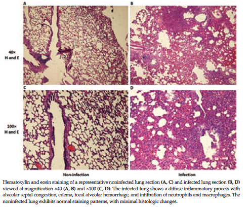
Figure 1. Histology of the Lung
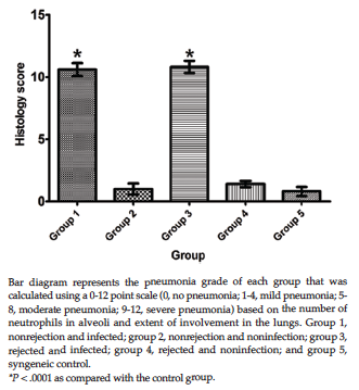
Figure 2. Histologic Score of the Lung
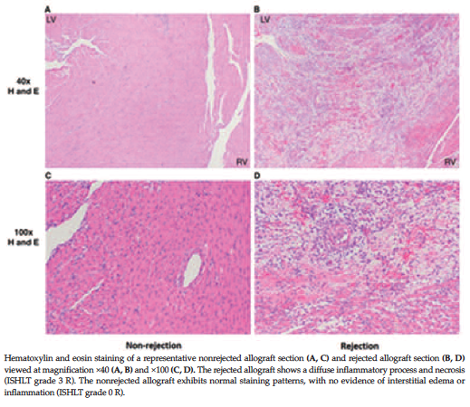
Figure 3. Histology of a Cardiac Allograft
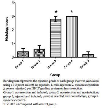
Figure 4. Histologic Score of the Cardiac Allograft