Volume: 12 Issue: 3 June 2014
FULL TEXT
Diaphragmatic hernias are a known complication of explanting a left ventricular assist device. The increasing use of left ventricular assist devices has resulted in an increased risk of this complication. We present the case of a patient who presented with diaphragmatic hernias on routine follow-up after a heart transplant. A left thoracotomy was performed to expose and reduce the hernia using a bovine pericardial patch to repair the defect. This biomaterial represents a viable alternative to traditional GORE-TEX patches with the probability of decreased infectious complications.
Key words : Diaphragm, Devices, Surgical complications
Case Report
At our institution, a 60-year-old man underwent a successful orthotropic heart transplant, secondary to ischemic heart failure. He was bridged with a HeartMate II Left Ventricular Assist System (Thoratec Corporation, Pleasanton, CA, USA) for 2 years before an organ became available. He had undergone a previous laparotomy, with a colon resection for ischemic colitis.
The patient’s postoperative recovery was uneventful, and was discharged home 12 days after having his operation. He returned for a scheduled, outpatient endomyocardial biopsy complaining of shortness of breath, increased fatigue, and intermittent epigastric abdominal pain. Subsequent fluoroscopy during his biopsy was performed concerning possible abdominal contents in his chest.
An expeditious computed topography scan confirmed the presence of a large diaphragmatic hernia (DH), with multiple loops of the bowel in his thoracic cavity (Figure 1). Of note, immunosuppression therapy, at that time, consisted of tacrolimus, mycophenolate mofetil, and prednisone.
He was taken to the operating suite to reduce the abdominal contents and repair his diaphragmatic defect. A seventh intercostal space left thoracotomy was done, providing adequate exposure of the hernia and facilitating reduction of the contents. At the time of the transplant, the diaphragmatic defect had been reapproximated with 0 monofilament absorbable sutures. After reducing the hernia, it was obvious that the sutures had pulled through the diaphragmatic tissue. Given the failure of the previous repair and the patient’s immunosuppressed status, the defect was closed using a bovine pericardium patch (Figure 2). His postoperative hospital course was unremarkable. The patient was discharged on postoperative day 8 on regular diet, no longer complaining of shortness of breath, abdominal pain, or fatigue.
Discussion
Diaphragmatic hernias are a well described and increasingly common postoperative complication of a heart transplant after explanting a left ventricular assist device.1 In no small part is this due to the splitting of the diaphragm to implant the left ventricular assist device. Often, the poor nutritional status, combined with the requirement for immunosuppression in the patient, increases the likelihood of primary diaphragm repair failure. Contemporary studies estimate the incidence of DHs at upwards of 15.9%.2 Furthermore, upwards of 30% of patients undergoing a heart transplant have had a prior left ventricular assist device, and this percentage is likely to become increased.3 As a result, the incidence of posttransplant DHs is likely to become a significant concern.
Health care providers managing this subset of patients must have an increased awareness of this entity as a diagnostic possibility. Suspicion must be raised in individual who complains of shortness of breath or abdominal pain postoperatively. Surgical repair of these defects is recommended upon their diagnosis, as they can lead to bowel strangulation, incarceration, necrosis, and perforation.4 Surgical principles of tension-free repair should be followed, and synthetic materials (eg, polytetrafluoroethylene and polypropylene) have been used successfully for this.2 The transplanted population, however, deserves special attention with reduction of potential long-term infectious complications. The use of an immunologically inert, biological material has certain advantages for this.
The use of a bovine pericardium has been previously evaluated by Zardo and associates who used it for chest wall reconstruction and diaphragmatic defect repairs secondary to tumor resection.5 It demonstrated high tensile strength, elasticity, and was easy to manipulate. The patch has good biocompatibility, allowing ingrowth of natural tissue and minimal risk of infection.5 These qualities, combined with the possibility of microtears in a reoperative setting, prompted the selection of a bovine pericardium to repair the DH in this patient. Although the long-term outcomes remain uncertain, this case demonstrates the successful use of a bovine pericardium to repair a DH. This material and technique should be in the armamentarium of the thoracic transplant surgeon as a viable alternative to using synthetic mesh in immunocompromised patients.
References:
- Phillips WS, Burton NA, Macmanus Q, Lefrak EA. Surgical complications in
bridging to transplantation: the Thermo Cardiosystems LVAD. Ann Thorac Surg.
1992;53(3):482-485; discussion 485-486.
CrossRef - PubMed - Chatterjee S, Williams NN, Ohara ML, Twomey C, Morris JB, Acker MA.
Diaphragmatic hernias associated with ventricular assist devices and heart
transplantation. Ann Thorac Surg. 2004;77(6):2111-2114.
CrossRef - PubMed - Kilic A, Ailawadi G. Left ventricular assist devices in heart failure.
Expert Rev Cardiovasc Ther. 2012;10(5):649-656.
CrossRef - PubMed - Mori T, Nagao G, Sugiyama M. Paraesophageal hernia repair. Ann Thorac
Cardiovasc Surg. 2012;18(4):297-305.
CrossRef - PubMed - Zardo P, Zhang R, Wiegmann B, Haverich A, Fischer S. Biological
materials for diaphragmatic repair: initial experiences with the PeriGuard
Repair Patch®. Thorac Cardiovasc Surg. 2011;59(1):40-44.
CrossRef - PubMed

Volume : 12
Issue : 3
Pages : 277 - 278
DOI : 10.6002/ect.2013.0068
From the 1Division of Cardiac Surgery, and the 2Division
of Thoracic Surgery, Wexner Medical Center, The Ohio State University, Columbus,
OH 43201, USA
Acknowledgements: The authors state that they have no conflicts of
interest to declare, and they received no funding for this case report.
Corresponding author: Kevin Ricci, Division of Cardiac Surgery, Wexner
Medical Center, The Ohio State University, N831 Doan Hall, 410 W. 10th Ave,
Columbus, OH 43201 USA
Phone: +1 (614) 293-8878
Fax: +1 (614) 293-2020
E-mail: kevin.ricci@osumc.edu
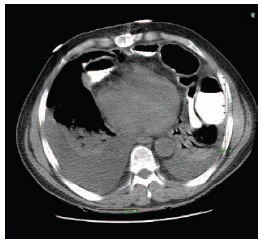
Figure 1. A Chest Computed Tomographic Image With Contrast Showing a Diaphragmatic Herniation of Small Bowel Into the Left Hemithoracic Cavity
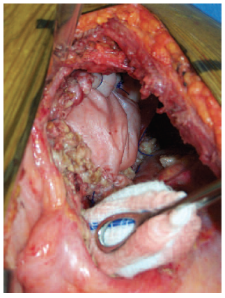
Figure 2. View of the Diaphragm From a Left Thoracotomy After a Hernia Repair With Bovine Pericardium