Volume: 12 Issue: 3 June 2014
FULL TEXT
Objectives: Complement-dependent cytotoxic crossmatch is an important indicator for kidney transplant. However, there is controversy about treatment for flow cytometry crossmatch-positive cases.
Materials and Methods: This was a retrospective study of 127 living-donor kidney transplant recipients from May 2007 to July 2011. We divided patients into 115 flow cytometry crossmatch T-cell and B-cell–negative cases, and 12 T-cell and B-cell–positive cases. Both groups were given 20 mg basiliximab the day of surgery and 4 days after surgery. Common oral immunosuppressive agents used were tacrolimus, mycophenolate mofetil, and methylprednisolone. Flow cytometry crossmatch T-cell and B-cell–negative recipients started immunosuppression 7 days before surgery, T-cell and B-cell–positive recipients started immunosuppression 14 days before surgery. T-cell and B-cell–positive patients also received 200 mg rituximab 1 week before surgery, had 3 plasma exchange sessions before transplant, and received intravenous immunoglobulin 20 g/day during surgery and after surgery for 5 days. We measured flow-panel reactive antibodies of T-cell and B-cell–positive patients just before surgery to check desensitization efficiency. We evaluated patient survival, graft survival, graft function, and frequency of rejection and infectious diseases.
Results: Patient survival and graft survival were 100% in both groups. Flow cytometry crossmatch T-cell and B-cell–positive cases had no rejection events, but T-cell and B-cell–negative groups developed rejection. There was no statistical difference in the incidence of infection and graft function. Flow-panel reactive antibody demonstrated improvement in all T-cell and B-cell–positive cases.
Conclusions: In living-donor kidney transplant, flow cytometry crossmatch T-cell and B-cell–positive patients are still considered to be at high risk. Although this is a short-term outcome, all T-cell and B-cell–positive patients in this study achieved excellent results with appropriate preoperative and postoperative treatment.
Key words : Chronic kidney disease, Desensitization, Rejection, Treatment
Introduction
Crossmatch tests before organ transplant have been integral to transplant since the studies of Patel and Terasaki in the late 1960s.1 Human leukocyte antigen (HLA) laboratories regularly perform crossmatches between donor cells and recipient serum before transplant. The flow cytometry crossmatch (FCXM) technique is more sensitive than complement-dependent cytotoxicity crossmatch assays for the detection of antibodies.2 Although complement-dependent cytotoxicity is an important method in planning kidney transplant, there is no established strategy for the indications or perioperative immuno-suppressive therapy for FCXM-positive patients.
Many reports describe a higher incidence of rejection or poor graft survival in FCXM-positive patients.3-10 However, in these reports the immuno-suppressive agents and preoperative treatment are different from the present situation. Good outcomes using rituximab, plasma exchange (PEX), and intravenous immunoglobulin (IVIG) have been reported in immunologically high-risk patients.11-15 We achieved better outcomes using a unique protocol that combined these methods including the use of retuximub, PEX, and IVIG in patients who were FCXM-positive for both T and B cells (FCXM T+B+). In the present study, we investigated the clinical course of FCXM T+B+ patients to validate our strategy.
Materials and Methods
We retrospectively reviewed 127 consecutive living-donor kidney transplant recipients from May 2007 to July 2011, when FCXM was introduced to our institution. All protocols, experimental studies, and clinical trials involving human subjects were approved by the ethics committee of the institution before the study began, and the protocols conformed to the ethical guidelines of the 1975 Helsinki Declaration. Written informed consent was obtained from every patient.
We excluded patients who were complement-dependent cytotoxicity positive, blood type incompatible, FCXM T-cell negative and B-cell positive, or undergoing an alternative immuno-suppressive protocol. We divided the patients into 2 groups: 115 FCXM T-B- cases and 12 FCXM T+B+ cases (Table 1). There were statistically significant differences between the groups in male-to-female ratio and number of HLA mismatches (Table 1). In the FCXM T+B+ group, 7 of 12 patients were transplants from the spouse, and 7 patients appeared to be sensitized by pregnancy (Table 2). The mean trough blood concentration of tacrolimus and doses of mycophenolate mofetil and methylprednisolone were similar between the groups (Table 1). The postoperative use of immunosuppressive drugs was similar between the groups. There were 2 male patients who had second transplant and there were 7 patients who were donor specific antibody (DSA) positive (Table 2). There was no significant difference in primary disease between the groups (Table 3). Both groups were administered 20 mg basiliximab on the day of surgery and 4 days after surgery. Common oral immunosuppressive agents used in the 2 groups were tacrolimus, mycophenolate mofetil, and methylprednisolone. These immuno-suppressive agents were started 7 days before surgery in FCXM T-B- recipients and 14 days before surgery in FCXM T+B+ recipients. The FCXM T+B+ patients also received 200 mg rituximab 1 week before surgery, 3 treatments with PEX before transplant, and 20 g/day of IVIG during and after surgery for 5 days. On the day of transplant surgery, we measured flow-panel reactive antibody (Flow-PRA) to confirm desensitization.
We performed a transplant kidney biopsy at 3 months and 1 year after surgery or whenever rejection was suspected. Diagnosis of rejection was based on the clinical course and findings obtained at biopsy, which were evaluated according to Banff diagnostic categories for renal allograft biopsies.16 All specimens were investigated by 2 experienced pathologists (A. Tsuchimoto and K. Masutani) using a double-headed light microscope.
We analyzed patient and graft survival and frequency of total rejection at different times (2 weeks, 3 months, 1 year, and 2 years after surgery). We evaluated the incidence of infectious diseases, serum creatinine level, and estimated glomerular filtration rate (eGFR) at 1 year after transplant and the attenuation ratio of fluorescein isothiocyanate (FITC) channels after desensitization of Flow-PRA from the FCXM T+B+ group.
Flow cytometry crossmatch
The FCXM and Flow-PRA testing were performed at the Center for Cellular and
Molecular Medicine. Recipient and donor blood samples were centrifuged (3000
rpm, 10 min) to obtain serum. The heparinized donor blood was separated using
medium (Lymphocyte Separation Medium, MP Biomedicals, Santa Ana, CA, USA) and
3-5 ×10⁶ mononuclear cells were obtained. There were 6 tubes prepared for flow
cytometry.
For flow cytometry of T and B cells, samples were set up for negative control (donor serum [100 μL] and donor mononuclear cells [100μL]), positive control (positive serum [100 μL] and donor mononuclear cells [100μL]), and samples (recipient serum [100 μL] and donor mononuclear cells [100μL]). Cells were incubated for 30 minutes at room temperature, washed 3 times with phosphate-buffered saline, and blocked with normal goat serum for 10 minutes at room temperature. Cells were labeled with FITC-labeled goat antihuman immunoglobulin G (IgG), T-cell phycoerythrin-Cy5 CD3 (PE Cy-5 Mouse Anti-Human, Becton Dickinson, Franklin Lakes, NJ), or B-cell phycoerythrin-labeled anti-CD19 (PE Mouse Anti-Human, Becton Dickinson) for 30 minutes at 4°C. Cells were washed 3 times with phosphate-buffered saline and analyzed by flow cytometry (BD FACS, Becton Dickinson). A positive result was defined by a shift of 15% (for T cells) or 20% (for B cells) from the negative control or by a significantly different waveform such as bimodal waveform. For the positive cases, we reexamined the samples to check reproducibility.
Flow-panel reactive antibody
Flow-PRA beads were vortexed before use. Test serum (20 μL) was incubated with
Flow-PRA HLA class I and class II beads in a 1.5 mL tube (Eppendorf, Hamburg,
Germany) or a 96-well plate for 30 minutes in the dark at 20°C to 25°C with
gentle shaking. Wash buffer (10×) was diluted in distilled water to make a 1×
solution. Wash buffer (1×) was added to each tube (1 mL) or each well of the
96-well plate (150 μL), vortexed, and centrifuged at 9000 × g for 2 minutes or
at 1500 × g for 10 minutes. Cells were aspirated and the supernatant was
discarded; this was done twice when tubes were used for the test. When the
96-well plate was used, the wells were washed twice with
200 μL wash buffer. The FITC-conjugated goat antihuman IgG (Fcγ) was diluted to
1 μL per test with wash buffer to make a 1× solution, and 100 μL was added to
beads, vortexed, and incubated for 30 minutes in the dark at 20°C to 25°C with
gentle shaking. The previous step was repeated twice when tubes were used for
the test. When the 96-well plate was used, the wells were washed twice with wash
buffer (100 μL and 200 μL). Fixing solution (1×) was added to the tube (0.5 mL)
or to each well of a 96-well plate (200 μL). The samples were used for analysis
or stored at 2°C to 5°C up to 24 hours before analysis. A positive result was
defined by a shift > 10% from the negative control or a multimodal waveform.
Statistical analyses
Data analyses were performed with statistical software (JMP8 for Windows, SAS
Institute, Inc., Cary, NC, USA). Differences between the 2 groups were evaluated
with t test for numerical data and the chi-square test for categorical data.
Kaplan-Meier method and log-rank test were used to evaluate differences between
the 2 groups. Statistical significance was defined by P ≤ .05.
Results
Patient and graft survival were 100% in both groups in the observation period (Table 4). The T-B- group developed rejection with a probability of 16.8% in 2 years. In contrast, the T+B+ group with desensitization developed no rejection. We also evaluated the incidence of cytomegalovirus and BK virus viremia that required treatment or other viral or bacterial infections that required hospital treatment including herpes zoster, bacterial pneumonia, urinary tract infection, and hemorrhagic cystitis. The frequency of infection was similar in the T-B- and T+B+ group (Table 4). All infectious diseases were cured by appropriate treatment. Serum creatinine level and eGFR at 1 year after transplant were similar between the groups (Table 4). In all T+B+ patients, we confirmed desensitization as expected by Flow-PRA on the day of transplant surgery followed by the elective operation. The mean FITC channels showed 29% ± 18% improvement in class I and 48% ± 15% improvement in class II after desensitization. In a typical case, the HLA class I, HLA class II, and DSA were positive before and negative after desensitization (Figure 1).
Discussion
Patients with a possibility of sensitization from various factors, such as pregnancy, blood transfusion, organ transplant, or infection may have existing antibodies. Such patients often are positive for FCXM, which has higher sensitivity than complement-dependent cytotoxicity. Many studies have reported a poor prognosis in FCXM T+B+ patients,3-10 and transplant was considered contraindicated in these patients in some treatment centers, depriving these patients from receiving a transplant. In many of these reports, there was no discussion of desensitization, immunosuppressive agents used, or perioperative treatment, which may have differed from the present study.
Some authors have reported good outcomes using rituximab, PEX, and IVIG, which are effective in the treatment of rejection in immunologically high-risk patients.11-15 These 3 methods were designed to inhibit existing antibodies. Rituximab is a chimeric anti-CD20 (anti-B cell) monoclonal antibody approved for the treatment of lymphoma. This antibody efficiently eliminates B cells because the CD20 antigen is expressed early in B-cell ontogeny but is absent on mature plasma cells.17 By eliminating B cells, rituximab inhibits new antibody production. Since 2006, our research group has adopted the desensitization protocol with rituximab for blood-type incompatible transplant. Based on our experience with no severe adverse events and secure deletion of CD19-positive cells or CD20-positive cells in the peripheral blood by rituximab, we determined a dose of 200 mg of rituximab and the timing for administration for T+B+ transplant patients.
The available IVIG products are derived from pooled human plasma from thousands of donors and have been used for the treatment of primary immunodeficiency, autoimmune, or inflammatory disorders. Although the mechanism of IVIG in desensitization is unclear, possible mechanisms of action include regulation of dendritic cell activation and monocyte and macrophage function, regulation of immune responses by neutralizing existing antibodies, and binding to complement to prevent complement-mediated antigen-antibody reactions.18 There is controversy about the timing and dose of IVIG. To prevent removal by PEX, we administered IVIG from the day of surgery for 5 days, after 3 times of PEX treatment were finished. The IVIG is an important component of this protocol, but it may be desirable to reduce the use of IVIG because IVIG is costly and large quantities of IVIG were used in these patients.
In the present protocol, Flow-PRA was measured before surgery but we performed the operation before checking the results. Although desensitization was successfully accomplished using our protocol, insufficient desensitization may occur. Therefore, it is important to observe the postoperative clinical course carefully based on the result of the Flow-PRA, and administer any necessary additional immuno-suppressive therapy. The complement-binding capacity of donor-specific anti-HLA antibodies is associated with an increased frequency of antibody-mediated rejection.19 Assessment of the complement-binding capacity of DSA may enable the early detection of antibody-mediated rejection for immunologically high-risk patients. Furthermore, it may be useful to evaluate the effectiveness of preoperative desensitization.
Although we reported only short-term outcomes, the T+B+ patients obtained a good quality of life with sufficient kidney function, without any episode of rejection after our simple desensitization protocol. This was good compared with T-B- patients who were considered immunologically low-risk patients. In Japan, there currently is a major shortage of deceased donors, and the present protocol provides an option to many sensitized patients who otherwise could not receive a transplant.
In summary, the essence of the present protocol is to inhibit formation of antibodies by rituximab, decrease antibody levels by PEX, and suppress antibodies by IVIG. This study had limited follow-up and a small sample size, and future studies may evaluate larger patient samples at long-term follow-up.
References:
- Patel R, Terasaki PI. Significance of the positive crossmatch test in
kidney transplantation. N Engl J Med. 1969;280(14):735-739.
CrossRef - PubMed - Bray RA, Lebeck LK, Gebel HM. The flow cytometric crossmatch. Dual-color
analysis of T cell and B cell reactivities. Transplantation. 1989;48(5):834-840.
CrossRef - PubMed - Graff RJ, Xiao H, Schnitzler MA, et al. The role of positive flow cytometry
crossmatch in late renal allograft loss. Hum Immunol. 2009;70(7):502-505.
CrossRef - PubMed - Rebibou JM, Bittencourt MC, Saint-Hillier Y, et al. T-cell flow-cytometry
crossmatch and long-term renal graft survival. Clin Transplant.
2004;18(5):558-563.
CrossRef - PubMed - El Fettouh HA, Cook DJ, Bishay E, et al. Association between a positive flow
cytometry crossmatch and the development of chronic rejection in primary renal
transplantation. Urology. 2000;56(3):369-372.
CrossRef - PubMed - Graff RJ, Buchanan PM, Dzebisashvili N, et al. The clinical importance of
flow cytometry crossmatch in the context of CDC crossmatch results. Transplant
Proc. 2010;42(9):3471-3474.
CrossRef - PubMed - Piatosa B, Rubik J, Grenda R. Is positive flow cytometric cross-match a risk
factor for early cadaveric kidney graft dysfunction? Transplant Proc.
2006;38(1):53-55.
CrossRef - PubMed - O'Rourke RW, Osorio RW, Freise CE, et al. Flow cytometry crossmatching as a
predictor of acute rejection in sensitized recipients of cadaveric renal
transplants. Clin Transplant. 2000;14(2):167-173.
CrossRef - PubMed - Ilham MA, Winkler S, Coates E, Rizzello A, Rees TJ, Asderakis A. Clinical
significance of a positive flow crossmatch on the outcomes of cadaveric renal
transplants. Transplant Proc. 2008;40(6):1839-1843.
CrossRef - PubMed - Takeda A, Uchida K, Haba T, et al. Acute humoral rejection of kidney
allografts in patients with a positive flow cytometry crossmatch (FCXM). Clin
Transplant. 2000;14(suppl 3):15-20.
CrossRef - PubMed - Goh A, Oei E, Vathsala A. Successful transplantation across positive B-cell
cross-match in deceased donor renal transplants. Transplant Proc.
2012;44(1):193-199.
CrossRef - PubMed - Montgomery RA, Lonze BE, King KE, et al. Desensitization in HLA-incompatible
kidney recipients and survival. N Engl J Med. 2011;365(4):318-326.
CrossRef - PubMed - Rehman S, Meier-Kriesche HU, Scornik J. Use of intravenous immune globulin
and rituximab for desensitization of highly human leukocyte antigen-sensitized
patients awaiting kidney transplantation. Transplantation. 2010;90(8):932-933.
CrossRef - PubMed - Montgomery RA, Zachary AA, Racusen LC, et al. Plasmapheresis and intravenous
immune globulin provides effective rescue therapy for refractory humoral
rejection and allows kidneys to be successfully transplanted into
cross-match-positive recipients. Transplantation. 2000;70(6):887-895.
CrossRef - PubMed - Vo AA, Peng A, Toyoda M, et al. Use of intravenous immune globulin and
rituximab for desensitization of highly HLA-sensitized patients awaiting kidney
transplantation. Transplantation. 2010;89(9):1095-1102.
CrossRef - PubMed - Sis B, Mengel M, Haas M, et al. Banff '09 meeting report: antibody mediated
graft deterioration and implementation of Banff working groups. Am J Transplant.
2010;10(3):464-471.
CrossRef - PubMed - Reff ME, Carner K, Chambers KS, et al. Depletion of B cells in vivo by a
chimeric mouse human monoclonal antibody to CD20. Blood. 1994;83(2):435-445.
PubMed - Jordan SC, Toyoda M, Vo AA. Regulation of immunity and inflammation by
intravenous immunoglobulin: relevance to solid organ transplantation. Expert Rev
Clin Immunol. 2011;7(3):341-348.
CrossRef - PubMed - Loupy A, Lefaucheur C, Vernerey D, et al. Complement-binding anti-HLA
antibodies and kidney-allograft survival. N Engl J Med. 2013;369(13):1215-1226.
CrossRef - PubMed

Volume : 12
Issue : 3
Pages : 227 - 232
DOI : 10.6002/ect.2013.0163
From the 1Department of Surgery and Oncology, Graduate School of Medical
Science, Kyushu University; the 2Center for Cellular and Molecular Medicine,
Kyushu University Hospital; and the 3Kidney Care Unit, Kyushu University
Hospital, Fukuoka, Japan
Acknowledgements: The authors received no funding for this study, and they
declare that they have no conflict of interest.
Corresponding author: Soushi Terasaka, Department of Surgery and Oncology,
Graduate School of Medical Sciences, Kyushu University, 3-1-1 Maidashi,
Higashi-ku, Fukuoka 812-8582, Japan
Phone: +81 92 642 5443
Fax: +81 92 642 5458
E-mail: terasakasoushi@me.com
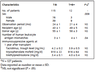
Table 1. Clinical Characteristics of Patients Who Had Living-Donor Kidney Transplant*
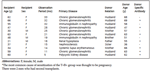
Table 2. Clinical Characteristics in T+B+ Patients Who Had Living-Donor Kidney Transplant*
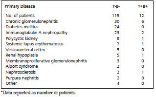
Table 3. Primary Kidney Disease in Patients Who Had Living-Donor Kidney Transplant*
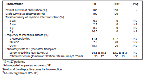
Table 4. Clinical Outcomes of Living-Donor Kidney Transplant*
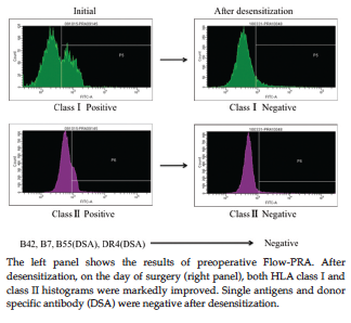
Figure 1. Flow-Panel Reactive Antibody (Flow-PRA) Tests For a Living-Donor Kidney Transplant Recipient of T+B+