Volume: 12 Issue: 3 June 2014
FULL TEXT
Objectives: The adenosine triphosphate assay using peripheral lymphocytes may be useful to evaluate the risks of acute rejection and infection in kidney transplant patients. We used the adenosine triphosphate assay to evaluate differences between recipients who were treated with cyclosporine- or tacrolimus-based immunosuppressive therapy.
Materials and Methods: Adenosine triphosphate levels were measured in peripheral CD4+ cells before and after transplant and were correlated with clinical outcomes in 45 kidney transplant recipients. These recipients received immunosuppressive therapy with either cyclosporine (23 patients) or tacrolimus (22 patients).
Results: Adenosine triphosphate levels were significantly lower in the cyclosporine- than tacrolimus-based therapy groups from 2 to 6 weeks after transplant. Adenosine triphosphate levels were similar between these groups before and 1 week after transplant. The frequency of cytomegalovirus infection was greater in the recipients who received cyclosporine (17 patients [74%]) than tacrolimus (6 patients [27%]; P ≤ .003). The frequency of acute rejection episodes was similar between the cyclosporine and tacrolimus groups.
Conclusions: These observations suggest that cyclosporine-based immunosuppressive therapy causes excessive immunosuppression compared with tacrolimus-based therapy, evidenced by the lymphocyte adenosine triphosphate levels. The adenosine triphosphate assay using peripheral CD4+ cells may be a useful method for predicting the occurrence of cytomegalovirus infections in kidney transplant recipients.
Key words : Chronic kidney disease, Cytomegalovirus, Immunosuppression, Rejection
Introduction
Patient-specific immunosuppressive therapy is an important issue to ensure successful kidney transplant.1-3 Periodic immune monitoring using each patient’s peripheral lymphocytes would be useful to individually optimize immunosuppressive drug therapy.4,5 The lymphocyte adenosine triphosphate (ATP) assay is an in vitro diagnostic method, used to evaluate the immune status, that uses samples of whole blood stimulated with phytohemagglutinin. The ATP assay has been developed with a commercially available assay kit that was approved by the United States Food and Drug Administration for evaluating the cellular immune response in patients undergoing organ transplant and receiving immunosuppressive agents. Therefore, the ATP assay appears useful for monitoring organ transplant recipients for the risk of acute rejection and infections.6
Kidney transplant recipients usually are given immunosuppressive therapy with either cyclosporine or tacrolimus, in combination with mycophenolate mofetil and methylprednisolone. In a study of ATP monitoring in kidney transplant recipients treated with cyclosporine or tacrolimus for > 5 years after transplant, there were no statistically significant differences in ATP levels between the cyclosporine- and tacrolimus-treated groups.7
In this study, we measured ATP levels in the peripheral CD4+ cells of kidney
transplant
recipients from before to 6 weeks after transplant to evaluate differences
between recipients who were treated with cyclosporine- or tacrolimus-based
immunosuppressive therapy. We compared the ATP levels in the CD4+ cells between
the recipients treated with these different immunosuppressive drugs and
evaluated the relation between ATP levels in CD4+ cells and the incidence of
cytomegalovirus (CMV) infection and acute rejection episodes.
Materials and Methods
Patients
This study was performed with 45 kidney transplant recipients who were treated
with either cyclosporine- or tacrolimus-based immunosuppressive therapy
(cyclosporine, 23 patients; tacrolimus, 22 patients) (Table 1). All recipients
received kidney allografts from living donors after blood sampling for an
analysis of the ATP levels in their peripheral CD4+ cells in vitro. Heparinized
venous blood (2 mL) was taken from the transplant recipients before and every
week until 6 weeks after transplant. All recipients underwent kidney transplant
between May 2010 and December 2011 at Niigata University Medical and Dental
Hospital. The study was approved by the Ethics Review Board of the Medical
Faculty of Niigata University, and all patients provided written informed
consent for participation. The protocol conformed with the ethical guidelines of
the 1975 Declaration of Helsinki.
Immunosuppressive therapy
After kidney transplant, the patients were treated with maintenance
immunosuppressive therapy that included a combination of either tacrolimus or
cyclosporine with basiliximab (20 mg) (Simulect, Novartis) on days 0 and 4, plus
methylprednisolone and mycophenolate mofetil (MMF) (250 mg). There were 2
patients who received tacrolimus-based immunosuppressive therapy and were not
given basiliximab. The starting dosages for tacrolimus were 0.05 mg/kg/day
intravenously or 0.2 mg/kg/day orally, and the starting dosages for cyclosporine
were 2 to 3 mg/kg/day intravenously or 8 mg/kg/day orally. The starting dosage
for methylprednisolone was 125 mg/day and for mycophenolate mofetil was 1000 or
2000 mg twice daily.
Cytomegalovirus infection
Infection episodes with CMV were diagnosed by the detection of serum CMV
immunoglobulin G (IgG) and immunoglobulin M (IgM) as CMV biomarkers. The CMV
infections in all recipients were treated with ganciclovir or valganciclovir.
Adenosine triphosphate assay
The immune cell function was measured using a commercially available assay
kit (ImmunKnow assay, Cylex Inc., Columbia, MD, USA). Peripheral blood samples
were collected into a sodium heparin-containing tube, and the intracellular ATP
level was measured. All blood samples were processed on the day of sample
collection. Anticoagulated whole blood (250 μL) was diluted with the provided
sample diluent to a final volume of 1000 μL. Samples were added to 96-well
plates and incubated (15-18 h) with phytohemagglutinin in an incubator (37°C; 5%
carbon dioxide). After enrichment of the CD4+ T cells by the addition of
magnetic particles coated with antihuman CD4 monoclonal antibody (Dynabeads,
Dynal, Oslo, Norway), the blood cells were washed and lysed to release the
intracellular ATP. The released ATP was measured with a luciferin/luciferase
assay in a luminometer. The patient’s immune response was expressed as the level
of ATP (ng/mL).
Statistical analyses
Data analyses were performed with statistical software (PASW statistics base
18.0, SPSS Japan Inc., Tokyo, Japan) and a spreadsheet program (Excel 2010,
Microsoft, Redmond, WA, USA). The kidney transplant recipients were grouped as
receiving cyclosporine- or tacrolimus-based immunosuppressive therapy, and the
median ATP level was compared between these 2 groups by Mann-Whitney U test. The
differences in the incidence of acute rejection and CMV infection episodes
between the 2 groups were analyzed by the chi-square test. Statistical
significance was defined by P ≤ .05.
Results
Adenosine triphosphate levels in the CD4+ cells of kidney transplant
recipients
The ATP levels in CD4+ cells had wide individual variation between kidney
transplant recipients before and after the operation. The mean ATP levels were
similar between the cyclosporine- and tacrolimus-treated groups before and 1
week after transplant. The ATP levels increased until 2 weeks after transplant
in both groups. In both the cyclosporine- and tacrolimus-treated groups, the ATP
levels were the highest at 2 weeks after transplant. The ATP levels decreased
from 3 to 6 weeks after the operation in both groups. The ATP levels were
significantly lower in the cyclosporine- than tacrolimus-treated group from 2 to
6 weeks after transplant (Figure 1).
Clinical importance of immune monitoring by assessing the adenosine
triphosphate level in CD4+ cells
The frequency of CMV reinfection after transplant was significantly greater in
the cyclosporine- than tacrolimus-treated group (Table 2). In the
cyclosporine-treated patients, primary CMV reinfection episodes occurred at 13,
16, 18, 20, 20, 22, 27, 27, 27, 29, 32, 34, 34, 41, 48, 52, and 92 days after
transplant. In the tacrolimus-treated group, CMV infections were primary CMV
infection episodes at 34, 34, 37, 41, 48, and 62 days after transplant (Table
2). No recipients experienced bacterial or fungal infections.
The frequency of acute rejection was similar between cyclosporine- and tacrolimus-treated groups (Table 2). The acute rejection episodes occurred at 7, 34, 55, and 63 days after transplant in the cyclosporine-treated group and 38 days after transplant in the tacrolimus-treated group (Table 2).
The cyclosporine- and tacrolimus-treated patients were divided into subgroups by the median value of their ATP levels, and the incidence of CMV infection or acute allograft rejection was compared between the high- and low-ATP subgroups. There were no significant differences found in the incidence of either infection or rejection episodes between the high- and low-ATP subgroups in both the cyclosporine- and tacrolimus-treated patients (data not shown).
Discussion
This study evaluated the relation between ATP levels in peripheral CD4+ cells and the incidence of CMV infection or rejection episodes in 45 kidney transplant recipients before and 1 to 6 weeks after kidney transplant. The data suggest that cyclosporine-based immunosuppressive therapy causes excessive immunosuppression compared with tacrolimus-based therapy, evidenced by the lymphocyte ATP levels. Low ATP levels (< 225 ng/mL) previously have been associated with an increased risk of CMV infection, and moderate to high ATP levels (> 225 ng/mL) have been associated with a reduced risk of CMV infection. High ATP levels (> 525 ng/mL) may be associated with an increased risk of acute rejection episodes but this was not observed in the present study.8
In a previous study, there were no statistically significant differences in the ATP levels (ImmuKnow kit) at 5 years after transplant between patients treated with cyclosporine or tacrolimus.7 In that study, the inclusion criteria for transplant recipients included recipients of a deceased-donor, living-related, or living-unrelated kidney, liver, or pancreas transplant, and the types of immunosuppressive therapies were not limited during their study. In addition, the use and dosage of immunosuppressive drugs were based on the standardized therapy at each center and varied within and between centers.7 These factors may have contributed to the difference between the previous and present observations.7
We observed no significant differences in the ATP levels between the cyclosporine- and tacrolimus-based immunosuppressive therapy groups before and 1 week after transplant. Therefore, the extent of immunosuppression in the cyclosporine- and tacrolimus-treated groups may be similar during the early phase after transplant. However, the ATP levels were significantly different between the cyclosporine- and tacrolimus-treated groups at 2 to 6 weeks after transplant (Figure 1). Furthermore, the frequency of CMV infection during these periods was significantly greater with cyclosporine- than tacrolimus-based immunosuppressive therapy (Table 2). The CMV infection episodes in these recipients occurred from day 13 to 92; therefore, the CMV infection risk increased approximately 2 weeks after transplant. These observations support the concept that the extent of immunosuppression from 2 weeks after transplant may be greater in the transplant recipients who received cyclosporine- than tacrolimus-based immunosuppressive therapy. There were no significant differences in the doses of the other immunosuppressive drugs or the frequency of using such drugs between the cyclosporine- and tacrolimus-treated groups (data not shown). The differences in the ATP levels between these 2 groups may have been caused by the different calcineurin inhibitors used.
In both the cyclosporine- and tacrolimus-based immunosuppressive therapy groups, the ATP levels were highest at 2 weeks after transplant, and then decreased from 2 to 6 weeks after transplant. These observations suggest that the risk for acute rejection episodes may be highest at 2 weeks after transplant. However, 4 of 5 acute rejection episodes (80%) occurred in these recipients at 34 to 63 days after transplant. In addition, there was no significant difference in frequency of rejection between the 2 groups (Table 2). Therefore, the ATP levels in peripheral CD4+ cells were not correlated with the risk of acute rejection episodes in these recipients.
The assay for ATP measurement uses phyto-hemagglutinin to estimate the activated T lympho-cytes.6-12 This assay also may assess cell-mediated immunity and can be used to titrate initial reduction and subsequent increase of immunosuppression in patients who have a viral infection after kidney transplant.9 High ATP values detected by the kit that was used in the present study may not confirm an increased risk of acute rejection episodes, but this assay has been used to assess the risk of infection in patients who have posttransplant lympho-proliferative disease caused by CMV infection.8 Assessing the CD4+ cell function is useful for predicting the risk of infection in kidney transplant recipients, but its use as a rejection indicator is unclear.6 Low ATP levels can identify patients at an increased risk for infection, but high ATP values do not correlate with rejection in kidney transplant recipients who receive antithymocyte globulin induction therapy.10 In addition, ATP levels < 100 ng/mL obtained with this kit within 1 month of infection may be an independent predictor for the development of infection syndromes in lung transplant recipients.11 Infection with BK virus may be associated with low ATP levels at 12 months after kidney transplant.12
In summary, the present study showed that ATP levels in peripheral CD4+ cells were lower in transplant recipients who received cyclosporine- than tacrolimus-based immunosuppressive, and the frequency of CMV infection was significantly higher in patients who had cyclosporine- than tacrolimus-based therapy. These data suggest that excessive immunosuppression is caused by cyclosporine- than tacrolimus-based therapy. However, the incidence of acute rejection episodes was similar between the cyclosporine- and tacrolimus-based therapy groups. The ATP level may be a useful biomarker to monitor the risk of CMV infection in kidney transplant recipients.
References:
- Hirano T. Cellular pharmacodynamics of immunosuppressive drugs for
individualized medicine. Int Immunopharmacol. 2007;7(1):3-22.
CrossRef - PubMed - Hirano T, Oka K, Takeuchi H, et al. Immunosuppressant pharmacodynamics
on lymphocytes from healthy subjects and patients with chronic renal
failure, nephrosis, and psoriasis: possible implications for individual
therapeutic efficacy. Clin Pharmacol Ther. 1997;62(6):652-664.
CrossRef - PubMed - Sugiyama K, Satoh H, Saito K, Takahashi K, Saito N, Hirano T.
Immunosuppressive efficacy of mycophenolate mofetil when compared with
azathioprine and mizoribine against peripheral lymphocytes from renal
transplant recipients. Transplant Int. 2005;18(5):590-595.
CrossRef - PubMed - Takeuchi H, Matsuno N, Hirano T, et al. Steroid withdrawal based on
lymphocyte sensitivity to endogenous steroid in renal transplant recipients.
Biol Pharm Bull. 2011;34(10):1578-1583.
CrossRef - PubMed - Bouma GJ, Hollander DA, van der Meer-Prins EM, et al. In vitro
sensitivity to prednisolone may predict kidney rejection after steroid
withdrawal. Transplantation. 1996;62(10):1422-1429.
CrossRef - PubMed - López-Hoyos M, Rodrigo E, Arias M. The usefulness of intracellular
adenosine-5’-triphosphate measurement in CD4+ cells in renal transplant [in
English, Spanish]. Nefrologia. 2013;33(3):381-388.
PubMed - Kowalski R, Post D, Schneider MC, et al. Immune cell function testing:
an adjunct to therapeutic drug monitoring in transplant patient management.
Clin Transplant. 2003;17(2):77-88.
CrossRef - PubMed - De Paolis P, Favarò A, Piola A, et al. "Immuknow" to measurement of
cell-mediated immunity in renal transplant recipients undergoing short-term
evaluation. Transplant Proc. 2011;43(4):1013-1016.
CrossRef - PubMed - Gautam A, Fischer SA, Yango AF, Gohh RY, Morrissey PE, Monaco AP. Cell
mediated immunity (CMI) and post transplant viral infections - role of a
functional immune assay to titrate immunosuppression. Int Immunopharmacol.
2006;6(13-14):2023-2026.
CrossRef - PubMed - Serban G, Whittaker V, Fan J, et al. Significance of immune cell
function monitoring in renal transplantation after Thymoglobulin induction
therapy. Hum Immunol. 2009;70(11):882-890.
CrossRef - PubMed - Husain S, Raza K, Pilewski JM, et al. Experience with immune monitoring
in lung transplant recipients: correlation of low immune function with
infection. Transplantation. 2009;87(12):1852-1857.
CrossRef - PubMed - Gralla J, Huskey J, Wiseman AC. Trends in immune function assay
(ImmuKnow; Cylex™) results in the first year post-transplant and
relationship to BK virus infection. Nephrol Dial Transplant.
2012;27(6):2565-2570.
CrossRef - PubMed

Volume : 12
Issue : 3
Pages : 195 - 199
DOI : 10.6002/ect.2013.0263
From the 1Department of Clinical Pharmacology, School of Pharmacy,
Tokyo University of Pharmacy and Life Sciences, Tokyo; the 2Division
of Pharmacy, Niigata University Medical and Dental Hospital, Niigata; and the
3Division of Urology, Graduate School of Medical and Dental Sciences,
Niigata University, Niigata, Japan
Acknowledgements: The authors have no conflicts of interest to disclose,
and there was no funding for the study.
Corresponding author: Kentaro Sugiyama, PhD, Department of Clinical
Pharmacology, School of Pharmacy, Tokyo University of Pharmacy and Life
Sciences, 1432-1 Horinouchi, Hachioji City, Tokyo 192-0392, Japan
Phone: +81 42 676 5111
Fax: +81 42 676 5798
E-mail: sugiyama@toyaku.ac.jp
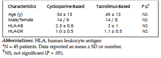
Table 1. Characteristics of Kidney Transplant Recipients With Different Immunosuppressive Therapy*
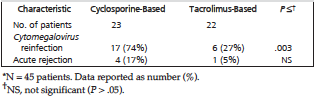
Table 2. Frequency of Cytomegalovirus Infection and Acute Rejection in Kidney Transplant Recipients With Different Immunosuppressive Therapy*
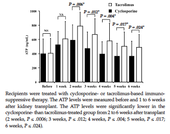
Figure 1. Adenosine Triphosphate (ATP) Levels in Peripheral CD4+ Cells in Kidney Transplant Recipients