Volume: 11 Issue: 6 December 2013
FULL TEXT
Objectives: In living-donor liver transplant recipients with hepatitis C virus infection, outcomes of recurrent hepatitis C virus infection and fibrosis progression are not well documented. We evaluated fibrosis progression, response to pegylated interferon treatment, and long-term graft survival in living-donor liver transplant recipients who had hepatitis C virus infection.
Materials and Methods: In 48 transplant recipients, including 29 recipients who had follow-up liver biopsy ≥ 6 months after transplant, histology and clinical courses were reviewed. Outcomes were evaluated for patients grouped into slow and rapid fibrosis groups. Treatment with pegylated interferon and ribavirin was assessed in 18 patients.
Results: Clinical features were similar between recipients with slow or rapid fibrosis. The time interval from transplant to recurrence of hepatitis C virus infection was significantly shorter in the recipients with rapid fibrosis. Recipients with rapid fibrosis had significantly greater confluent necrosis, acidophil bodies, and fibrosis score than recipients with slow fibrosis. Graft survival rates were similar between patients with slow or rapid fibrosis. Cumulative proportion of long-term graft survival was 60% at 7 years after transplant. Sustained virologic response was noted in 5 of 18 patients (28%) who received pegylated interferon and ribavirin.
Conclusions: In recipients of living-donor liver transplant with early recurrence of hepatitis C have worse fibrosis progression but graft survival was not affected. Therapy with pegylated interferon and ribavirin achieved sustained virologic response only in a small proportion of the patients.
Key words : Cirrhosis, Fibrosis, Interferon, Ribavirin
Introduction
Hepatitis C virus is the most common indication for liver transplant in the United States and Europe. Histologic evidence of hepatitis C virus recurrence occurs in > 90% patients within 5 years of transplant and has a varied clinical course.1,2 Liver transplant recipients who are infected with hepatitis C virus have worse graft outcomes with living-donor than deceased-donor grafts, and this has raised concerns about using living-donor grafts for these recipients.3-7 Furthermore, response to treatments based on pegylated interferon (PEG-IFN) in patients with living-donor liver transplant is unknown.
The primary aim of this study was to describe the clinical, virologic, and histologic outcome in liver transplant recipients infected with hepatitis C virus and who received a transplant from a living donor. The secondary aim was to assess the response to a low-accelerated dosage regimen of PEG-IFN and ribavirin in patients with histologic evidence of recurrent hepatitis C virus infection.
Materials and Methods
Patient population
A retrospective review of medical records was performed for consecutive adult
living-donor liver transplant recipients who had liver transplant for cirrhosis
related to chronic hepatitis C virus infection at Mount Sinai Hospital Medical
Center between January 3, 1999, and February 8, 2005. Inclusion criteria for
liver transplant recipients included (1) hepatitis C virus infection, defined by
serologic and virologic testing available before transplant, and (2) survival >
6 months after transplant, to exclude death from complications of transplant.
Liver transplant recipients were excluded from the study when there was (1)
human immunodeficiency virus or hepatitis B virus coinfection; (2) domino liver
transplant; or (3) concurrent liver disease, such as primary sclerosing
cholangitis, autoimmune hepatitis, or primary biliary cirrhosis that might have
an effect on the clinical course after liver transplant. Patients having liver
transplant for hepatocellular carcinoma were not excluded. The study was
approved by the institutional review board of Mount Sinai Hospital Medical
Center. All protocols conformed to the ethical guidelines of the 1975 Helsinki
Declaration.
Evaluation
Data were abstracted from the clinical records including information about
immunosuppression, indications for liver biopsy, episodes of rejection,
virologic studies, hepatitis C virus genotypes and RNA measurements, treatment
for recurrent hepatitis C virus infection, and the laboratory model for
end-stage liver disease (MELD) score calculated at the time of transplant.8
Biliary complications were noted from laboratory, ultrasonography, and magnetic
resonance imaging, and confirmed by reports from percutaneous intervention,
endoscopy, and surgery.
All causes of graft loss were included, and death was regarded as a graft loss. Long-term graft survival was evaluated at ≥ 6 months after transplant to avoid inclusion of early graft loss from complications associated with the transplant procedure. Patient survival was defined as the time from the initial transplant until death or last known follow-up. Patients lost to follow-up were censored on the date when they were last known to be alive for analysis of patient and graft survival. Death from hepatitis C virus was defined as death resulting from graft failure secondary to either severe recurrent hepatitis C or the cholestatic variant of recurrent hepatitis C virus. Death not related to hepatitis C virus included death from other causes such as infection, perioperative complications, recurrent hepatocellular carcinoma, or biliary complications. A separate analysis was performed, including all recipients who received the low-escalating dosage regimen protocol, to evaluate treatment outcome with PEG-IFN.
Treatment
The following protocol was used for enrolling recipients who had hepatitis C
virus infection. Diagnosis of hepatitis C was based on detectable serum
hepatitis C viral RNA by polymerase chain reaction (COBAS Hepatitis C Virus
Amplification and Detection, version 2.0, Roche Diagnostic, Indianapolis, IN,
USA) (sensitivity, 60 IU/mL) and histologic evidence of hepatitis C virus (liver
biopsy showing fibrosis > 1 on a modified Ishak score9 or lobular
hepatitis). There were 18 transplant recipients who received a low-escalating
dosage regimen of PEG-IFN and ribavirin starting with PEG-IFN-α-2a (90 μg
subcutaneously, once weekly) or PEG-IFN-α-2b (0.75 μg/kg subcutaneously, once
weekly) and ribavirin (7 mg/kg orally, twice daily). The dosage was increased to
a maximum of PEG-IFN-α-2a (180 μg subcutaneously, once weekly) or PEG-IFN-α-2b
(1.5 μg/kg subcutaneously, once weekly) and ribavirin (14 mg/kg orally, twice
daily). The subjects received therapy for 24 weeks (genotypes 2 or 3) or 48
weeks (genotypes 1 or 4). Treatment response was analyzed at 12 weeks (early
virologic response); 24 weeks (genotypes 2 or 3) or 48 weeks (genotypes 1 or 4)
(end of treatment response); and 72 weeks (sustained virologic response).
Patients who had therapy continued beyond the typical duration of therapy (24
weeks for genotypes 2 or 3; 48 weeks for genotypes 1 or 4) and had negative
viral load at the end of this duration were considered to have attained
end-of-treatment response but were not assessed as having sustained virologic
response. Data of patients in the present study who required supportive
treatment with erythropoietin and/or filgrastim were retrieved.
Liver allograft histology
Archived biopsy specimens were obtained for of those reviewed. All histology
assessments were performed by the same liver pathologist (MIF) who was blinded
to patient identity. Indications for liver biopsy included abnormal elevation of
liver enzymes. Slides stained with hematoxylin-eosin were blindly reviewed
without background clinical information and graded for necrosis, inflammation,
rejection, and cholestasis. Slides stained with Masson trichrome were reviewed
to assess fibrosis staging. Necrosis and inflammation were graded with the
hepatitis activity index.9 Scores included assessment of the presence
and degree of periportal or interface hepatitis, confluent necrosis, focal
necrosis, and inflammation and portal inflammation. Acute cellular rejection was
graded with the Banff rejection consensus criteria.10 The presence or
absence of mixed portal inflammation, bile duct damage, and endotheliitis were
assessed and a grade was assigned.
The presence and degree of fibrosis were assessed with modified Ishak and METAVIR staging systems.10 Because protocol liver biopsies were not performed, we used the most recently obtained liver biopsy with highest fibrosis stage and calculated the fibrosis progression rate (FPR) per year based on the fibrosis stage calculated from the Ishak score (range, 1 to 6) divided by the interval time (y) since the living-donor liver transplant. Based on the median fibrosis progression rate (1.27), transplant recipients were defined as having slow fibrosis (fibrosis progression rate ≤ 1.27) or rapid fibrosis (fibrosis progression rate > 1.27).
Statistical analyses
Statistical analyses were performed with SPSS software (SPSS: An IBM Company,
version 17.0, IBM Corporation, Armonk, NY, USA). Descriptive statistics were
calculated, including frequency and percentage for categorical variables and
median or mean and standard deviation for continuous variables. Categorical
variables were compared with the chi-square or the Fisher exact test. Continuous
variables were compared with Mann-Whitney U test. Survival curves were
calculated with Kaplan-Meier method and comparisons were made with log-rank
test, with linear trends for factor levels pooled over strata. Statistical
significance was defined by P ≤ .05.
Results
In 56 living-donor liver transplant recipients who had hepatitis C virus infection, 8 patients were excluded because of graft loss within 6 months of liver transplant unrelated to hepatitis C virus recurrence (4 patients), domino liver transplant (2 patients), concurrent human immunodeficiency virus infection (1 patient), and concurrent primary sclerosing cholangitis (1 patient). The 48 liver transplant recipients included in the study were mostly white men (Table 1). Most transplants were from living-related donors who, on average, were younger than the recipients (Table 1). A substantial frequency of cytomegalovirus (CMV) antibody was noted in donors, but no donors tested positive for antihepatitis B core antigen (Table 1). A substantial frequency of recipients had CMV antibody and/or hepatocellular carcinoma before transplant (Table 1). Half of the recipient group had ≥ 1 rejection episode, mostly mild (Table 1).
Fibrosis progression and graft survival
In the 48 recipients, 126 biopsies were performed during the first year after
liver transplant. There were 19 recipients who were excluded from the analysis
because follow-up biopsy after 6 months was not available or not performed. In
the 29 transplant recipients who had follow-up liver biopsy ≥ 6 months after
transplant, clinical features were similar between patients with slow or rapid
fibrosis (Table 2). Recipients with rapid fibrosis had significantly greater
mean fibrosis progression rate, mean confluent necrosis, acidophil bodies, and
mean fibrosis score than recipients with slow fibrosis (Table 3). The other
histologic parameters examined were similar between recipients who had slow or
rapid fibrosis (Table 3). A trend for earlier recurrence of hepatitis C was
noted in the in the group of patients with rapid fibrosis (5 ± 3 vs 12 ± 12
months; P = .11) (Table 2), but it did not show statistical significance.
Graft survival was similar between recipients who had slow or rapid fibrosis
(data not shown).
Of the 29 recipients, a single patient had spontaneous clearance of his HCV infection and in the rest 28 recipients the median time of histologic recurrence of HCV infection was 6 months (mean ± SD, 8.5 ± 8.7 months). Recipients with early histologic recurrence (defined as ≤ 6 months since their transplant) had a significantly higher fibrosis progression rate as compared to those with later recurrence (2.21 ± 1.41 vs 1.04 ± 0.28, P = .009). We have shown this relation more clearly in the Figure 1 that shows the aggregation of higher rate of FPR toward the left of the X-axes, suggesting earlier the recurrence worse the FPR. However, despite these differences in their FPR, long-term graft survival between those with early versus later recurrence was not affected over a median follow up of 76 months by Kaplan-Meier survival analysis (log rank, P = .99).
Posttransplant biliary complications
Biliary complications were assessed 48 recipients with living-donor liver
transplants with recurrent hepatitis C, 33 of them (68.8%) had duct-duct
anastomosis, and 15 (31.3%) had Roux-en-Y anastomosis. Biliary complications
(anastomotic strictures and bile leaks) were frequent and was noted in 20 of the
48 recipients (41.7%) (Table 4). Anastomotic stricture was noted in 11 who had a
duct-to-duct anastomosis (33.35) compared with 3 who had a Roux-en-Y anastomosis
(20%) (P = .49). Bile leak was noted in 9 recipients (18.8%), 6 with
Roux-en-Y anastomosis (40%) compared to 3 with duct-to-duct anastomosis (9.1%) (P
= NS). Overall long-term graft survival was not affected by bile duct stricture
(P = .42) or biliary complication (P = .47) on Kaplan-Meier
survival analyses (Figures 2A and 2B).
Long-term graft survival
In 48 transplant recipients who were included in the assessment of long-term
graft survival, most recipients were alive at mean follow-up of 5 years (Table
4). The most common causes of death included recurrent hepatocellular carcinoma,
metastatic cancer, and sepsis (Table 4). Cumulative proportion of long-term
graft survival was 60% at 7 years after transplant (Table 4). Overall long-term
graft survival in recipients who had hepatocellular carcinoma before liver
transplant was not significantly different from recipients without HCC.
Therapy with pegylated interferon and ribavirin
In 18 transplant recipients with recurrent hepatitis C virus infection who were
treated with a low-escalating dose regimen of PEG-IFN and ribavirin, all
recipients received immunosuppression with tacrolimus with or without other
immunosuppressive drugs (Table 5). The mean interval from the living-donor liver
transplant to the histologic diagnosis of recurrence was 24 weeks and the mean
interval from transplant to treatment initiation was 83 weeks (Table 5). Few
recipients who had PEG-IFN and ribavirin developed sustained virologic response
(Table 5).
Response to therapy
Hepatitis C virus RNA was not detected at 24 weeks in 9 patients (50%), at 48
weeks in 7 patients (39%), and at 72 weeks in 5 patients (28%). Therefore,
sustained virologic response was noted in 5 patients (28%). In 13 patients who
had paired liver biopsies before treatment and at follow-up, mean fibrosis score
was significantly worse at follow-up than before treatment (Table 6). There were
no differences in mean hepatitis activity index before and after treatment
(Table 6).
In the 5 patients who had sustained virologic response, 4 patients had paired liver biopsies before and after treatment; fibrosis score after treatment was worse in 3 patients and unchanged in 1 patient. In the 3 patients with worse fibrosis score, 2 patients had concomitant bile duct stricture that required multiple biliary interventions, and one patient had moderate steatohepatitis.
In 7 of the 18 patients (39%), therapy was discontinued owing to adverse
events. Therapy was stopped in 3 patients because of sepsis, and 1 of the 3
patients died because of complications of severe sepsis. Acute severe cellular
rejection was noted at
9 months after transplant in 1 patient and treatment was discontinued; this
patient had negative polymerase chain reaction test for hepatitis C virus when
rejection occurred, but hepatitis C virus relapsed at 12 months. In 2 patients,
treatment was discontinued because of severe constitutional symptoms. Metastatic
hepatocellular carcinoma was noted early in 1 patient, and treatment was
discontinued.
Discussion
There is limited information available about the long-term outcome and treatment of recurrent chronic hepatitis C virus in recipients of living-donor liver transplant. A primary finding of the current study was the excellent 1-year graft survival frequency with living-donor liver transplant in patients with cirrhosis associated with hepatitis C virus, albeit with some overall reduction in long-term graft survival. The reduction in graft survival was independent of the fibrosis progression rate, biliary complications, and hepatocellular carcinoma status before transplant.
A recent meta-analysis evaluated long-term survival in deceased-donor and living-liver transplant recipients who had been infected with hepatitis C virus; deceased-donor and living-donor liver transplants were equivalent in patient survival, long-term graft survival, hepatitis C virus recurrence, and acute rejection rate. Graft survival was lower at 1 and 3 years after living-donor liver transplant, but graft survival at 2, 4, and 5 years after transplant were similar between deceased-donor and living-donor transplants.11
The effect of hepatitis C virus recurrence on the graft and the underlying evolution of the histologic parameters, especially the severity and progression of liver fibrosis, have been studied previously.6,12-14 Additional studies have addressed demographic, clinical, perioperative, biochemical, and viral factors associated with the accelerated histologic and clinical course of recurrent hepatitis C virus infection after liver transplant.15,16 Factors that may affect the severity of hepatitis C virus recurrence include donor age,6,12 degree and specific type of immunosuppression,17-19 development and treatment of acute cellular rejection,15,20 development of CMV infection,21 and type of liver transplant (deceased-donor or living-donor transplant).22-24 However, much information about these risk factors for severe recurrence was determined from studies with deceased-donor liver transplant and may not be applicable to living-donor liver transplant recipients. In the present study, we could show a clear relation between earlier histologic recurrence of hepatitis C and worsening fibrosis progression in recipients with living-donor liver transplants; however, long-term graft survival was not affected by this trend. The long-term survival also was not affected by the presence or absence of bile duct problems.
Another finding of the present study was that a low-accelerating dosage regimen of combination therapy yielded a sustained virologic response rate similar to that reported in deceased-donor liver transplant patients. A previous study with an early intervention strategy reported sustained virologic response rate 34.7%, but eligibility for that study was low (56%).25 In the present study, eligibility for PEG-IFN and ribavirin also was low (50%). Combination therapy in liver transplant patients may be associated with frequent and severe adverse events, including the frequent need for erythropoietin and/or filgrastim.26-29 All patients in the present study required supportive treatment with erythropoietin and/or filgrastim. The frequency of treatment discontinuation because of adverse events was 39% in the present study, higher than 16% reported in immunocompetent subjects with significant severity.30 The role of combination therapy with PEG-IFN and protease inhibitors is not established; despite preliminary reports of benefit, wider application has been limited by toxicity, and safer regimens that are not based on interferon are being evaluated.31,32
In the present study, patients with paired biopsies before and after transplant had no improvement in the hepatitis activity index and had deterioration of mean fibrosis score (Table 6). Previous studies with PEG-IFN therapy have reported improvement or no improvement in fibrosis score in recipients of deceased-donor liver transplant.33-35 In the present study, worsening fibrosis may have occurred because of multiple underlying causes such as bile duct stricture and steatohepatitis after transplant.
The limitations of the current study are those limitations inherent to any retrospective, single-center study. Donor selection criteria and recipient selection criteria are center-specific and introduce bias that may limit applicability of these results to other centers. On the other hand, single-center analyses do provide some clarity to certain variables by permitting observations with consistent donor and recipient selection, operative experience, and postoperative management, or by identifying and testing variables not available in registries. Additionally, small samples, particularly in subgroup analyses, have limited statistical power but permit meaningful observations that can be used to develop future areas of investigation. Knowledge gained from results of such retrospective studies on treatment of hepatitis C in living-donor liver transplant recipients with hepatitis C can be used for future randomized, multicenter trials. Additionally, our knowledge on fibrosis progression in recipients with recurrent hepatitis C is still evolving as opposed to the more commonly performed deceased-donor liver transplants especially in the Western world. More such series of center-specific studies, and subsequent meta-analyses, may afford the best alternative to building the knowledge that will ultimately help us define the natural history of hepatitis C more definitively in recipients with living-donor liver transplant recipients.
In conclusion, long-term graft survival in living-donor liver transplant recipients who were infected with hepatitis C virus was less than previously reported graft survival after deceased-donor liver transplant. However, lack of direct comparison with recipients of deceased-donor liver transplant precludes a definitive conclusion. Rapid fibrosis may occur in subjects with early recurrence of hepatitis C virus infection, but long-term graft survival was not affected. Biliary complications, if well treated, do not affect long-term graft survival. A low-accelerating dose regimen of PEG-IFN and ribavirin is associated with a low frequency of sustained virologic response, similar to that reported with deceased-donor liver transplant recipients.
References:
- Brown RS. Hepatitis C and liver transplantation. Nature.
2005;436(7053):973-978.
CrossRef - PubMed - Charlton M. Liver biopsy, viral kinetics, and the impact of viremia on
severity of hepatitis C virus recurrence. Liver Transpl. 2003;9(11):S58–S62.
CrossRef - PubMed - Forman LM, Lewis JD, Berlin JA, Feldman HI, Lucey MR. The association
between hepatitis C infection and survival after orthotopic liver
transplantation. Gastroenterology. 2002;122(4):889-896.
CrossRef - PubMed - Prieto M, Berenguer M, Rayón JM, et al. High incidence of allograft
cirrhosis in hepatitis C virus genotype 1b infection following
transplantation: relationship with rejection episodes. Hepatology.
1999;29(1):250-256.
CrossRef - PubMed - Berenguer M, Ferrell L, Watson J, et al. HCV-related fibrosis
progression following liver transplantation: increase in recent years. J
Hepatol. 2000;32(4):673-684.
CrossRef - PubMed - Wali M, Harrison RF, Gow PJ, Mutimer DJ. Advancing donor liver age and
rapid fibrosis progression following transplantation for hepatitis C. Gut.
2002;51(2):248-252.
CrossRef - PubMed /li> - Poynard T, Bedossa P, Opolon P. Natural history of liver fibrosis
progression in patients with chronic hepatitis C. The OBSVIRC, METAVIR,
CLINIVIR, and DOSVIRC groups. Lancet. 1997;349(9055):825-832.
CrossRef - PubMed - Wiesner RH, Sorrell M, Villamil F; International Liver Transplantation
Society Expert Panel. Report of the first International Liver
Transplantation Society expert panel consensus conference on liver
transplantation and hepatitis C. Liver Transpl. 2003;9(11):S1-S9.
CrossRef - PubMed - Ishak K, Baptista A, Bianchi L, et al. Histological grading and staging
of chronic hepatitis. J Hepatol. 1995;22(6):696-699.
CrossRef - PubMed - Demetris A, Adams D, Bellamy C, et al. Update of the International Banff
Schema for Liver Allograft Rejection: working recommendations for the
histopathologic staging and reporting of chronic rejection. An International
Panel. Hepatology. 2000;31(3):792-799.
CrossRef - PubMed - Hu A, Liang W, Zheng Z, Guo Z, He X. Living donor vs. deceased donor
liver transplantation for patients with hepatitis C virus-related diseases.
J Hepatol. 2012;57(6):1228-1243. doi: 10.1016/j.jhep.2012.07.015.
CrossRef - PubMed - Berenguer M, Prieto M, San Juan F, et al. Contribution of donor age to
the recent decrease in patient survival among HCV-infected liver transplant
recipients. Hepatology. 2002;36(1):202-210. Erratum in Hepatology.
2003;37(2):489.
CrossRef - PubMed - Neumann UP, Berg T, Bahra M, et al. Fibrosis progression after liver
transplantation in patients with recurrent hepatitis C. J Hepatol.
2004;41(5):830-836.
CrossRef - PubMed - Yilmaz N, Shiffman ML, Stravitz RT, et al. A prospective evaluation of
fibrosis progression in patients with recurrent hepatitis C virus following
liver transplantation. Liver Transpl. 2007;13(7):975-983.
CrossRef - PubMed - Sreekumar R, Gonzalez-Koch A, Maor-Kendler Y, et al. Early
identification of recipients with progressive histologic recurrence of
hepatitis C after liver transplantation. Hepatology. 2000;32(5):1125-1130.
CrossRef - PubMed - Testa G, Crippin JS, Netto GJ, et al. Liver transplantation for
hepatitis C: recurrence and disease progression in 300 patients. Liver
Transpl. 2000;6(5):553-561.
CrossRef - PubMed - Papatheodoridis GV, Davies S, Dhillon AP, et al. The role of different
immunosuppression in the long-term histological outcome of HCV reinfection
after liver transplantation for HCV cirrhosis. Transplantation.
2001;72(3):412-418.
CrossRef - PubMed - Samonakis DN, Triantos CK, Thalheimer U, et al. Immunosuppression and
donor age with respect to severity of HCV recurrence after liver
transplantation. Liver Transpl. 2005;11(4):386-395.
CrossRef - PubMed - Berenguer M, Royuela A, Zamora J. Immunosuppression with calcineurin
inhibitors with respect to the outcome of HCV recurrence after liver
transplantation: results of a meta-analysis. Liver Transpl.
2007;13(1):21-29.
CrossRef - PubMed - Samonakis DN, Mela M, Quaglia A, et al. Rejection rates in a randomised
trial of tacrolimus monotherapy versus triple therapy in liver transplant
recipients with hepatitis C virus cirrhosis. Transpl Infect Dis.
2006;8(1):3-12.
CrossRef - PubMed - Burak KW, Kremers WK, Batts KP, et al. Impact of cytomegalovirus
infection, year of transplantation, and donor age on outcomes after liver
transplantation for hepatitis C. Liver Transpl. 2002;8(4):362-369.
CrossRef - PubMed - Shiffman ML, Stravitz RT, Contos MJ, et al. Histologic recurrence of
chronic hepatitis C virus in patients after living donor and deceased donor
liver transplantation. Liver Transpl. 2004;10(10):1248-1255.
CrossRef - PubMed - Schmeding M, Neumann UP, Puhl G, Bahra M, Neuhaus R, Neuhaus P.
Hepatitis C recurrence and fibrosis progression are not increased after
living donor liver transplantation: a single-center study of 289 patients.
Liver Transpl. 2007;13(5):687-692.
CrossRef - PubMed - Selzner N, Girgrah N, Lilly L, et al. The difference in the fibrosis
progression of recurrent hepatitis C after live donor liver transplantation
versus deceased donor liver transplantation is attributable to the
difference in donor age. Liver Transpl. 2008;14(12):1778-1786. doi:
10.1002/lt.21598.
CrossRef - PubMed - Castells L, Vargas V, Allende H, et al. Combined treatment with
pegylated interferon (alpha-2b) and ribavirin in the acute phase of
hepatitis C virus recurrence after liver transplantation. J Hepatol.
2005;43(1):53-59.
CrossRef - PubMed - Toniutto P, Fabris C, Fumo E, et al. Pegylated versus standard
interferon-alpha in antiviral regimens for post-transplant recurrent
hepatitis C: Comparison of tolerability and efficacy. J Gastroenterol
Hepatol. 2005;20(4):577-582.
CrossRef - PubMed - Oton E, Barcena R, Garcia-Garzon S, et al. Pegylated interferon and
ribavirin for the recurrence of chronic hepatitis C genotype 1 in transplant
patients. Transplant Proc. 2005;37(9):3963-3964.
CrossRef - PubMed - Ross AS, Bhan AK, Pascual M, Thiim M, Benedict Cosimi A, Chung RT.
Pegylated interferon alpha-2b plus ribavirin in the treatment of post-liver
transplant recurrent hepatitis C. Clin Transplant. 2004;18(2):166-173.
CrossRef - PubMed - Saab S, Kalmaz D, Gajjar NA, et al. Outcomes of acute rejection after
interferon therapy in liver transplant recipients. Liver Transpl.
2004;10(7):859-867.
CrossRef - PubMed - Bárcena R, Gil-Grande L, Moreno J, et al. Partial splenic embolization
for the treatment of hypersplenism in liver transplanted patients with
hepatitis C virus recurrence before peg-interferon plus ribavirin.
Transplantation. 2005;79(11):1634-1635.
CrossRef - PubMed - Charlton M. Telaprevir, boceprevir, cytochrome P450 and
immunosuppressive agents--a potentially lethal cocktail. Hepatology.
2011;54(1):3-5. doi: 10.1002/hep.24470.
CrossRef - PubMed - Werner CR, Egetemeyr DP, Lauer UM, et al. Telaprevir-based triple
therapy in liver transplant patients with hepatitis C virus: a 12-week pilot
study providing safety and efficacy data. Liver Transpl.
2012;18(12):1464-1470. doi: 10.1002/lt.23542.
CrossRef - PubMed - Bizollon T, Pradat P, Mabrut JY, et al. Benefit of sustained virological
response to combination therapy on graft survival of liver transplanted
patients with recurrent chronic hepatitis C. Am J Transplant.
2005;5(8):1909-1913.
CrossRef - PubMed - Firpi RJ, Abdelmalek MF, Soldevila-Pico C, et al. Combination of
interferon alfa-2b and ribavirin in liver transplant recipients with
histological recurrent hepatitis C. Liver Transpl. 2002;8(11):1000-1006.
CrossRef - PubMed - Bizollon T, Ahmed SN, Radenne S, et al. Long term histological
improvement and clearance of intrahepatic hepatitis C virus RNA following
sustained response to interferon-ribavirin combination therapy in liver
transplanted patients with hepatitis C virus recurrence. Gut.
2003;52(2):283-287.
CrossRef - PubMed

Volume : 11
Issue : 6
Pages : 522 - 529
DOI : 10.6002/ect.2013.0054
From the 1Division of Liver Diseases, Department of Medicine,
Mount Sinai Medical Center, New York, NY; the 2Division of Surgery,
Methodist University Hospital Transplant Institute, University of Tennessee
Health Sciences Center, Memphis, TN; the 3Department of Pathology,
Mount Sinai Medical Center, New York, NY; Mount Sinai Medical Center, New York,
NY; and the 4Recanati-Miller Transplantation Institute, Mount Sinai
Medical Center, New York, NY, USA
Acknowledgements: The authors declare that no funding was received and
they have no conflicts of interest to declare. Sanjaya K. Satapathy performed
the study concept development and design, data collection, data analysis,
statistical analysis, article drafting, critical revision, and approval of the
article. Maria Isabel Fiel performed the study concept development and design,
article drafting, critical revision, and approval of the article. Jason M.
Vanatta performed article drafting, critical revision, and approval of the
article. Juan Del Rio Martin and Thomas D. Schiano performed the study concept
develop him ment and design, article drafting, critical revision, and approval
of the article.
Corresponding author: Sanjaya K. Satapathy, University of Tennessee
Health Sciences Center, Methodist University Hospital Transplant Institute, 1211
Union Avenue, Suite #340, Memphis, TN 38104 USA
Phone: +1 901 516 8954
Fax: +1 901 516 8993
E-mail: ssatapat@uthsc.edu
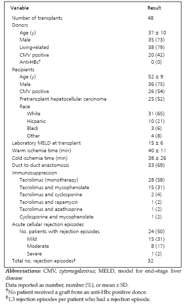
Table 1. Characteristics of Living-Donor Liver Transplant Donors and Recipients
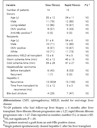
Table 2. Relation Between Fibrosis Formation and Characteristics of Patients Before and After Liver Transplant*
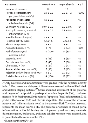
Table 3. Relation Between Fibrosis Formation and Histologic Characteristics Assessed at the Stage of Maximum Fibrosis
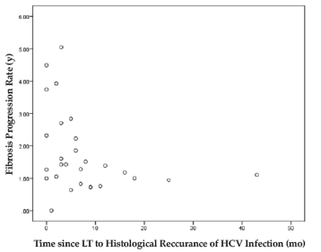
Figure 1. Relation Between Fibrosis Progression Rate and Time Since Liver Transplant of Histologic Recurrence of Hepatitis C Infection in the Allograft.
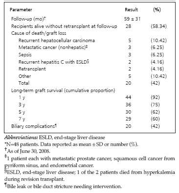
Table 4. Follow-Up in Recipients of Liver Transplant Who Had Hepatitis C Virus Infection*
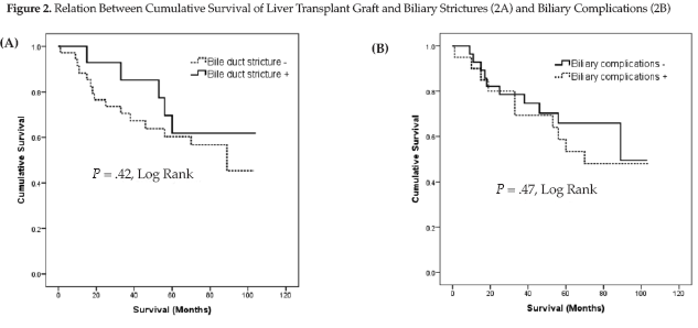
Figure 2. Relation Between Cumulative Survival of Liver Transplant Graft and Biliary Strictures (2A) and Biliary Complications (2B)
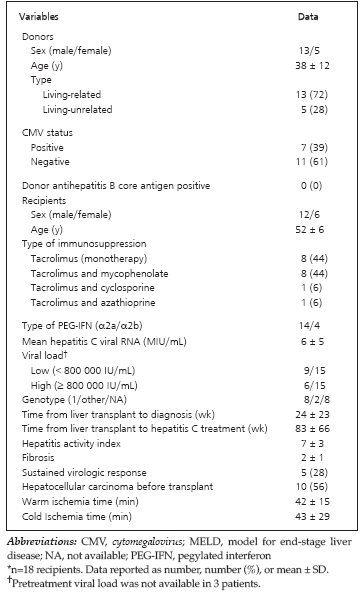
Table 5. Clinical Characteristics of Liver Transplant Recipients With Recurrent Hepatitis C Virus Infection Who Were Treated With Pegylated Interferon and Ribavirin*
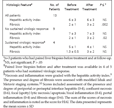
Table 6. Histologic Changes After Treatment of HCV-Positive Living Donor Liver Transplant Recipients With Pegylated Interferon and Ribavirin*