Volume: 11 Issue: 6 December 2013
FULL TEXT
Objectives: We sought to develop a B-cell in vitro culture system and test B cells isolated from sensitized kidney recipients and healthy controls, and assess the effectiveness of proteasome inhibitors and mycophenolic acid on antibody secretion and cell apoptosis.
Materials and Methods: CD19+ B cells and CD19+CD27+ memory B-cell subsets were detected from peripheral blood mononuclear cells obtained from 6 MICA-sensitized kidney recipients and 6 healthy controls. Peripheral blood B cells were isolated and cultured with CpG2006, PMA, MICA antigen, B-cell activating factor, CD40 ligand (CD40L), human recombinant IL-2 (rhuIL-2), rhuIL-10, rhuIL-4, and rhuIL-21. After culturing for 7 days, we tested several variables of B-cell activity including differentiation, apoptosis, and IgM production. We also assessed the effects of 2 immunosuppressive drugs (mycophenolic acid and bortezomib) on antibody secretion and cellular apoptosis.
Results: Kidney recipients had a lower ratio of CD19+ B cells in peripheral blood mononuclear cells than did healthy controls. However, the percentage of CD19+CD27+ B cells was higher in kidney recipients than in healthy controls. In the cell stimulation culture system, the ratio of CD19+ B cells, CD19+CD27+ B cells, and CD19+CD138+ B cells increased after culturing for 7 days compared with unstimulated controls. In addition, the percentage of apoptotic B cells decreased, and antibody production increased in sensitized transplant patients and healthy controls. Treatment with bortezomib or mycophenolic acid induced B-cell apoptosis and inhibited secretion of antibodies.
Conclusions: This study describes establishment of a B-cell in vitro culture system, showing that B cells may be stimulated to secrete antibodies. The study also provides an assay for studying B cells in vitro. This study provides information suggesting that bortezomib and mycophenolic acid can inhibit B-cell antibody secretion.
Key words : B cells, Humoral rejection, Kidney transplant, Immunoglobulin production
Introduction
Antibodies against major histocompatibility complex class I chain-related gene A (MICA) antigen and human leukocyte antigen after transplant remain barriers to long-term survival after kidney transplants.1-3 Multiple protocols have been developed to facilitate successful transplant of sensitized transplant candidates including treatment of antithymocyte globulin, interleukin-2 receptor antagonists, intravenous immunoglobulin, and anti-CD20 antibody injection.4 However, the effectiveness of these approaches to improve long-term survival is unclear owing to the high incidence of acute humoral rejection during the early stages after transplant,5 as well as antibody-mediated injury owing to formation of transplant glomerulopathy.6,7 The most widely used desensitization protocols currently are not sufficient to prevent antibody production by antibody-secreting B cells.8,9
Bortezomib is a proteasome inhibitor first approved by the United States Food and Drug Administration in 2003 to treat multiple myeloma. The drug inhibits tumor proliferation by blocking proteasome activity and inducing apoptosis. Previously, it had been evaluated in several trials against various hematologic malignancies, including multiple myeloma. Studies also have demonstrated an inhibitory effect of bortezomib on the formation of antibodies in treating acute graft-versus-host disease, including kidney transplant.10-12 Perry and associates found that bortezomib can induce apoptosis of normal human plasma cells, thereby preventing alloantibody production.13 Flechner and associates also found that bortezomib exhibits an antihumoral rejection effect in antibody-mediated rejection.14 These studies provide information on the reduction of antibody production; however, it is currently unclear whether bortezomib has a direct effect on B cells. Therefore, we evaluated the potential biological effects of bortezomib together with the commonly used antihumoral rejection immunosuppressive drug, mycophenolic acid (MPA), directly on purified B cells.
Materials and Methods
Ethical considerations
This study was approved by the Research Subjects Review Board at First
People’s Hospital. Written, informed consent was obtained from all participants.
The study was performed in accordance with the ethical guidelines of the 1975
Helsinki Declaration. Research data were coded so that the subjects could not be
identified, directly or through linked identifiers, in compliance with the
Department of Health and Human Services Regulations for the Protection of Human
Subjects.
Blood samples
Major histocompatibility complex class I chain-related gene A is an
endothelial cell surface antigen. It has been reported that presensitization of
kidney transplant recipients against MICA antigens is associated with an
increased frequency of graft loss, and may contribute to allograft loss among
recipients who are well-matched for human leukocyte antigen.1 Terasaki and
associates also provided evidence showing that similar to human leukocyte
antigen antibodies, MICA antibodies are associated with graft failures.3
However, there has not been an optimal in vitro system for stimulating B cells
to secrete antibodies.
In this study, we assessed the serum from 68 long-term survival kidney recipients using Luminex technology (One Lambda, Inc, Canoga Park, CA, USA), and found 11 subjects who were MICA positive. Six MICA-sensitized kidney transplant recipients agreed to join the study, and 6 health volunteer subjects not receiving a transplant were used as controls (anti-MICA antibodies were present in the recipients’ serum, as detected by Luminex single antigen beads based upon the manufacturer’s instructions). The method used is described in our previous study.15 All recipients survived for at least 10 years after the transplant and were taking immunosuppressive agents (Table 1) at the time of blood sample collection. Approximately 20 to 50 mL of heparin-treated (50 U/mL) peripheral blood was obtained from each individual.
Antibodies and cytokines
Flow cytometry analysis was performed according to standard protocols.
Antihuman CD19-APC, antihuman CD27-PE, and antihuman CD138-PerCP-Cy5 antibodies
were obtained from BD Biosciences (BD Pharmingen, San Jose, CA, USA). The
cytokines used for cell stimulation were human recombinant IL-2 (rhuIL-2;
PeproTech, Rocky Hill, NJ, USA), rhuIL-10 (PeproTech), rhuIL-4 (PeproTech),
rhuIL-21 (BD Biosciences), CD40L (Alexis Biochemicals, San Diego, CA, USA), BAFF
(ProSpec-Tany TechnoGene Ltd., Rehovot, Israel), and MICA-specific antigen (One
Lambda, Inc).
Isolation of human B lymphocytes
Peripheral blood mononuclear cells were isolated by Ficoll-Paque density
gradient centrifugation. B lymphocytes were enriched by negative selection using
magnetic beads (B Cell Isolation Kit, Miltenyi Biotec, Cologne, Germany)
according to the manufacturer’s instructions. Briefly, cells were incubated with
the biotin-labeled antibody mixture for 10 minutes and then mixed with magnetic
microbeads coupled with antibodies against biotin for an additional 15 minutes.
The labeled cells were removed by passing them through a magnetic cell sorter
column type LS (Miltenyi Biotec) that resulted in an enriched population of B
cells. The purity of the isolated B cells was analyzed by flow cytometry using
an APC-conjugated monoclonal antibody against CD19 (eBioscience, Inc., San
Diego, CA, USA), and the purity was found to be greater than 95%.
B-cell culture and stimulation
Cells were cultured in RPMI 1640 medium (Invitrogen Corporation, Carlsbad,
CA, USA) containing 10% heat-inactivated fetal bovine serum (Sigma-Aldrich Corp.
St. Louis, MO, USA) and 100 U/mL penicillin/streptomycin (Invitrogen) at a
density of 1 × 10⁵ cells/200 μL in round-bottomed 96-well plates
(Costar 3799, Corning Costar, Sigma-Aldrich Corp. St. Louis, MO, USA). For
B-cell stimulation, we added CpG 2006 (10 ng/mL; synthesized in Sangon Biotech
[Shanghai Sangon Biotechnology, Shanghai, China]), MICA antigen (5 μL/well; One
Lambda), PMA (10 ng/mL; Sigma-Aldrich), rhuIL-2, rhuIL-10 (50 ng/mL), rhuIL-4
(50 ng/mL), rhuIL-21 (10 ng/mL), CD40L (500 ng/mL), and BAFF (75 ng/mL) to the
culture system, and the cells were cultured for 7 days. Different dosages of
bortezomib (initial concentration: 200 ng/mL; Millennium) and MPA (initial
concentration: 50 ng/mL; Sigma-Aldrich) were then added to the cell culture
system to determine the direct inhibitory effects on B cells.
Apoptosis assays
After 7 days of culture, the cells were centrifuged at 1500 rpm for 5
minutes at 4°C and then the supernatant was collected. Cell pellets were
resuspended in binding buffer for apoptosis assays. Cells were stained according
to the instruction of the FITC-labeled Annexin V Apoptosis Detection Kit I (BD
Pharmingen). Briefly, 5 μL Annexin V and 5 μL PI in working solution were added
in cell suspension and incubated for 15 minutes at room temperature. The cells
then were washed and resuspended in binding buffer for flow cytometry analysis.
Results were analyzed using FloJo software (Tree Star, Inc. Ashland, OR, USA).
ELISA assay
Antibody concentrations were detected using an ELISA according to the
manufacturer’s protocol. Briefly, supernatants recovered from cell stimulation
experiments were tested for IgM levels with a standard sandwich ELISA. Plates
were coated overnight with a goat anti-IgM (Southern Biotech, Birmingham, AL,
USA) diluted in 10 mM Tris pH 9.0 and then blocked with 2% bovine serum albumin
(BSA; Sigma-Aldrich) and 0.025% Tween-20 (Sigma-Aldrich) in phosphate-buffered
saline (PBS-T). Fifty microliters of supernatants or standard human serum
(Southern Biotech) was serially diluted for 60 minutes at 37°C. After washing
with PBS-T, biotin-labeled goat anti-IgM (Southern Biotech) diluted in 1%
BSA/PBS-T was incubated for 60 minutes at 37°C. After extensive washing,
streptavidin horseradish peroxidase (Pierce Protein Biology Products [Thermo
Fisher Scientific Inc., Rockford, IL, USA]) diluted in PBS-T that contained 1%
BSA was added and incubated for 60 minutes at 37°C. TMB High Sensitivity
Substrate solution was added, and the reaction was terminated by the addition of
2N H2SO4 (Sigma-Aldrich) and measured at a wavelength of OD450 nm in an ELISA
reader (Bio-Rad Laboratories B.V., Veenendaal, The Netherlands). Data were
analyzed using Microplate Manager software version 4 (Bio-Rad).
Statistical analyses
Results were analyzed using GraphPad software (La Jolla, CA, USA). A t test
and 1-way analysis of variance were used to analyze the data. A P value < .05
was considered statistically significant.
Results
The constituent ratio of the B-cell subpopulation in peripheral blood in
kidney recipients and healthy controls
Peripheral blood mononuclear cells from kidney recipients had a lower ratio
of CD19+ B cells compared with those from healthy controls.
Additionally, kidney recipients had a higher percentage of CD19+CD27+
B cells compared with healthy controls. The average percentage of CD19+
B cells in PBMC samples was significantly lower in MICA-sensitized transplant
patients than it was in healthy controls (3.58 ± 0.80% vs 8.53 ± 1.04%; P
< .01). In CD19+ B cells, the percentage of memory B cells (CD19+CD27+)
in long-term survival sensitized kidney recipients and healthy controls was
49.55% ± 8.42% and 26.33% ± 5.49% (Figure 1; P < .01).
The in vitro B-cell culture system: effects of stimuli on purified B-cell
differentiation
Peripheral blood B cells from healthy controls were used to establish an in
vitro B-cell culture system. Different B-cell stimuli (toll-like receptor-9
ligand ODN-2006 CpG, MICA antigen, PMA, rhuIL-2, rhuIL-10, rhuIL-4, rhuIL-21,
CD40L, and BAFF) were add to the culture system to stimulate the B cells. One
major limitation for the in vitro differentiation of B cells is the high rate of
spontaneous cell death that occurs as the cells acquire a fully differentiated
phenotype.16 In addition, the proliferation of B cells could be
associated with cell death, especially in conditions where the stimulation
signal is not optimal. Therefore, to further address these issues, we assessed
the percentage of different B-cell phenotypes. After 7 days of cell stimulation,
the ratio of CD19+ B cells, CD19+CD27+ B cells,
and CD19+CD138+ B cells increased compared with controls
(Figure 2).
Effects of stimuli on purified B-cell apoptosis and immunoglobulin
production
The effects of the stimuli on apoptosis and immunoglobulin production of B
cells purified from healthy controls and transplant patients were next evaluated
by flow cytometry and ELISA. After stimulation the B cells from healthy controls
and transplant patients had a low percentage of apoptosis; however, the average
percentage of apoptosis of B cells from transplant patients was significantly
higher than that of cells from healthy controls (Figure 3; P < .01). In
addition, IgM is the first antibody produced by B cells after antigen
stimulation, and we found that production of IgM was higher in stimulated B
cells from transplant patients than from healthy controls (Figure 4).
Bortezomib and mycophenolic acid induce B-cell apoptosis
One possible explanation for the inhibition of antibody production is the
induction of apoptosis. To test this hypothesis, B cells from healthy controls
were cultured for 7 days with stimulus in the presence or absence of increasing
concentrations of MPA and bortezomib (50, 100, and 150 ng/mL; and 200, 500, 1000
ng/mL). B-cell apoptosis was measured by Annexin-V-FITC and PI staining followed
by flow cytometry analysis. We found that the percentage of B-cell apoptosis
increased in a drug dose-dependent manner (Figure 5). We then fixed the dose of
MPA (100 ng/mL) and bortezomib (1000 ng/mL) and treated B cells from both
transplant patients and healthy controls. We found that the percentage of viable
cells decreased in both groups, and that the percentage of prophase and terminal
apoptosis cells in the 2 groups increased compared with untreated controls
(Figure 6; P < .01). Therefore, MPA and bortezomib induced a
significantly higher percentage of apoptosis in B cells from both patient groups
compared with controls.
Effects of immunosuppression on immunoglobulin production
Mycophenolic acid (100 ng/mL) and bortezomib (1000 ng/mL) then were applied
to the culture system to determine the effects on antibody secretion. After 7
days’ cell culture, bortezomib and MPA decreased production of IgM in the in
vitro stimulation environment (Figure 7).
Discussion
In this study, peripheral blood B cells were purified from 6 MICA-sensitized patients’ and 6 healthy controls’ peripheral blood and were assessed by flow cytometry. We found that the sensitized patients had fewer CD19+ B cells in the total PBMC population than did the healthy controls. Therefore, immunosuppressive agents that are administered to transplant patients seem to alter the constitution of B-cell subpopulations. In our study, all the transplant patients were taking cyclosporine, mycophenolate mofetil, and prednisone, and therefore, B-cell proliferation and cell differentiation might be influenced by the immunosuppressive agents, which may be a benefit for long-term survival. However, more patient data and control groups are needed to confirm this hypothesis. In the total population of B cells, the percentage of CD19+CD27+ B cells was higher in transplant patients than in healthy controls. The CD19+CD27+ B cells are the subset of memory B cells, and therefore, these results suggest that B cells from sensitized patients have the potential ability to secrete antibodies.
In our in vitro B-cell culture system, cell differentiation and function were regulated by multiple pathway stimuli. The stimuli we used in this study have separate and unique functions for B-cell activation. Toll-like receptor-9 ligand ODN-2006 CpG binds with Toll-like receptor-9 to trigger the TLR-9–My88-NF-kB pathway to promote B-cell proliferation and differentiation.17 Interleukin-10 has been shown to increase Toll-like receptor-9 expression18 and stimulate immunoglobulin production19-21 in B cells. Although IL-2 is considered to be a key factor for T-cell proliferation after activation, it also has been shown to sustain B-cell proliferation. Interleukin-21 is mainly produced by follicular helper T cells and is required for plasma cell differentiation and immunoglobulin production in germinal centers.22-24 Therefore, the addition of IL-21 in the culture system most likely promotes the activation of B cells. CD40 signaling is an important factor for the maturation and proliferation of B cells.25,26 PMA and MICA antigen also can activate the NF-k B pathway for cell proliferation and differentiation. In this study, we stimulated 2 signaling pathways using different combinations of stimuli. CpG2006 combined with IL-2, IL-6, IL-10, and BAFF promote B-cell proliferation,27,28 while CD40L, together with IL-4 and IL-21, induces the differentiation of B cells into antibody secreting cells. Our results show that the levels of IgM in the supernatant of stimulated B cells and the number of viable cells dramatically increased upon stimulation.
In the in vitro B-cell culture system, the average percentage of apoptosis of B cells from healthy controls and transplant patients was 9.73% ± 1.46% and 21.53% ± 3.31%. Because transplant patients are under immunosuppressive regimens, the B-cell population is likely to be compromised, which may alter the apoptosis of B cells under stimulation compared with healthy controls. In the present study, different dosages of MPA and bortezomib were added to the culture system, which resulted in a dose-dependent increase in the average percentage of B cells undergoing apoptosis. At a clinically relevant fixed dose (MPA, 100 ng/mL; and bortezomib, 1000 ng/mL), bortezomib and MPA effectively induced apoptosis of B cells. In addition, both immunosuppressive agents decreased the production of IgM in the stimulation environment, which is consistent with previous studies.29,30 Other studies also have shown that a decrease in donor-specific antibodies strongly correlates with successful treatment of antibody-mediated rejection.8,31,32 Our results suggest that bortezomib and MPA can inhibit B-cell antibody secretion.
Antibodies secreted by antibody-producing B cells in vivo are driven by multiple interactions, including BCR-antigen interactions, costimulatory stimulations, and cytokines. To date, an optimal in vitro system for stimulating B cells to secrete antibodies has not been established. Therefore, optimizing such a system using stimuli to trigger cultured B cells would be a valuable tool in this research setting. In this study, we successfully stimulated B cells with a combination of antigens and cytokine cocktails using purified B cells from MICA-sensitized kidney recipients and healthy controls.
Conclusions
In this study, peripheral blood B cells were isolated from sensitized kidney recipients and found to display a subset bias. In our established B-cell in vitro culture system, stimulation promoted antibodies secretion in vitro. We also found that bortezomib exhibited inhibitory effects on B-cell survival and function, which may provide an experimental basis for future studies.
References:
- Zou Y, Stastny P, Süsal C, Döhler B, Opelz G. Antibodies against MICA
antigens and kidney-transplant rejection. N Engl J Med.
2007;357(13):1293-1300.
CrossRef - PubMed - Zou Y, Yang X, Jiang X, et al. High levels of soluble Major
Histocompatibility Complex class I related chain A (MICA) are associated
with biliary cast syndrome after liver transplantation. Transpl Immunol.
2009;21(4):210-214.doi: 10.1016/j.trim.2009.06.003.
CrossRef - PubMed - Terasaki PI, Ozawa M, Castro R. Four-year follow-up of a prospective
trial of HLA and MICA antibodies on kidney graft survival. Am J Transplant.
2007;7(2):408-415.
CrossRef - PubMed - Emami S, Huang E, Kuo HT, Kamgar M, Bunnapradist S. Multivariate
analysis of antibody induction therapy and their associated outcomes in live
donor kidney transplantation in the recent era. Clin Transplant.
2012;26(2):351-358. doi:10.1111/j.1399-0012.2011.01517.x.
CrossRef - PubMed - Stegall MD, Gloor J, Winters JL, Moore SB, Degoey S. A comparison of
plasmapheresis versus high-dose IVIG desensitization in renal allograft
recipients with high levels of donor specific alloantibody. Am J Transplant.
2006;6(2):346-351.
CrossRef - PubMed - Gloor JM, Sethi S, Stegall MD, et al. Transplant glomerulopathy:
subclinical incidence and association with alloantibody. Am J Transplant.
2007;7(9):2124-2132.
CrossRef - PubMed - Cosio FG, Gloor JM, Sethi S, Stegall MD. Transplant glomerulopathy. Am J
Transplant. 2008;8(3):492-496. doi: 10.1111/j.1600-6143.2007.02104.x.
CrossRef - PubMed - Lefaucheur C, Nochy D, Andrade J, et al. Comparison of combination
Plasmapheresis/IVIg/anti-CD20 versus high-dose IVIg in the treatment of
antibody-mediated rejection. Am J Transplant. 2009;9(5):1099-1107. doi:
10.1111/j.1600-6143.2009.02591.x.
CrossRef - PubMed - Everly MJ, Terasaki PI. The state of therapy for removal of alloantibody
producing plasma cells in transplantation. Semin Immunol.
2012;24(2):143-147. doi: 10.1016/j.smim.2011.08.014.
CrossRef - PubMed - Nencioni A, Grünebach F, Patrone F, Ballestrero A, Brossart P.
Proteasome inhibitors: antitumor effects and beyond. Leukemia.
2007;21(1):30-36.
CrossRef - PubMed - Liang Y, Mao X, Liu H. Proteasome inhibitor clioquinol as a candidate
drug in prophylaxis and treatment of acute graft-versus-host disease. Med
Hypotheses. 2011;76(3):400-402. doi: 10.1016/j.mehy.2010.11.002.
CrossRef - PubMed - Everly MJ, Terasaki PI, Hopfield J, Trivedi HL, Kaneku H. Protective
immunity remains intact after antibody removal by means of proteasome
inhibition. Transplantation. 2010;90(12):1493-1498. doi:
10.1097/TP.0b013e3181ff87b1.
CrossRef -
PubMed - Perry DK, Burns JM, Pollinger HS, et al. Proteasome inhibition causes
apoptosis of normal human plasma cells preventing alloantibody production.
Am J Transplant. 2009;9(1):201-209. doi: 10.1111/j.1600-6143.2008.02461.x.
CrossRef - PubMed - Flechner SM, Fatica R, Askar M, et al. The role of proteasome inhibition
with bortezomib in the treatment of antibody-mediated rejection after
kidney-only or kidney-combined organ transplantation. Transplantation.
2010;90(12):1486-1492. doi: 10.1097/TP.0b013e3181fdd9b0.
CrossRef - PubMed - Li Z, Luo M, Qiu J, et al. Detection of antibodies against major
histocompatibility complex class I-related chain A in long-term renal graft
recipients. Exp Clin Transplant. 2012;10(3):239-242.
CrossRef - PubMed - Cassese G, Arce S, Hauser AE, et al. Plasma cell survival is mediated by
synergistic effects of cytokines and adhesion-dependent signals. J Immunol.
2003;171(4):1684-1690.
PubMed - Huggins J, Pellegrin T, Felgar RE, et al. CpG DNA activation and
plasma-cell differentiation of CD27- naive human B cells. Blood.
2007;109(4):1611-1619.
CrossRef - PubMed - He B, Qiao X, Cerutti A. CpG DNA induces IgG class switch DNA
recombination by activating human B cells through an innate pathway that
requires TLR9 and cooperates with IL-10. J Immunol. 2004;173(7):4479-4491.
PubMed - Agematsu K, Nagumo H, Oguchi Y, et al. Generation of plasma cells from
peripheral blood memory B cells: synergistic effect of interleukin-10 and
CD27/CD70 interaction. Blood. 1998;91(1):173-180.
PubMed - Nagumo H, Agematsu K. Synergistic augmentative effect of interleukin-10
and CD27/CD70 interactions on B-cell immunoglobulin synthesis. Immunology.
1998;94(3):388-394.
CrossRef - PubMed - Gantner F, Hermann P, Nakashima K, Matsukawa S, Sakai K, Bacon KB.
CD40-dependent and -independent activation of human tonsil B cells by CpG
oligodeoxynucleotides. Eur J Immunol. 2003;33(6):1576-1585.
CrossRef - PubMed - Dienz O, Eaton SM, Bond JP, et al. The induction of antibody production
by IL-6 is indirectly mediated by IL-21 produced by CD4+ T cells. J Exp Med.
2009;206(1):69-78. doi: 10.1084/jem.20081571.
CrossRef - PubMed - Matsui W, Huff CA, Vala M, Barber J, Smith BD, Jones RJ. Anti-tumour
activity of interferon-alpha in multiple myeloma: role of interleukin 6 and
tumor cell differentiation. Br J Haematol. 2003;121(2):251-258.
CrossRef - PubMed - Good KL, Bryant VL, Tangye SG. Kinetics of human B cell behavior and
amplification of proliferative responses following stimulation with IL-21. J
Immunol. 2006;177(8):5236-5247.
PubMed - Xu H, Yan J, Huang Y, et al. Costimulatory blockade of CD154-CD40 in
combination with T-cell lymphodepletion results in prevention of allogeneic
sensitization. Blood. 2008;111(6):3266-3275.
CrossRef - PubMed - Fecteau JF, Roy A, Néron S. Peripheral blood CD27+ IgG+ B cells rapidly
proliferate and differentiate into immunoglobulin-secreting cells after
exposure to low CD154 interaction. Immunology. 2009;128(suppl 1):e353-365.
doi: 10.1111/j.1365-2567.2008.02976.x.
CrossRef - PubMed - Rauch M, Tussiwand R, Bosco N, Rolink AG. Crucial role for BAFF-BAFF-R
signaling in the survival and maintenance of mature B cells. PLoS One.
2009;4(5):e5456. doi: 10.1371/journal.pone.0005456.
CrossRef - PubMed - Sarantopoulos S, Stevenson KE, Kim HT, et al. Altered B-cell homeostasis
and excess BAFF in human chronic graft-versus-host disease. Blood.
2009;113(16):3865-3874. doi: 10.1182/blood-2008-09-177840.
CrossRef - PubMed - Sharma KG, Radha R, Pao A, et al. Mycophenolic acid and intravenous
immunoglobulin exert an additive effect on cell proliferation and apoptosis
in the mixed lymphocyte reaction. Transpl Immunol. 2010;23(3):117-120. doi:
10.1016/j.trim.2010.04.009.
CrossRef - PubMed - Lee CF, Eldeen FZ, Chan KM, et al. Bortezomib is effective to treat
acute humoral rejection after liver transplantation. Transplant Proc.
2012;44(2):529-531. doi: 10.1016/j.transproceed.2012.01.051.
CrossRef - PubMed - Everly MJ, Everly JJ, Arend LJ, et al. Reducing de novo donor-specific
antibody levels during acute rejection diminishes renal allograft loss. Am J
Transplant. 2009;9(5):1063-1071. doi: 10.1111/j.1600-6143.2009.02577.x.
CrossRef - PubMed - Hachem RR, Yusen RD, Meyers BF, et al. Anti-human leukocyte antigen
antibodies and preemptive antibody-directed therapy after lung
transplantation. J Heart Lung Transplant. 2010;29(9):973-980. doi:
10.1016/j.healun.2010.05.006.
CrossRef - PubMed

Volume : 11
Issue : 6
Pages : 482 - 488
DOI : 10.6002/ect.2012.0281
From the 1Department of Urology, First People’s Hospital, School
of Medicine, Shanghai Jiao Tong University; the 2Department of
Urology, Shanghai East Hospital, School of Medicine, Tong Ji University; the
3Shanghai Institute of Immunology, School of Medicine, Shanghai Jiao
Tong University; and the 4Department of Urology, Shanghai No. 10
Hospital, Shanghai, PR China
Acknowledgements: The project was supported by The National Natural
Science Foundation of China (NSFC, 30972997) and doctorial innovation fund of
Shanghai Jiao Tong University. No conflicts of interest exist for any author.
Corresponding author: Jianxin Qiu MD, PhD No.100, Hai Ning Rd, Shanghai
200080, PR China
Phone: +86 21 6324 0090
Fax: +86 21 6324 0825
E-mail: jasonqiu@medmail.com.cn.
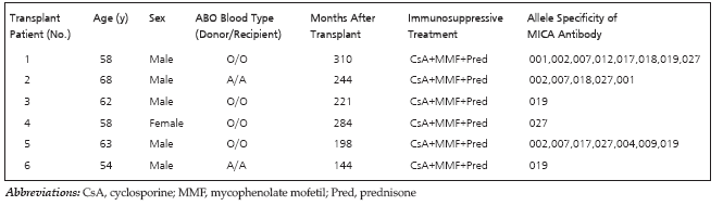
Table 1. Patient Characteristics
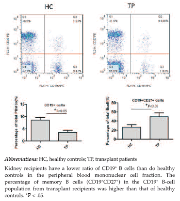
Figure 1. Constituent Ratio of CD19+ and CD19+CD27+ B Cells in Peripheral Blood of Kidney Recipients and Healthy Controls
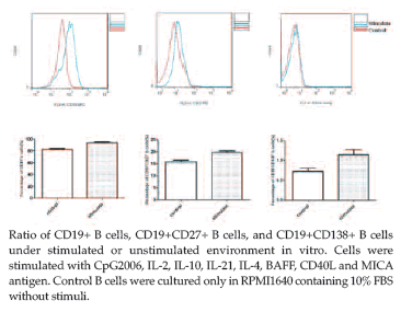
Figure 2. Effects of Stimuli on Purified B-Cell Differentiation
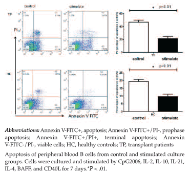
Figure 3. Effects of Stimuli on Purified B-Cell Apoptosis
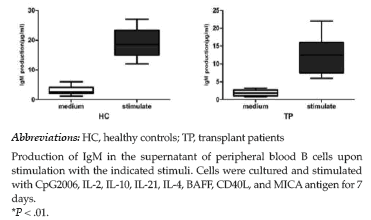
Figure 4. Effects of Stimuli on Purified B-Cell Immunoglobulin Production
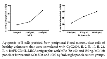
Figure 5. Tendency of Different Dosages of Bortezomib and Mycophenolic Acid Induce B-Cell Apoptosis
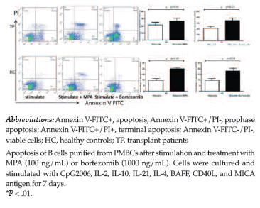
Figure 6. B-Cell Apoptosis Under Fixed Dosage of Bortezomib and Mycophenolic Acid
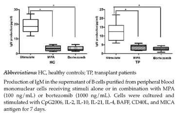
Figure 7. Effects of Immunosuppression on Immunoglobulin Production