Volume: 10 Issue: 3 June 2012
FULL TEXT
Posterior reversible encephalopathy syndrome is characterized by varying neurologic symptoms associated with brain vasogenic edema. Posterior reversible encephalopathy syndrome can be associated with severe hypertension (eg, in eclampsia or HELLP syndrome), but it also has been observed without hypertension and in several clinical conditions including infections and autoimmune disorders. The literature offers several reports of posterior reversible encephalopathy syndrome detected or induced after bone-marrow and solid-organ transplant, or induction by immunosuppression. We describe what is, to the best of our knowledge, the first case of man who successfully underwent a kidney transplant with preexisting posterior reversible encephalopathy syndrome induced by Goodpasture's syndrome.
Key words : Goodpasture's syndrome, Immunosuppression, Kidney transplant, PRES, Transplant
Introduction
Posterior reversible encephalopathy syndrome (PRES) is characterized by varying neurologic symptoms associated with brain vasogenic edema, typically involving the watershed zones of the parietal or occipital lobes.1
Posterior reversible encephalopathy syndrome can be associated with severe hypertension, for example, in eclampsia or HELLP syndrome, but has also been recognized in the absence of hypertension and in a variety of clinical conditions including infection or autoimmune disorders. However, the literature also reports several instances of PRES detected or induced after bone-marrow and solid-organ transplant, for example, kidney transplant, and induction by immunosuppression with calcineurin inhibitors.1
In this report, we describe what we believe is the first case of a patient who was successfully kidney transplanted with preexisting PRES induced by Goodpasture’s syndrome.
Case report
We report a 23-year-old man who underwent an ABO-compatible living kidney transplant. Nearly 12 months earlier, he had been admitted to our emergency department owing to generalized seizures followed by hemoptysis. On admission, he presented with reduced alertness, with an elevated blood pressure of 160/85 mm Hg, and a heart rate of 80 beats/min. His temperature was 37.5°C.
His lungs were clear to auscultation; oxygen saturation was 94%; however; a computed tomography (CT) of the lungs showed alveolar effusions with diffuse bleeding. An emergency cranial CT scan excluded bleeding or ischemia, while a subsequent cranial magnetic resonance image (MRI) showed diffuse, high-intensity lesions, with disordered diffusion of the cerebellum, and the parietal and occipital lobes suspicious for PRES (Figures 1 and 2).
Laboratory tests showed an elevated creatinine level 583 µmol/L (6.6 mg/dL), urea 12.5 mmol/L (35 mg/dL), sodium 144 mmol/L, and potassium 5.5 mmol/L. The white blood cell count was 1 × 10⁹ cells/L, lactate dehydrogenase was 8.5 µkat/L (509 U/L), and C-reactive protein was 76 nmol/L (8 mg/dL). The hemoglobin level (10.1 g/L), platelets (86 × 109 cells/L), and haptoglobin (< 2 µmol/L) were decreased, while his fragmentocytes were not elevated.
Because of an anuric kidney, a biopsy was done confirming the clinical suspicion of Goodpasture’s syndrome. Corresponding antiglomerular basement membrane antibodies were elevated over 200 U/mL without any elevation of other immunologic parameters.
Although dialysis and plasma exchange were started immediately, the patient still had repeat, generalized seizures, right-sided hemiplegic symptoms, accompanied by a complete loss of vision and intermittent hypertension (up to > 200/100 mm Hg). An examination of his cerebrospinal fluid was unremarkable.
A repeat MRI showed the familiar lesions of the first scan, with new lesions at the frontal lobe, which were typical for PRES (Figures 1 and 2). An accompanied electroencephalogram showed signs of severe encephalopathy that were detectable for the next months.
A stable clinical status was reached after 12 plasma exchanges, and immunosuppressive medication consisting of 1 gram of cyclophosphamide monthly (cumulative dosage, 6 g) could be started. After this, his vision and neurologic symptoms normalized completely; however, dialysis was necessary owing to an ongoing anuric kidney until transplant.
After transplant, a stable renal function with a baseline creatinine level of 106 to 123 µmol/L (1.2 to 1.4 mg/dL) could be reached. An immunosuppressive regimen composed of low-dose tacrolimus, mycophenolate mofetil, and prednisolone was started.
A follow-up neurologic examination, including an electroencephalogram, 6 and 12 months after transplant showed nothing abnormal. An MRI scan no longer showed the typical lesions for PRES (Figures 3 and 4).
Discussion
Different mechanisms in the pathogenesis of PRES are discussed. One theory assumes that abrupt rising of blood pressure results in a loss of cerebral autoregulation, with disturbance of the blood-brain barrier, which leads to vasogenic cerebral edema. Another theory assumes that neurotoxicity caused by drugs leads to endothelial dysfunction or injury of the blood-brain barrier.1, 2 The cause of PRES in our case was thought to be caused by endothelial injury owing to antiglomerular basement membrane antibodies of Goodpasture’s syndrome.
Typical signs for PRES that may develop immediately or after several days are headaches, a change of vision, paresis, nausea, altered mental status, and generalized seizures (as was the case in our patient). Moderate-to-severe blood pressure elevation could be observed in 70% to 80% of affected patients; however, in up to 20% of patients, normal blood pressures have been described. There are no characteristic laboratory findings specific for PRES.1
Computed tomographic/magnetic resonance scans in patients with PRES typically show focal regions of symmetric hemispheric edema that affect (in most cases) the parietal and occipital lobes.1, 3 However, the frontal lobes, the inferior temporal-occipital junction, and the cerebellum also can be affected (as was the case in our patient) (Figures 1 and 2).3
In contrast, malignant hypertension and hypertensive encephalopathy is marked by retinal hemorrhage, exudates, and/or papilledema. Neurologic signs are based on intracerebral or subarachnoid bleeding, lacunar infarcts, or hypertensive encephalopathy, which is linked to insidious headaches with nonlocalizing symptoms such as restlessness and confusion.4
Posterior reversible encephalopathy syndrome has been well-described after bone marrow or stem cell transplant, often owing to preconditioning regimens. However, PRES also is reported with an incidence between 0.4% to 6% after solid organ transplant.5, 6 Onset of PRES occurs earlier after liver transplant and is uncommon after heart or lung transplant. In contrast, kidney transplant recipients appear to develop PRES late after transplant and are commonly seen with coexistent severe hypertension.1, 6 Immunosuppressive drugs and transplant rejection are risk factors for PRES.
Posterior reversible encephalopathy syndrome can be seen in varying percentages after transplant.6 However, there are no data on patients with preexistent PRES and subsequent kidney transplant.
We decided on a low-dose immunosuppressant regimen with tacrolimus, and a strict adjustment of blood pressure to levels of less than 130/80 mm Hg. Neurologic follow-ups are conducted every 6 months, with an electroencephalogram every 12 months.
There are reports indicating that cyclosporine rather than tacrolimus induces more endothelial injury in patients with PRES.6 However, there are no reports regarding mycophenolate mofetil.
In this case, the decision for kidney transplant was based on the clinical status, age, and quality of life of our patient, and the likeliness of another PRES episode. We believe that PRES is not a contraindication for transplant after immunosuppression, especially in young patients.
Our case is one of a patient with preexisting PRES caused by Goodpasture’s syndrome after a kidney transplant. The decision to perform a transplant in affected patients should be based on age, quality of life, the likeliness of another episode, and should be accompanied by consequent follow-up examinations.
References:
- Bartynski WS. Posterior reversible encephalopathy syndrome, part 1:
fundamental imaging and clinical features. AJNR Am J Neuroradiol.
2008;29(6):1036-1042.
CrossRef - PubMed - Preul C, Gerth J, Lang S, et al. Cerebral involvement in a patient with
Goodpasture’s disease due to shortened induction therapy: a case report. J Med
Case Reports. 2009;3:120.
CrossRef - PubMed - Bartynski WS. Posterior reversible encephalopathy syndrome, part 2:
controversies surrounding pathophysiology of vasogenic edema. AJNR Am J
Neuroradiol. 2008;29(6):1043-1049.
CrossRef - PubMed - Vaughan CJ, Delanty N. Hypertensive emergencies. Lancet.
2000;356(9227):411-417.
CrossRef - PubMed - Yamada SM, Kitagawa R, Teramoto A. A case of reversible posterior
leukoencephalopathy syndrome with acute hypotension. Neurol Sci.
2011;32(1):165-168.
CrossRef - PubMed - Bartynski WS, Tan HP, Boardman JF, Shapiro R, Marsh JW. Posterior reversible
encephalopathy syndrome after solid organ transplantation. AJNR Am J Neuroradiol.
2008;29(5):924-930.
CrossRef - PubMed

Volume : 10
Issue : 3
Pages : 299 - 301
DOI : 10.6002/ect.2011.0177
From the 1Department of Nephrology, and the 2Department of Neurology, Klinikum
rechts der Isar, Technische Universitaet Muenchen, Germany
Corresponding author: Tobias Lahmer, MD, Department of Nephrology, Klinikum
rechts der Isar, TU Muenchen, Ismaninger Str. 22, 81675 Munich, Germany
Phone: +49 89 4140 2231
Fax: +49 89 4140 4878
E-mail:
Tobias.Lahmer@lrz.tu-muenchen.de
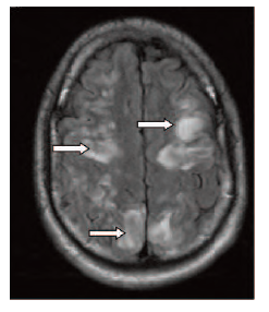
Figure 1. T2-weighted MRI scan of the parietal and occipital lobes with vasogenic edema areas (arrows).
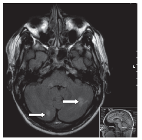
Figure 2. T2-weighted MRI scan of the cerebellum with vasogenic edema areas (arrows).
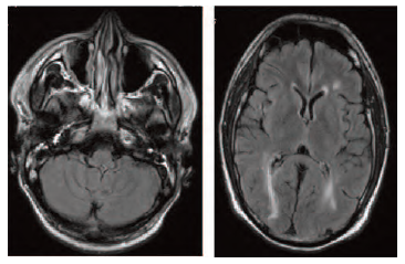
Figure 3-4. Follow-up T2-weighted MRI scans of the cerebellum and parietal lobes without vasogenic edema areas.