Volume: 8 Issue: 2 June 2010
FULL TEXT
Proteinuria Among Renal Transplant Patients and Its Relation to Hepatitis C Virus and Graft Outcome: A Single Center Experience
Objectives: Chronic hepatitis C virus has been associated with glomerular disease in native and transplanted kidneys. Reports suggest that hepatitis C virus-infected renal recipients may develop de novo glomerulonephritis. We evaluated the presence of hepatitis C virus at transplant, the occurrence of proteinuria in Egyptian renal transplant patients, and its possible link with graft survival.
Materials and Methods: Three hundred seventeen patients with end-stage renal disease receiving transplants in Mansoura Urology and Nephrology Center were retrospectively evaluated between 2000 and 2003. Their sera were assayed for anti-hepatitis C virus-antibodies at transplant. The relation between hepatitis C virus and development of posttransplant proteinuria was evaluated, along with possible effects of proteinuria on long-term graft survival.
Results: Two hundred seventy-three recipients fulfilled the inclusion criteria, 169 were positive and 104 were negative for hepatitis C virus-antibodies by ELISA. Mean duration of posttransplant follow-up was 87.73 ± 26.79 and 84.29 ± 28.55 months for both groups. Groups were comparable regarding the incidence and quantity of hepatitis C virus-positive patients and 0.4 grams/day (P = .09 of proteinuria).
In both hepatitis C virus-positive and negative groups, those with nephrotic range proteinuria showed worse graft survival (P = .001) and higher frequency of chronic allograft nephropathy (P = .05) compared with nonproteinuric patients.
Conclusions: There is a high prevalence of hepatitis C virus in our end-stage renal disease patients awaiting renal transplant. The incidence and quantity of proteinuria is similar in both hepatitis C virus-positive and hepatitis C virus-negative transplant recipients. Nephrotic range proteinuria is associated significantly with a higher incidence of chronic allograft nephropathy. Independent from serology, it is associated with poorer graft outcome.
Key words : HCV, Kidney transplant, Egyptian
The proportion of anti-hepatitis C virus-antibodies in renal transplant patients varies from 6% to 64% depending on geographic areas (1). Hepatitis C virus infection has now been shown to be responsible for extrahepatic diseases in nonimunocompromised individuals, especially in glomerulonephritis (2). However, little is known about its effect in renal transplant, although some reports suggest that hepatitis C virus-infected kidney recipients may develop recurrent or de novo glomerulonephritis due to cryoglobulinemia or the deposition of complexes containing viral antigen and anti-hepatitis C virus-antibodies (3). Other reports indicate extrahepatic syndromes associated with hepatitis C also can occur after organ transplant (4).
In Egypt, hepatitis C virus-infection has reached epidemic proportions, with up to 21.9% of the population affected. This has been partly attributed to the routine parenteral administration of anti-Schistosomal drugs during the 1960s (5). The current study was therefore done to estimate the prevalence of hepatitis C virus among our renal transplant recipients, and to investigate any association between pretransplant anti-hepatitis C virus- antibodies, and the occurrence of posttransplant proteinuria. Finally, the study also attempts to clarify any effects of both hepatitis C virus and proteinuria on patient and graft survival.
Materials and Methods
Of 317 patients with end-stage renal disease, 273 fulfilled our inclusion criteria and were included in the study. Prior to the study, the study protocol was approved by our local institutional ethics committee, and the protocol conforms with the ethical guidelines of the 1975 Helsinki Declaration. Written, informed consent was obtained from all subjects. Forty-four patients were excluded from the study; 11 being HBsAg positive, 10 patients died and/or had graft failure within 6 months posttransplant, and 23 patients were lost during follow-up. Pretransplant anti-hepatitis C virus status was available for all patients (using third generation ELISA). Participants all received transplants at Mansoura Urology and Nephrology Center between April 2000 and January 2003. Two hundred eleven patients (77.7%) received transplants from a living-related donor, while the remaining patients (22.7%) received transplants from living-unrelated donors. All were adults (age, > 18 years), HBsAg negative, and lived with a functioning graft for 6 months posttransplant.
Pretransplant workup
All patients in our units who received regular hemodialysis were screened
monthly (serum creatinine levels before and after dialysis, electrolyte levels,
Kt/V for dialysis efficiency and liver function tests: Serum bilirubin, total
proteins, albumin, serum alanine aminotransferase (ALT), serum aspartate
aminotransaminase (AST), serum alkaline phosphatase (ALP), prothrombin time,
fasting and postprandial blood glucose, serum uric acid, and a complete blood
count).
Screening were carried out every 3 months for hepatitis C virus-antibodies (Murex anti-hepatitis C virus, England), hepatitis B virus markers (ELISA technique Behring, Germany), cytomegalovirus, IGM antibody (Microparticle enzyme immunoassay (IMX) (Abbott Laboratories, Abbott Park, IL, USA), and antibodies to human immunodeficiency virus 1 and 2 (HIV) using Murex HIV1 and 2 enzyme immunoassay test.
Cryoglobulin assay
The sample (10 mL of whole blood) was brought to the laboratory immediately
after drawing. The specimen was not refrigerated before the test. Tubes for
collection were not anticoagulated, because the use of plasma may result in the
development of cold-precipitable fibrinogen, cryofibrinogen, or heparin-precipitable
protein. The specimen was incubated for at least 30 minutes to 1 hour at 37ºC in
a heat block or water bath before being centrifuged at room temperature. Fresh
serum was placed into the appropriately labeled tubes; 1 being placed in the
refrigerator for a minimum of 4 days, the other left at room temperature.
Posttransplant workup: Maintenance immunosuppression
After renal transplant, all recipients were immunosuppressed with triple
immunosuppression (prednisolone, cyclosporine, and azathioprine) according to
the standard protocol described by Ghoniem and associates (1993) (6). These were
then evaluated for graft function (serum creatinine, creatinine clearance,
urinalysis, graft ultrasound, and renal isotopic scanning). After discharge,
they were subjected to thorough clinical examination and laboratory
investigation (urinalysis, 24-hour urinary protein measured by a timed endpoint
method (12 months after transplant) (Fujita et al., 1983 [7], and corrected to
urinary protein in mg/hour/m2). Proteinuria was defined as urinary protein above
1 gram/day at 2 successive assays. Nephrotic range proteinuria was defined as
daily protein excretion
≥
3.5 gram/1.73 m2/surface area, while persistent proteinuria was defined
as protein that remaining above 1 g/day over 3 months after the onset. Transient
proteinuria was not considered for the analysis.
Liver function tests were checked daily during hospitalization then rechecked monthly for patients with normal liver function and upon each visit for those with hepatic dysfunction. Azathioprine dosage was reduced by 50% whenever the leucocytes fell below 3000/cm.
In the past, doubling of serum transaminases and/or rising of serum bilirubin was managed by replacing azathioprine with cyclophosphamide (1 mg/kg/d). Azathioprine was reintroduced 1 month after normalization of liver function tests. It was permanently discontinued if this reintroduction was associated with hepatic dysfunction. Cyclophosphamide is no longer used; instead, patients with hepatic dysfunction are administered mycophenolate mofetil (1.5 g/d).
Rejection of the graft (acute or chronic allograft nephropathy) was diagnosed and graded by graft biopsy using Banff classification 1997 (8).
Antirejection therapy
Our current protocol is to treat a first episode of acute, predominantly
cellular rejection with a steroid pulse. A biopsy should almost always precede
the administration of any therapy, unless absolutely contraindicated. Pulse
methylprednisolone, 500 mg was given intravenously for 3 to 5 days.
Antibody therapies, intravenous immune globulin, OKT3, in addition to corticosteroids and plasmapheresis were used to treat antibody-mediated rejection.
Viral screening was carried out at 6 monthly intervals and whenever indicated (HBsAg, cytomegalovirus, and anti-HIV 1 and 2 and anti-hepatitis C virus-antibodies).
Statistical analyses
Quantitative data are displayed in terms of mean ± SD for normally distributed
data, Median and range for nonnormally distributed data used the unpaired t test and the Mann-Whitney
U test for comparison. Qualitative data were described in
cross tabulation with the chi-square and Fisher exact tests used for the
comparison of frequencies. A P value < .05 was considered statistically
significant. Graft and patients survival rates were assessed using the Kaplan-Meier
method (the long-rank test). All analyses were carried out using statistical
analyses for Windows (Statistical Product and Service Solutions, version 17.0,
SSPS Inc, Chicago, IL, USA).
Results
Prevalence of hepatitis C virus among renal transplant patients
A total of 317 patients were transplanted during the study period. The study
excluded patients who were HBsAg positive, acquired the infection after
transplant (11 patients), died, had graft failure within 6 months posttransplant
(10 patients), or were lost during follow-up (23 patients). The remaining 273
recipients were living with functioning grafts for at least 6 months. Of these,
169 patients (62%) were positive for the hepatitis C virus-antibody, and the
remaining 104 (38%) were negative. Polymerase chain reaction was carried out for
146 patients, with 39% of them testing positive.
The 2 groups of renal allograft recipients were comparable with respect to age, sex, donor relation, HLA mismatches, and original kidney disease. Anti-hepatitis C virus-positive recipients received significantly more blood transfusion and had significantly longer duration of hemodialysis (see Table 1).
Both groups were comparable with regard to the incidence and quantity of proteinuria, C3 level, reactivity of rheumatoid factor, incidence of acute and chronic allograft glomerulopathy, incidence of de novo, and recurrent glomerulonephritis and serum creatinine at the start and at the end of the study.
Those positive for hepatitis C virus had a statistically significant lower level of C 4 (47 patient vs 9, P = .001), and a higher incidence of posttransplant chronic hepatitis (72 patients vs 19 patients, P = .001) (see Table 2).
Hepatitis C virus and proteinuria
Ninety patients (33%) showed persistent proteinuria, of which 56 (62%) were
hepatitis C virus-positive while 34 (38%) were hepatitis C virus-negative (by
dipstick examination). Of these, 47 patients (17%) displayed nephrotic range
proteinuria; 28 (60%) being hepatitis C virus-positive and 19 (40%) hepatitis C
virus- negative.
When hepatitis C virus-positive and -negative patients with nephrotic range proteinuria were compared, there was no statistically significant difference (regarding the original kidney disease, number of acute rejection episodes, levels of C3 and C4, reactivity of rheumatoid factor, incidence of posttransplant chronic hepatitis, de novo and recurrent glomerulonephritis, or graft function at the start and at the end of the study). Despite being greater in hepatitis C virus-positive patients, the frequency of chronic allograft nephropathy did not reach a statistically significant level (Table 3).
When hepatitis C virus-positive patients with nephrotic range proteinuria were compared with nonproteinuric patients, the number of acute rejection episodes, recurrent and de novo glomerulonephritis, chronic allograft nephropathy, and serum creatinine at the end of the study, and the frequencies of graft failure were all statistically greater in proteinuric patients (18/28 vs 22/141) (Table 4).
The same finding was observed in hepatitis C virus-negative patients when a comparison was made between nephrotic range proteinuric and nonproteinuric patients. Serum creatinine was significantly higher at the end of the study in proteinuric patients compared with nonproteinuric (6.08 ± 3.54 vs 3.6 ± 3.02), and a statistically significant difference in the frequency of graft failure between both groups was observed (12/19 vs 19/70; P = .003 ).
Graft survival
There was no significant difference regarding graft survival between anti-hepatitis
C virus-positive and negative patients as shown in Figure 1a (P = .25). However,
graft survival was significantly lower in patients with nephrotic range
proteinuria irrespective of their serology (Figure 1b).
Discussion
Hepatitis C virus infection is the main cause of chronic liver disease among renal transplant recipients, being the fourth-most prevalent cause of mortality (9). The proportion of anti-hepatitis C virus antibodies in renal transplant patients varies from 6% to 64%, depending on geographic areas (1). In Egypt, hepatitis C virus has reached epidemic proportions, with up to 21.9% of the population affected (5). To our knowledge, there are no clear data about the prevalence of hepatitis C virus among Egyptian renal transplant recipients.
The present study was a single center study of a homogenous group of living kidney transplant recipients, with a mean duration of 86.4 ± 27.4 months follow-up. Our first aim was to determine the prevalence of hepatitis C virus antibodies among the renal transplant recipients. One of the most-striking findings of our study is the inordinately high prevalence of hepatitis C virus antibody reactivity in our end-stage renal disease patients awaiting renal transplant. Sixty percent of them tested positive for anti-hepatitis C virus by third generation ELISA (169/273). Hestin and associates (10) described a lower prevalence of hepatitis C virus (9.6 %) in renal transplant patients, which can be explained as follows: First, there is a low prevalence of hepatitis C virus in Europe (antibodies range from 0.1% to 1.5% with a north-to-south gradient (11) compared to the high prevalence of hepatitis C virus in the general population in Egypt.
Second, we previously reported a higher prevalence of hepatitis C virus among patients with glomerulopathy (up to 38%) compared to the general population (12).
Third, other factors may increase prevalence such as duration of hemodialysis and frequency of blood transfusion. In our study, we found a positive correlation between receiving blood transfusions while on hemodialysis and the risk of hepatitis C virus infection, as patients positive for anti-hepatitis C virus antibodies receive significantly more blood units and significantly longer duration of hemodialysis compared with those negative for the antibodies. Finally, other factors may be specific to the Egyptian population including routine antischistosomal therapy (5).
Hepatitis C virus infection may have a significant negative impact on graft survival of renal transplant recipients (13) or may have no impact at all (13-15). Lower graft survival results from lowered patient survival, hepatitis C virus-related glomerulonephritis, acute rejection, or chronic allograft nephropathy. We found no significant difference in graft survival between hepatitis C virus-positive and hepatitis C virus-negative groups after 87.73 ± 26.79 months (range, 19-123 months) and 84.29 ± 28.55 months (range, 11-123 months) of follow-up for each group respectively. We do believe, however, that longer duration of follow-up is necessary for comprehensive conclusions to be drawn.
The underlying cause of proteinuria, not the proteinuria, per se, could be the factor contributing to transplant dysfunction and failure. Persistent proteinuria is almost invariably a sign of structural renal disease even when renal function is normal. Fontan and associates (1999) (16) observed that when proteinuria was categorized according to its persistence, persistent but nontransient proteinuria was associated with poor patient survival. Roodnat and associates (1999) (17) also found increased risk of death and cardiovascular risk when proteinuria was present.
Our second aim of the study was to investigate whether there is an association between pretransplant hepatitis C virus status and posttransplant proteinuria. In our study, no statistical difference was observed regarding the quantity of proteinuria, level of C3, rheumatoid factor positivity, incidence of de novo and recurrent glomerulonephritis when hepatitis C virus-positive and negative proteinuric patients were compared. Chronic allograft glomerulopathy, despite of being higher in hepatitis C virus-positive patients, did not reach statistical significance.
It recently has been argued (18) that the allograft is particularly vulnerable to the adverse effect of proteinuria for several reasons. Protein-loaded epithelial cells express more MHC-2 antigens, rendering them potentially susceptible to immune reactions. It also has been shown that proximal tubular cells from patients with cyclosporine nephrotoxicity express more endothelin I, a potent agonist in the generation of renal fibrosis (19).
Our data are not in agreement with those described by Hestin and associates (1998) (10), who reported a significant difference in the cumulative probability of posttransplant proteinuria between recipients with and without anti-hepatitis C virus antibodies (45.1% in hepatitis C virus-positive and 13.1% in hepatitis C virus-negative patients at 5 years). The histology of biopsies from 26/44 recipients with proteinuria, in his study, showed de novo glomerular lesions were more frequent in hepatitis C virus-positive patients. This inconsistency between Hestin’s results and ours may be due to factors related to the viral titer, difference in viral genotyping, and the different sources of renal allograft (deceased donor allograft recipients have a significantly higher incidence of rejection episodes and received more profound immunosuppressive during the posttransplant course compared with patients receiving living-donated grafts).
Our third aim was to clarify the influence of hepatitis C virus and proteinuria on both patients and graft survival. In our study, renal transplant recipients with nephrotic range proteinuria, irrespective of their serology, showed statistically significant de novo, recurrent graft nephropathy, and chronic allograft nephropathy when compared with nonproteinuric patients. This suggests that proteinuria, per se, is a marker of underlying graft damage.
Our findings are supported by what was reported previously by Roodnat and associates (2001) (17) where a relative risk (RR) of graft failure was found to be elevated in proteinuric patients (RR 2.03) as compared to nonproteinuric patients. Interestingly, patient death was also higher in graft recipients with proteinuria (RR 1.98) as compared to nonproteinuric patients.
In agreement with our results, Hohage and associates examined 327 patients transplanted between 1980 and 1990 (20). Proteinuria at 6 months after transplant was noted in 25.5% of the patients. Nonproteinuric patients had a 5-year graft survival of 85.6% as compared with much-lower graft survival in proteinuric patients (58.9%). No correlation was found with regard to sex or age of recipient, duration of hemodialysis, age of the donor, cold ischemia time, or mismatches. It appears that mild proteinuria at 6 months after transplant is a predictor of decreased long-term graft function.
In our study, we did not find a significant influence of hepatitis C virus infection on patients or graft survival, which is in agreement with some earlier reports (21-23). However, we feel that more long-term follow-ups are needed to confirm or disprove this observation.
The association between hepatitis C virus and essential mixed cryoglobulinemia has been known very early (2). We previously reported an incidence of 54% in patients with hepatitis C virus-associated nephropathy (12). Surprisingly, none of our cases in this study had cryoglobulins. We considered possible explanations for our failure to detect cryoglobulins, which is consistent with some reports about cases of membranoproliferative glomerulonephritis in patients with replicative hepatitis C virus in which cryoglobulinemia was absent (3, 24). Firstly, cryoglobulins appear only many years after chronic hepatitis C virus infection, owing to continued exposure to the antigen (25), so we may speculate that long-term follow-up for these patients is needed to identify possible development of cryoglobulins. Secondly, it is possible that in immunosuppressed transplant patients, hepatitis C virus can produce glomerular disease in the absence of cryoglobulins. Thirdly, it has been reported previously that immunosuppression increases hepatitis C virus viremia and decreases immunoglobulin synthesis (26)
It is recognized that our study has certain limitations, being a retrospective study with all the drawbacks of such studies; methodologic deficiencies compared to controlled trials, imprecise and incomplete data gathering, and lack of randomization. Hopefully, these limitations will be avoided in a future work.
In conclusion, there is a high prevalence of hepatitis C virus in our end-stage renal disease patients awaiting renal transplant. No significant difference was found regarding the incidence and quantity of proteinuria in our hepatitis C virus-positive and hepatitis C virus-negative renal transplant recipients. Nephrotic range proteinuria is associated significantly with a higher incidence of chronic allograft nephropathy and associated with poorer graft outcome.
References:
- Legendre C, Garrigue V, Le Bihan C, et al. Harmful long-term impact of hepatitis C virus infection in kidney transplant recipients. Transplantation. 1998;65(5):667-670.
- Johnson RJ, Gretch DR, Yamabe H, et al. Membranoproliferative glomerulonephritis associated with hepatitis C virus infection. N Engl J Med. 1993;328(7):465-470.
- Roth D, Cirocco R, Zucker K, et al. De novo membranoproliferative glomerulonephritis in hepatitis C virus-infected renal allograft recipients. Transplantation. 1995;59(12):1676-1682.
- Baid S, Cosimi AB, Tolkoff-Rubin N, Colvin RB, Williams WW, Pascual M. Renal disease associated with hepatitis C infection after kidney and liver transplantation. Transplantation. 2000;70(2):255-261.
- Frank C, Mohamed MK, Strickland GT, et al. The role of parenteral antischistosomal therapy in the spread of hepatitis C virus in Egypt. Lancet. 2000;355(9207):887-891.
- Ghoneim MA, Sobh MA, Shokeir AA, Bakr MA, el-Sherif A, Foda MA. Prospective randomized study of triple vs conventional immunosuppression in living donor kidney transplantation. Transplant Proc. 1993;25(3):2243-2245.
- Fujita Y, Mori I, Kitano S. Determination of proteins by using the color reaction with pyrocatechol violet-molybdenum(VI) complex. Chem Pharm Bull. 1984;32(10):4161-4164.
- Racusen LC, Solez K, Colvin RB, et al. The Banff 97 working classification of renal allograft pathology. Kidney Int. 1999;55(2):713-723.
- Cisterne JM, Rostaing L, Izopet J, et al Epidemiology of HCV infection: disease and renal transplantation. Nephrol Dial Transplant. 1996;11(suppl 4):46-47.
- Hestin D, Guillemin F, Castin N, Le Faou A, Champigneulles J, Kessler M. Pretransplant hepatitis C virus infection: a predictor of proteinuria after renal transplantation. Transplantation. 1998;65(5):741-744.
- Degos F. Natural history of hepatitis C virus infection. Nephrol Dial Transplant. 1996;11(suppl 4):16-18.
- Sabry AA, Sobh MA, Irving WL, et al. A comprehensive study of the association between hepatitis C virus and glomerulopathy. Nephrol Dial Transplant. 2002;17(2):239-245.
- Legendre C, Garrigue V, Le Bihan C, et al. Harmful long-term impact of hepatitis C virus infection in kidney transplant recipients. Transplantation. 1998;65(5):667-670.
- Mathurin P, Mouquet C, Poynard T, et al. Impact of hepatitis B and C virus on kidney transplantation outcome. Hepatology. 1999;29(1):257-263.
- First MR, Vaidya PN, Maryniak RK, et al. Proteinuria following transplantation. Correlation with histopathology and outcome. Transplantation. 1984;38(6):607-612.
- Pérez Fontán M, Rodríguez-Carmona A, García Falcón T, Valdés F. Early proteinuria in renal transplant recipients treated with cyclosporin. Transplantation. 1999;67(4):561-568.
- Roodnat JI, Mulder PG, Rischen-Vos J, et al. Proteinuria after renal transplantation affects not only graft survival but also patient survival. Transplantation. 2001;72(3):438-444.
- Rosenkranz AR, Mayer G. Proteinuria in the transplanted patient. Nephrol Dial Transplant. 2000;15(9):1290-1292.
- Peddi VR, Dean DE, Hariharan S, Cavallo T, Schroeder TJ, First MR. Proteinuria following renal transplantation: correlation with histopathology and outcome. Transplant Proc. 1997;29(1-2):101-103.
- Hohage H, Kleyer U, Brückner D, August C, Zidek W, Spieker C. Influence of proteinuria on long-term transplant survival in kidney transplant recipients. Nephron. 1997;75(2):160-165.
- Pol S, Legendre C, Saltiel C, et al. Hepatitis C virus in kidney recipients. Epidemiology and impact on renal transplantation. J Hepatol. 1992;15(1-2):202-206.
- Orloff SL, Stempel CA, Wright TL, et al. Long-term outcome in kidney transplant patients with hepatitis C (HCV) infection. Clin Transplant. 1995;9(2):119-124.
- Roth D, Zucker K, Cirocco R, et al. The impact of hepatitis C virus infection on renal allograft recipients. Kidney Int. 1994;45(1):238-244
- Davis CL, Gretch DR, Perkins JD, et al. Hepatitis C--associated glomerular disease in liver transplant recipients. Liver Transpl Surg. 1995;1(3):166-175.
- Trendelenburg M, Schifferli JA. Cryoglobulins in chronic hepatitis C virus infection. Clin Exp Immunol. 2003;133(2):275-282.
- Morales JM. Hepatitis C virus infection and renal disease after renal transplantation. Transplant Proc. 2004;36(3):760-762.

Volume : 8
Issue : 2
Pages : 91 - 97
From the Nephrology and Internal Medicine Department, Mansoura Urology and
Nephrology Center, Mansoura University, Egypt
Acknowledgement: The authors are very grateful to Michelle Chambers, Primary
Care Mental Health Worker, Nottingham City NHS, UK for the revision of the
manuscript.
Address reprint requests to: Dr. Alaa Sabry, Assistant Professor of Nephrology,
Urology and Nephrology Center, Mansoura University, Egypt
Phone: +20502262222
Fax: +20502263717
E-mail:
asabry2040@yahoo.comr
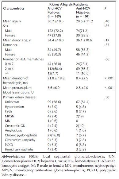
Table 1. Pretransplant characteristics of 273 patients included in the study.
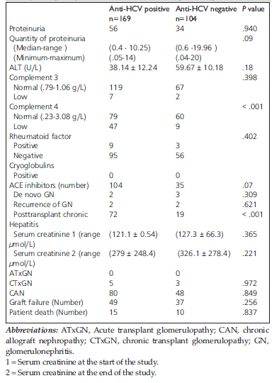
Table 2. Comparison between anti-HCV positive and anti HCV negative kidney transplant patients (86.4 ± 27.4 months) posttransplant.
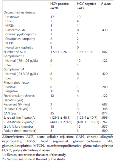
Table 3. Comparison between HCV positive and negative kidney transplant patients with nephrotic range proteinuria.
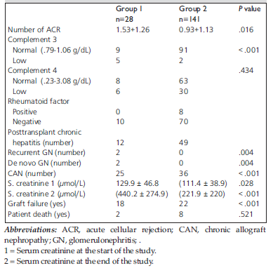
Table 4. Comparison between anti-HCV positive kidney transplant patients with nephrotic range proteinuria (group 1) and those without proteinuria (group 2) .
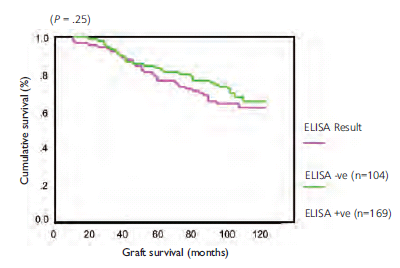
Figure 1a. Graft survival according to anti-HCV antibodies.
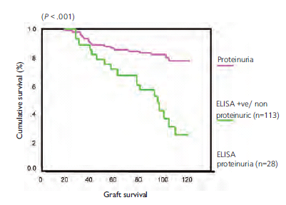
Figure 1b. Graft survival in anti-HCV antibodies according to proteinuria.