Volume: 8 Issue: 2 June 2010
FULL TEXT
Clinical Relevance of Curcumin-induced Immunosuppression in Living-Related Donor Renal Transplant: An In Vitro Analysis
Objectives: In this study, we assessed the immunosuppressive potential of curcumin, a pharmacologically safe and cost-effective naturally occurring polyphenolic phytochemical, on the induction of Th1 cytokines that are frequently overexpressed in patients experiencing rejection after renal transplant.
Materials and Methods: Peripheral blood lymphocytes obtained from 68 renal transplant recipients and 17 healthy controls were treated with curcumin before stimulation with phorbol myristate acetate and were analyzed with flow cytometry for interferon-γ and interleukin 4 positive cells.
Results: Patients experiencing acute rejection exhibited a high level of interferon-γ (38.3% ± 11.2%) and a low level of interleukin 4 (4.2% ± 2.0%) in their activated peripheral blood lymphocytes. The use of curcumin dose-dependently decreased interferon-γ induction in cultures from healthy controls (28.1% ± 4.8% - 10.7% ± 5.3%, P < .001), patients experiencing acute rejection (38.3% - 18.3%, P < .001), and those experiencing chronic rejection (40.6% - 12.9%, P = .01) when compared with corresponding untreated cultures. In contrast, curcumin exerted only a marginal effect on interleukin 4 expression. Interestingly, curcumin was found to inhibit nuclear factor kappa beta activation by blocking the degradation of the inhibitory unit I kappa B alpha. We also noted the synergistic inhibitory effect of in vitro treatment with curcumin in combination with cyclosporine on the peripheral blood lymphocytes of patients experiencing acute rejection.
Conclusions: These data provide a rationale for the use of curcumin as an affordable, pharmacologically safe, adjuvant immunosuppressant when used with cyclosporine and suggest that curcumin can effectively suppress Th1 cytokine induction after renal transplant.
Key words : Peripheral blood lymphocytes, acute rejection, chronic rejection, interferon-γ , flow cytometry
Despite improvements in histocompatibility leukocyte antigen tissue typing technologies and the advent of newer immunosuppressive drugs, acute rejection remains an important cause of early and late allograft failure (1-5). Because immunologic processes such as antigen recognition and the proliferation of lymphoid cells mark the beginning of graft dysfunction, cytokines and antibodies, which are the products of those processes and are produced by alloreactive T cells, appear first in the circulation and have a major role in graft tolerance and rejection (4, 6, 7). Case control studies involving solid-organ transplant (8, 9) or murine cardiac allograft experiments (10) have revealed that an increase in the production of type 1 cytokines after transplant is associated with allograft rejection and that a decrease in those levels often correlates with graft acceptance. However, an increase in the level of type 2 cytokines such as interleukin 4 and interleukin 10, either by direct action or by down-regulation of the expression of type 1 cytokines, has been reported to promote tolerance (11, 12).
Recently, our group has noted a positive correlation between transplant rejection and the increased occurrence of interferon-γ –positive CD8+ T cells in the peripheral blood of patients after living-related donor renal transplant (13). Despite the patients’ treatment with immunosuppressive therapy, the percentage of interferon-γ –producing cells increased significantly in those experiencing acute rejection episodes. Similar observations were made earlier by other investigators (14). Currently, immunosuppressive regimens using drugs such as cyclosporine, tacrolimus, sirolimus, steroids, and mycophenolate mofetil are the best options for the suppression of alloreactive cells (15-17). However, most of those compounds are expensive and are associated with significant adverse effects such as nephrotoxicity, anemia, leucopenia, thrombocytopenia, hypercholesterolemia, arthralgias, extremity edema, opportunistic infections, and impaired wound healing (18, 19). Although regimens based on mycophenolate mofetil have reduced the reliance on calcineurin inhibitors, there have been sporadic reports of the association of mycophenolate mofetil with cytomegalovirus infection (20). Therefore, it is essential to develop alternate therapeutic protocols in combination with newer and relatively less toxic agents to reduce or eliminate the use of calcineurin inhibitors for the long-term maintenance of renal allografts.
Curcumin (1,7-Bis(4-hydroxy-3-methoxyphenyl)-1,6-heptadiene-3,5-dione), a naturally occurring polyphenolic phytochemical isolated from the rhizome of the medicinal plant Curcuma longa, has been reported to cause the concentration-dependent inhibition of T-lymphocyte proliferation in cells stimulated by phorbol myristate acetate and anti-CD 28 antibody in vitro (21, 22). Curcumin has been shown to suppress IL-12 production in macrophages (23) and to inhibit IL-12 signaling in T cells; effects that lead to the increased production of IL-4 and to reciprocally decreased interferon-γ production by T cells (24). Furthermore, curcumin-treated T cells inhibit IL-12–induced T-cell proliferation and Th1 differentiation (25). Recently, curcumin was shown to enhance the immunosuppressive activity of cyclosporine in a rat cardiac allograft model (26); this suggests the possible use of curcumin as an effective adjuvant immunosuppressant when administered with cyclosporine. Other investigators have shown that curcumin can block immunologically induced renal ischemic injury in animal models (27-29) and thus, may be a useful renoprotective agent. These observations indicate the potential anti-Th1 activity of curcumin, which could be beneficial in renal recipients who otherwise exhibit elevated Th1 cytokine levels during rejection episodes.
Despite convincing indications for the potential use of curcumin to prevent the immunologic rejection of renal allografts, little information about the possible therapeutic use of that agent in clinical settings exists. Because the infiltration of lymphocytes into the graft epithelial compartment is a key factor in the progression of clinical renal allograft rejection, we studied the effects of curcumin on the peripheral blood lymphocytes of patients who underwent renal transplant. In our investigation, we monitored the frequency of peripheral blood lymphocytes expressing interferon-γ and IL-4, the signature cytokines of type 1 and type 2 immune responses in those subjects, and we tested the immunosuppressive effects of curcumin in patients experiencing acute or chronic rejection, the peripheral blood lymphocytes of whom were refractory to conventional immunosuppressive therapy.
Materials and Methods
Patients
Sixty-eight patients with end-stage renal disease who underwent renal transplant
at the All India Institute of Medical Sciences Hospital, and 17 healthy subjects
were included in this study, which was approved by the Institutional Ethics
Committee of the hospital in confirmation with the ethical guidelines of the
1975 Helsinki Declaration. Informed consent was obtained from each patient
studied. Of those 68 patients, 9 experienced acute rejection episodes, 6
exhibited chronic rejection, and the remaining 53 had a well-functioning graft.
Table 1 summarizes the demographic data of the subjects enrolled in our study.
Peripheral blood samples were collected before the transplant, before the
initiation of the immunosuppressive regimen, and on days 3, 7, 15, 30, 60, and
90 after transplant. In addition, samples were collected immediately after the
confirmation of an acute rejection episode. All patients were monitored
regularly (1-3 months) and were evaluated for the development of acute rejection
defined by stringent clinicopathologically proven biopsy data and a decrease in
renal function (ie, an increase in the serum creatinine level to ≥
176.8 µmol/L
[≥
2.0 mg/dL]).
All patients received immunosuppressive therapy 48 hours before undergoing renal transplant: A triple drug regimen consisting of cyclosporine (6 mg/kg body weight), azathioprine (1.5-2 mg/kg body weight), and prednisolone (50 mg tapered rapidly to 30 mg/kg body weight). Azathioprine was replaced with mycophenolate mofetil (30 mg/kg body weight) in patients who could afford the cost. Acute rejection was defined by clinical parameters that included increasing serum creatinine levels in the absence of other pathologic conditions. Acute rejection episodes that were subsequently confirmed by renal biopsy were treated with intravenously administered methylprednisolone at a dosage of 0.5 g/d for 3 days. Refractory cases were treated with either muromonab-CD3 or antithymocyte globulin (if the patient could afford it) after a repeat biopsy had confirmed ongoing acute rejection. Immunosuppressive therapy was tapered off in patients with a smooth clinical course, which was defined as the absence of acute rejection episodes. After 6 months, cyclosporine was administered at a dose of 3 to 4 mg/kg, and the dose of prednisolone was reduced to 15 mg/kg. However, the azathioprine dose remained unaltered. Graft failure was defined as requirement of maintenance dialysis or death.
Cell isolation
Peripheral blood lymphocytes were isolated from heparinized blood by Ficoll
Hypaque gradient centrifugation and were suspended in RPMI-1640 (Caisson
Laboratories, UT, USA) supplemented with glutamine (Sigma, St. Louis, USA),
HEPES (Sigma), antibiotics (Penicillin, Streptomycin, Biological Industries,
Israel), and 10% heat inactivated fetal calf serum (Biological Industries). The
viability of the cells was measured with the trypan blue dye exclusion test; the
result was usually > 98%. Those cells were used for subsequent flow-cytometry–based
intracellular cytokine assays. The cells were gated for lymphocytes on forward-
and side-scatter parameters as per the standard protocol.
In vitro stimulation of lymphocytes
Peripheral blood lymphocytes from healthy volunteers were pretreated with
various concentrations (1, 5, 10, 20 µM) of curcumin (Sigma) for 3 hours or with
cyclosporine (1 µg/mL, Novartis, NJ, USA) to reveal dose kinetics. Following
determination of the optimal dose, cells were pretreated with 10 µM of curcumin
before stimulation. For polyclonal stimulation, the cells were treated for 6
hours with phorbol myristate acetate (5 ng/mL, Sigma), ionomycin (2 mM, Sigma),
and monensin (2 µM, Sigma), and were processed for intracellular cytokine
staining.
Intracellular cytokine staining
After completion of the experiments, the cells were processed for 3-color
fluorochrome staining as described earlier (13). Briefly, the cells were stained
with anti-interferon-γ
-fluorescein isothiocyanate and anti-IL-4-phycoerythrin
or matched isotype controls (0.2 µg/mL each) to reveal the intracellular
expression of interferon-γ
and IL-4. Stained cells were acquired on BD FACS
Calibur (BD Biosciences, CA, USA) and were subsequently analyzed with Cell quest
Pro software (BD Biosciences). Ten thousand events were recorded for each sample.
All antibodies used in the assay were obtained from BD Biosciences.
Preparation of protein extracts
Protein extracts were prepared from treated and untreated lymphocytes as
described previously (30). Briefly, 2 × 10⁶ peripheral blood lymphocytes were
washed with cold phosphate buffered saline and were suspended for 30 minutes in
0.4 mL of hypotonic buffer containing protease inhibitors. The cells were lysed
with 12.5 µL of 10% Nonidet P-40 (Sigma), the homogenate was centrifuged, and
supernatant containing the cytoplasmic extracts was stored frozen at -80°C. The
nuclear pellet was resuspended in 25 µL of ice-cold nuclear extraction buffer.
After 30 minutes of intermittent mixing, the extract was centrifuged, and the
supernatants containing nuclear extracts were secured. The protein content was
measured with the Bradford Protein Assay (BioRad, USA). Extracts that were not
used immediately were stored at -80°C.
Electrophoretic mobility shift assay
Nuclear factor kappa beta (NF-ΚB
) activation was analyzed with an
electrophoretic mobility shift assay as described previously (30). In brief, 8
µg of nuclear extracts prepared from curcumin-treated or untreated cells were
incubated with 32P end-labeled 45-mer double-stranded NF-ΚB
oligonucleotide
from human immunodeficiency virus–1 long terminal repeat (5’-TTGTTACAAGGGACTTTCCGCTGGGGACTTTCCAGGGAGGCGTGG-3’;
the underlining indicates the NF-ΚB
binding site) for 15 minutes at 37°C,
and the deoxyribonucleic acid (DNA)-protein complexes were resolved in a 6.6%
native polyacrylamide gel. The radioactive bands from the dried gels were
visualized and quantitated with the PhosphorImager (Fujifilm FLA-5100) and Multi
Gauge software.
Western blot for I kappa B alpha
Cytoplasmic extracts were resolved in 10% SDS-polyacrylamide gel. The proteins
were electrotransferred to a polyvinylidene difluoride membrane. The membrane
was blocked with 10% skimmed milk at 40ºC for 30 minutes, was then washed 4
times with phosphate buffered saline at 5-minute intervals, and was incubated
overnight with rabbit primary antibody against I kappa B alpha (IΚB
α
).
Then, the membrane was washed again, incubated with horseradish peroxidase-conjugated
secondary antibody for 2 hours at room temperature, and examined via enhanced
chemiluminescence (ECL, GE Healthcare, USA).
Statistical analyses
Statistical analyses were performed with SPSS software (Statistical Product and
Service Solutions, version 11.5, SSPS Inc, Illinois, USA), and P values were
estimated with the Wilcoxon signed rank test. A value of P < .05 was considered
statistically significant.
Results
Expression of interferon-γ
and IL-4 in peripheral blood lymphocytes of
renal transplant recipients
To monitor the immune status of patients who underwent living-related donor
renal transplants, pretransplant and posttransplant frequencies of peripheral
blood lymphocytes expressing interferon-γ
and IL-4 were assessed and
compared with those in healthy controls. The pretransplant frequency of
interferon-γ
expression by activated peripheral blood lymphocytes was
significantly lower in renal transplant patients (14.2% ± 4.5%) than in healthy
controls (28.1% ± 7.0%, P = .0001). Fifty-three patients with a well-functioning
graft (nonrejectors) exhibited a persistently low frequency of
interferon-γ
– positive peripheral blood lymphocytes 7 days after transplant
(Figure 1A). The mean frequencies of interferon-γ
expression in that group
during the pretransplant and 7-day posttransplant periods were 14.2% ± 4.53% and
11.9% ± 3.8%, respectively. Nine patients who experienced acute rejection
episodes (rejectors) had a significantly higher number of interferon-γ
–producing
peripheral blood lymphocytes (38.3% ± 11.2%) than that revealed by their mean
expression levels before transplant (15.2% ± 4.9% P = .001). However, the
frequency of IL-4 expression had not changed significantly (Figure 1B) from
pretransplant levels in the acute rejector group at the time of rejection (3.9%
± 0.98% vs 4.2% ± 1.0%) or in the nonrejector group 7 days after transplant
(5.4% ± 0.73% vs 4.8% ± 1.03%).
Curcumin dose-dependently decreases the frequency of interferon-γ
and IL-4
expression in the peripheral blood lymphocytes of healthy subjects
To determine the immunosuppressive effect of curcumin on peripheral blood
lymphocytes, we studied normal peripheral blood lymphocytes derived from healthy
subjects. Using various dosages of curcumin to treat the peripheral blood
lymphocytes of healthy subjects reduced the frequency of interferon-γ
– and
IL-4–expressing peripheral blood lymphocytes in a dose-dependent manner (Figure
2). Curcumin treatment resulted in the decrease of interferon-γ
+ peripheral
blood lymphocytes by 36% (range, 28.1%-17.9%; P < .01) and by 61% (range,
28.1%-10.7%; P < .001), respectively, when a dose of either 1 µM or 10 µM was
used. However, the decrease in IL-4–expressing cells was only marginal (range,
4.5%-3.6%). Curcumin treatment at a dose higher than 10 µM did not result in
further inhibition (data not shown). Hence, for all subsequent experiments,
curcumin at a dose of 10 µM was used. The inhibitory effects of curcumin were
further compared with those of the standard immunosuppressant cyclosporine.
Peripheral blood lymphocytes of healthy individuals were pretreated with either cyclosporine (1 µg/mL) or curcumin (10 µM) and were then checked for the expression of interferon-γ in peripheral blood lymphocytes in the absence and presence of stimulation. The results showed extremely low frequencies of interferon-γ –expressing cells after cyclosporine treatment in the phorbol-myristate-acetate–stimulated peripheral blood lymphocytes of healthy controls (Table 2). Similarly, cultures treated with curcumin also showed inhibition of interferon-γ –expressing cells similar to that produced by cyclosporine (Table 2).
Curcumin decreases the frequency of interferon-γ
expression in the
peripheral blood lymphocytes of patients experiencing acute or chronic rejection
Because we had observed the inhibitory effect of curcumin on interferon-γ
and IL-4 in healthy individuals, we investigated whether a similar effect was
also possible in patients experiencing acute rejection. The data from that
investigation revealed a significant reduction of interferon-γ
expression
in peripheral blood lymphocytes in curcumin-treated cells from such patients
(38.3%-18.3%, P = .001) (Figure 3A). However, no significant alteration was
observed in the IL-4 expression after curcumin treatment.
We specifically monitored the effect of curcumin on peripheral blood lymphocytes isolated at various stages of renal transplant (ie, before transplant, after transplant but before rejection, and at the time of rejection) to determine its prophylactic potential in preventing rejection episodes at an early stage. Although curcumin treatment had only a marginal effect on the frequency of interferon-γ –expressing peripheral blood lymphocytes before transplant (Figure 3B), that effect was more pronounced before rejection as well as during rejection episodes; the frequency of interferon-γ expressing peripheral blood lymphocytes decreased by 65%, from 30.8% to 10.8% (P = .001) and by 62%, from 36.5% to 8.7% (P ≤ .001), respectively. Again, when curcumin-treated and untreated samples were compared, changes in the expression of IL-4 were not significantly relevant (5.45% vs 4.26%).
Six patients experienced chronic rejection 2 years after renal transplant. An analysis of the expression of interferon-γ and IL-4 in those patients showed a 70% down-regulation in the frequency of interferon-γ –expressing lymphocytes in curcumin-treated as opposed to untreated samples (a decrease from 40.6% to 12.9%, P = .01; Figure 3C). However, the difference in the expression of IL-4 in the curcumin-treated group (4.7%) versus the untreated group (3.2%) was less pronounced (P = .248).
Effect of curcumin on the activation of NF-ΚB
Curcumin-pretreated peripheral blood lymphocytes from 5 healthy donors were
stimulated with both phorbol myristate acetate and ionomycin, and their nuclear
and cytoplasmic protein extracts were then assayed for NF-ΚB
DNA binding
activity (by electrophoretic mobility shift assay) and IΚB
α
degradation (by immunoblotting). In contrast with unstimulated cells, cells
stimulated with both phorbol myristate acetate and ionomycin demonstrated strong
DNA binding; this indicates the induction of NF-ΚB
activity. However, DNA
binding was absent in cells treated with curcumin and then stimulated with
phorbol myristate acetate and ionomycin (Figure 4A). The induction of NF-ΚB
DNA binding was accompanied by a reduced expression of IΚB
α
, because
of the stimulation-induced degradation of the later (Figure 4B). After curcumin
treatment, the degradation of IΚB
α
was found to be inhibited; this
confirms blocked NF-ΚB
activation.
Effect of curcumin on the cyclosporine-induced inhibition of interferon-γ
expression
We also investigated whether the inhibitory effect of curcumin is additive or
synergistic to the immunosuppressive effect of cyclosporine. Peripheral blood
lymphocytes from 5 healthy individuals and 5 rejectors were pretreated with
curcumin (10 µM) in the presence or absence of cyclosporine (1 µg/mL) to
determine the inhibition kinetics of interferon-γ
expression. A pronounced
reduction in the expression of interferon-γ
was noted in the peripheral
blood lymphocytes that were obtained from healthy individuals and were treated
with either cyclosporine or curcumin alone or in combination (Figure 5A).
Treatment with curcumin alone inhibited interferon-γ
expression by 71% in
those peripheral blood lymphocytes, and cyclosporine alone inhibited
interferon-γ
expression up to 90%, but cotreatment with those 2 agents did
not produce an additional inhibitory effect. In comparison, peripheral blood
lymphocytes from acute rejectors only partially responded to cyclosporine
treatment: There was a 32% decrease in the number of interferon-γ
–expressing
cells. However, when those cells were treated with curcumin alone or in
combination with cyclosporine, we noted a profound decrease in interferon-γ
expression that ranged from 60% (range, 38.8%-15.5%) in curcumin-treated cells
to 90% (range, 38.8%-3.9%) in cells treated with a combination of curcumin and
cyclosporine (Figure 5B). This suggests that the combination of curcumin and
cyclosporine produces a synergistic inhibitory response.
Discussion
In our investigation, we used the polyclonal activation of peripheral blood lymphocytes to identify their cytokine secretory potential and propensity to produce either type 1 or type 2 cytokines. Earlier studies have clearly shown that such nonspecific stimulation optimizes the secretory potential of T lymphocytes but does not alter the profile of cytokine secretion from that exhibited by antigen-challenged physiologically stimulated cells (10, 31, 32). Using the polyclonal stimulation technique, we demonstrated in another investigation that the estimation of interferon-γ –producing cells could be used as a predictive marker for acute rejection episodes, because the frequency of interferon-γ producing cells occurred consistently higher in patients experiencing renal allograft rejection despite treatment with immunosuppressive regimens (13). We observed that curcumin, a pharmacologically safe anti-inflammatory molecule, exerts a potent immunosuppressive action against the increased expression of the type 1 cytokine interferon-γ during acute rejection. Our observations also suggested that curcumin only marginally and inconsistently inhibits the type 2 cytokine IL-4. Interestingly, the degree of the inhibitory effect of curcumin was much less in patients before they underwent transplant surgery, which correlates well with the reduced immunity caused by the repeat hemodialysis required in end-stage renal disease. This finding may reflect chronic malnutrition and a compromised immune status, both of which are common in such patients.
Studies by our group and those of others have indicated an increased production of type 1 cytokines after transplant; a finding directly associated with allograft rejection (decreased production of those cytokines is associated with graft tolerance) (8, 9, 13). Lymphocytes, including variety of effector cells such as T cells (both CD4+ and CD8+), produce significant numbers of type 1 cytokines (33). This is important because those cytokines constitute a major cell population in graft infiltrates during rejection episodes (34). Therefore, we evaluated the effect of curcumin on the overall production of interferon-γ in peripheral blood lymphocytes, as defined by forward- and side-scatter parameters of flow cytometry, to determine the benefit (especially in a clinical setting) of curcumin in patients who have undergone renal transplant.
Our study of healthy volunteers and patients experiencing rejection episodes showed the consistent dose-dependent inhibition of phorbol-myristate-acetate–induced interferon-γ expression in peripheral blood lymphocytes and revealed that IL-4 was only marginally expressed, and that curcumin did not significantly affect the expression of IL-4. To our knowledge, this is the first report that shows the specific curcumin-induced inhibition of interferon-γ production during renal transplant rejection, and that information has a potential therapeutic value. Our data are supported by a recent study demonstrating the differentially modulatory effect of curcumin on the expression profile of Th1 cells and peripheral blood mononuclear cells (35). In that study, relative quantification with reverse transcription real-time polymerase chain reaction showed that low concentrations of curcumin significantly down-regulated mitogen-induced granulocyte macrophage colony-stimulating factor messenger ribonucleic acid in a dose-dependent and time-dependent manner. Evidence suggests that curcumin modulates both the proliferation and activation of T cells (35-37) and suppresses cytokine synthesis (23). It also has been demonstrated that curcumin inhibits mitogen-stimulated splenic lymphocyte proliferation (22). The specific inhibition of type 1 cytokines, interferon-γ , IL-12, tumor necrosis factor-α , and IL-1 by curcumin also has been reported in other disease models (38). In addition, curcumin has been shown to inhibit the production of type 1 cytokines by suppressing the production of interleukin-12 from monocytes and macrophages (23). Because the minor contamination of other cells in peripheral blood lymphocytes isolated by Lymphoprep (Sigma) can occur, the indirect inhibition of interferon-γ by curcumin through other cells such as monocytes and macrophages cannot be ruled out. A published study of atopic asthmatic patients indicated that curcumin can also inhibit the expression of IL-4 and IL-5, both of which are type 2 cytokines (39).
Our results demonstrate that curcumin blocked NF-ΚB activation and inhibited the degradation of IΚB α , an essential event responsible for the release and subsequent nuclear translocation of the active functional NF-ΚB p50/p65 heterodimer and downstream gene activation. Available literature on both human and animal models strongly supports the suggestion that the inhibition of interferon-γ expression is mediated by targeting inducible transcription factor NF-ΚB . Earlier studies by Ranjan and colleagues (22) showed that the inhibition of phorbol-myristate-acetate–induced lymphocyte proliferation was mediated via the suppression of NF-ΚB by low doses of curcumin. It has been demonstrated that NF-ΚB expression and activation in T cells are essential for acute allograft rejection (40, 41) and that the inhibition of that expression in animal models resulted in prolonged renal allograft survival (41-43). Lymphocyte infiltration and accumulation are key features of acute allograft rejection. Data on murine allograft models clearly indicate that the expression of cell adhesion molecules and cellular infiltration during rejection episodes are mediated by NF-ΚB (44, 45). Hence, the inhibition of NF-ΚB and other related signaling pathways by curcumin is well established (46) and has been found to be executed by the direct inhibition of an upstream kinase, IkappaB kinase (IΚB α , IKK) (30), the enzyme responsible for the phosphorylation and degradation of IΚB α . These results suggest that the curcumin-mediated inhibition of cytokine production and the reduced infiltration of cytotoxic T cells in allograft rejection could provide an important rationale for the therapeutic use of curcumin in renal transplant patients. A recent study of deceased-donor renal transplants reported the beneficial effects of curcumin in reducing acute rejection and neurotoxicity in clinical settings (47).
It should be noted that activated NF-ΚB has been implicated in the expression of interferon-γ during chronic rejection (48), a finding that indicates a common mechanism of interferon-γ inhibition in both chronic and acute allograft rejection. Our data provide evidence that curcumin treatment effectively abrogates interferon-γ expression in patients undergoing chronic rejection as it does in those experiencing acute rejection episodes.
The prospective analysis of renal transplant recipients in our study showed that despite their having been treated with high doses of cyclosporine or its analog, a significantly increased number of interferon-γ producing cells was identified. Using curcumin to treat the peripheral blood lymphocytes from those patients resulted in the significant inhibition of interferon-γ production; this suggests that curcumin therapy could overcome cyclosporine resistance and provide a cumulative suppressive effect. Recently, curcumin has been shown to enhance the immunosuppressive activity of cyclosporine in rat cardiac allografts (26). Interestingly, both curcumin and cyclosporine have been shown to inhibit NF-ΚB, but via 2 mutually exclusive pathways. Although curcumin inhibits IKK activity (30), cyclosporine acts as an uncompetitive inhibitor of proteasome activity (49), which is essential for NF-ΚB activation.
Current literature indicates that the use of curcumin in patients who have undergone organ transplant might have other advantages. Curcumin has been shown to attenuate cyclosporine-induced renal dysfunction and oxidative stress (50), both of which are routinely observed in cyclosporine- and azathioprine-treated renal transplant patients (51, 52). Curcumin also has been shown to block fibrosis in patients with anti-thy1 glomerulonephritis (53), to reduce ischemia-reperfusion injury (27), and to synergize with mycophenolate mofetil (28). Those findings suggest the protective role of curcumin as an adjunct immunosuppressant that may help to reduce or replace the use of cyclosporine as an effective immunosuppressant.
Much of the knowledge about clinical renal transplant is based on experimental studies performed in murine and other animal-model systems, and the results of those investigations may not be applicable to humans. Thus, the in vitro approach used in our study of living-related donor transplants presents findings more pertinent to the treatment of human patients. The results of our study and the investigations of others, as well as the long history of curcumin use in the Indian system of medicine for wound healing, suggest that using curcumin as an alternate immunosuppressive regimen could regulate type I cytokine expression and promote allograft tolerance by inhibiting a subset of NF-ΚB controlled genes that cause allograft rejection (41, 44, 45). Although further research is needed to clarify the benefits of curcumin-mediated inhibition of NF-ΚB activation, our study and overwhelming evidence from other investigations (54, 55) on the safety of curcumin in humans provide strong support for an in vivo pilot trial of the use of curcumin in renal transplant recipients (especially those who cannot afford the cost of relatively more expensive immunosuppressive drugs, such as cyclosporine derivatives).
References:
- Rodrigues MA, Hutchinson IV, Morris PJ. Alloreactive T suppressor cells in the rat. I. Evidence of three distinct subsets of splenic suppressor T cells resistant to cyclosporine. Transplantation. 1989;47(5):847-852.
- Ratner LE, Hadley GA, Hanto DW, Mohanakumar T. Immunology of renal allograft rejection. Arch Pathol Lab Med. 1991;115(3):283-287.
- Almond PS, Matas A, Gillingham K, et al. Risk factors for chronic rejection in renal allograft recipients. Transplantation. 1993;55(4):752-756; discussion 756-757.
- Kaminski ER, Kaminski A, Bending MR, et al. In vitro cytokine profiles and their relevance to rejection following renal transplantation. Transplantation. 1995;60(7):703-706.
- Baker RJ, Hernandez-Fuentes MP, Brookes PA, Chaudhry AN, Cook HT, Lechler RI. Loss of direct and maintenance of indirect alloresponses in renal allograft recipients: implications for the pathogenesis of chronic allograft nephropathy. J Immunol. 2001;167(12):7199-7206.
- Lehmann M, Graser E, Risch K, et al. Anti-CD4 monoclonal antibody-induced allograft tolerance in rats despite persistence of donor-reactive T cells. Transplantation. 1997;64(8):1181-1187.
- Halloran PF, Urmson J, Ramassar V, et al. Lesions of T-cell-mediated kidney allograft rejection in mice do not require perforin or granzymes A and B. Am J Transplant. 2004;4(5):705-712.
- Dallman MJ. Cytokines and transplantation: Th1/Th2 regulation of the immune response to solid organ transplants in the adult. Curr Opin Immunol. 1995;7(5):632-638.
- Strom TB, Roy-Chaudhury P, Manfro R, et al. The Th1/Th2 paradigm and the allograft response. Curr Opin Immunol. 1996;8(5):688-693.
- Stinn JL, Taylor MK, Becker G, et al. Interferon-gamma-secreting T-cell populations in rejecting murine cardiac allografts: assessment by flow cytometry. Am J Pathol. 1998;153(5):1383-1392.
- Takeuchi T, Lowry RP, Konieczny B. Heart allografts in murine systems. The differential activation of Th2-like effector cells in peripheral tolerance. Transplantation. 1992;53(6):1281-1294.
- Alard P, Lantz O, Perrot JY, Chavanel G, Senik A, Charpentier B. A possible role for specific "anergy" in immunologic hyporeactivity to donor stimulation in human kidney allograft recipients. Transplantation. 1993;55(2):277-283.
- Basak U, Mitra DK, Panigrahi A, et al. Clinical relevance of monitoring cytokine production following living donor renal transplantation. Transplant Proc. 2003;35(1):404-406.
- Benvenuto R, Bachetoni A, Cinti P, et al. Enhanced production of interferon-gamma by T lymphocytes cloned from rejected kidney grafts. Transplantation. 1991;51(4):887-890.
- Mayer AD, Dmitrewski J, Squifflet JP, et al. Multicenterrandomized trial comparing tacrolimus (FK506) and cyclosporine in the prevention of renal allograft rejection: a report of the European Tacrolimus Multicenter Renal Study Group. Transplantation. 1997;64(3):436-443.
- Ponticelli C. Calcineurin-inhibitors in renal transplantation. Too precious to be abandoned. Nephrol Dial Transplant. 2000;15(9):1307-1309.
- Samaniego M, Becker BN, Djamali A. Drug insight: maintenance immunosuppression in kidney transplant recipients. Nat Clin Pract Nephrol. 2006;2(12):688-699.
- Grinyó JM, Cruzado JM. Mycophenolate mofetil and sirolimus combination in renal transplantation. Am J Transplant. 2006;6(9):1991-1999.
- Mulay AV, Cockfield S, Stryker R, Fergusson D, Knoll GA. Conversion from calcineurin inhibitors to sirolimus for chronic renal allograft dysfunction: a systematic review of the evidence. Transplantation. 2006;82(9):1153-1162.
- Song AT, Abdala E, Bonazzi PR, Bacchella T, Machado MC. Does mycophenolate mofetil increase the risk of cytomegalovirus infection in solid organ transplant recipients?--A mini-review. Braz J Infect Dis. 2006;10(2):132-138.
- Ranjan D, Johnston TD, Wu G, Elliott L, Bondada S, Nagabhushan M. Curcumin blocks cyclosporine A-resistant CD28 costimulatory pathway of human T-cell proliferation. J Surg Res. 1998;77(2):174-178.
- Ranjan D, Chen C, Johnston TD, Jeon H, Nagabhushan M. Curcumin inhibits mitogen stimulated lymphocyte proliferation, NFkappaB activation, and IL-2 signaling. J Surg Res. 2004;121(2):171-177.
- Kang BY, Song YJ, Kim KM, Choe YK, Hwang SY, Kim TS. Curcumin inhibits Th1 cytokine profile in CD4+ T cells by suppressing interleukin-12 production in macrophages. Br J Pharmacol. 1999;128(2):380-384.
- Natarajan C, Bright JJ. Curcumin inhibits experimental allergic encephalomyelitis by blocking IL-12 signaling through Janus kinase-STAT pathway in T lymphocytes. J Immunol. 2002;168(12):6506-6513.
- Kim HY, Park EJ, Joe EH, Jou I. Curcumin suppresses Janus kinase-STAT inflammatory signaling through activation of Src homology 2 domain-containing tyrosine phosphatase 2 in brain microglia. J Immunol. 2003;171(11):6072-6079.
- Chueh SC, Lai MK, Liu IS, Teng FC, Chen J. Curcumin enhances the immunosuppressive activity of cyclosporine in rat cardiac allografts and in mixed lymphocyte reactions. Transplant Proc. 2003;35(4):1603-1605.
- Shoskes DA. Effect of bioflavonoids quercetin and curcumin on ischemic renal injury: a new class of renoprotective agents. Transplantation. 1998;66(2):147-152.
- Shoskes DA, Jones EA, Shahed A. Synergy of mycophenolate mofetil and bioflavonoids in prevention of immune and ischemic injury. Transplant Proc. 2000;32(4):798-799.
- Jones EA, Shoskes DA. The effect of mycophenolate mofetil and polyphenolic bioflavonoids on renal ischemia reperfusion injury and repair. J Urol. 2000;163(3):999-1004.
- Bharti AC, Donato N, Singh S, Aggarwal BB. Curcumin (diferuloylmethane) down-regulates the constitutive activation of nuclear factor-kappa B and IkappaBalpha kinase in human multiple myeloma cells, leading to suppression of proliferation and induction of apoptosis. Blood. 2003;101(3):1053-1062.
- Openshaw P, Murphy EE, Hosken NA, e al. Heterogeneity of intracellular cytokine synthesis at the single-cell level in polarized T helper 1 and T helper 2 populations. J Exp Med. 1995;182(5):1357-1367.
- Picker LJ, Singh MK, Zdraveski Z, et al. Direct demonstration of cytokine synthesis heterogeneity among human memory/effector T cells by flow cytometry. Blood. 1995;86(4):1408-1419.
- Mosmann TR, Li L, Hengartner H, Kagi D, Fu W, Sad S. Differentiation and functions of T cell subsets. Ciba Found Symp. 1997;204:148-154; discussion 154-158.
- Obata F, Yoshida K, Ohkubo M, et al. Contribution of CD4+ and CD8+ T cells and interferon-gamma to the progress of chronic rejection of kidney allografts: the Th1 response mediates both acute and chronic rejection. Transpl Immunol. 2005;14(1):21-25.
- Gertsch J, Güttinger M, Heilmann J, Sticher O. Curcumin differentially modulates mRNA profiles in Jurkat T and human peripheral blood mononuclear cells. Bioorg Med Chem. 2003;11(6):1057-1063.
- Yasni S, Yoshiie K, Oda H, Sugano M, Imaizumi K. Dietary Curcuma xanthorrhiza Roxb. increases mitogenic responses of splenic lymphocytes in rats, and alters populations of the lymphocytes in mice. J Nutr Sci Vitaminol (Tokyo). 1993;39(4):345-354.
- Sikora E, Bielak-Zmijewska A, Piwocka K, Skierski J, Radziszewska E. Inhibition of proliferation and apoptosis of human and rat T lymphocytes by curcumin, a curry pigment. Biochem Pharmacol. 1997;54(8):899-907.
- Zhang M, Deng CS, Zheng JJ, Xia J. Curcumin regulated shift from Th1 to Th2 in trinitrobenzene sulphonic acid-induced chronic colitis. Acta Pharmacol Sin. 2006;27(8):1071-1077.
- Kobayashi T, Hashimoto S, Horie T. Curcumin inhibition of Dermatophagoides farinea-induced interleukin-5 (IL-5) and granulocyte macrophage-colony stimulating factor (GM-CSF) production by lymphocytes from bronchial asthmatics. Biochem Pharmacol. 1997;54(7):819-824.
- Csizmadia V, Gao W, Hancock SA, et al. Differential NF-kappaB and IkappaB gene expression during development of cardiac allograft rejection versus CD154 monoclonal antibody-induced tolerance. Transplantation. 2001;71(7):835-840.
- Finn PW, He H, Ma C, et al. Molecular profiling of the role of the NF-kappaB family of transcription factors during alloimmunity. J Leukoc Biol. 2002;72(5):1054-1062.
- Finn PW, Stone JR, Boothby MR, Perkins DL. Inhibition of NF-kappaB-dependent T cell activation abrogates acute allograft rejection. J Immunol. 2001;167(10):5994-6001.
- Azuma H, Tomita N, Kaneda Y, et al. Transfection of NFkappaB-decoy oligodeoxynucleotides using efficient ultrasound-mediated gene transfer into donor kidneys prolonged survival of rat renal allografts. Gene Ther. 2003;10(5):415-425.
- Vos IH, Govers R, Gröne HJ, et al. NFkappaB decoy oligodeoxynucleotides reduce monocyte infiltration in renal allografts. FASEB J. 2000;14(5):815-822.
- Suzuki J, Morishita R, Amano J, Kaneda Y, Isobe M. Decoy against nuclear factor-kappa B attenuates myocardial cell infiltration and arterial neointimal formation in murine cardiac allografts. Gene Ther. 2000;7(21):1847-1852.
- Aggarwal BB, Kumar A, Bharti AC. Anticancer potential of curcumin: preclinical and clinical studies. Anticancer Res. 2003;23(1A):363-398.
- Shoskes D, Lapierre C, Cruz-Correa M, et al. Beneficial effects of the bioflavonoids curcumin and quercetin on early function in cadaveric renal transplantation: a randomized placebo controlled trial. Transplantation. 2005;80(11):1556-1559. Erratum in: Transplantation. 2006;82(5):715.
- Lee JR, Seok CJ, Kim JS, Chang JM, Seo JW. Expression of NF-kappaB and cytokines in chronic rejection of transplanted murine heart. J Korean Med Sci. 2001;16(4):397-406.
- Meyer S, Kohler NG, Joly A. Cyclosporine A is an uncompetitive inhibitor of proteasome activity and prevents NF-kappaB activation. FEBS Lett. 1997;413(2):354-358.
- Tirkey N, Kaur G, Vij G, Chopra K. Curcumin, a diferuloylmethane, attenuates cyclosporine-induced renal dysfunction and oxidative stress in rat kidneys. BMC Pharmacol. 2005;5:15.
- McGrath LT, Treacy R, McClean E, Brown JH. Oxidative stress in cyclosporin and azathioprine treated renal transplant patients. Clin Chim Acta. 1997;264(1):1-12.
- Zhong Z, Arteel GE, Connor HD, et al. Cyclosporin A increases hypoxia and free radical production in rat kidneys: prevention by dietary glycine. Am J Physiol. 1998;275(4 Pt 2):F595-F604.
- Gaedeke J, Noble NA, Border WA. Curcumin blocks fibrosis in anti-Thy 1 glomerulonephritis through up-regulation of heme oxygenase 1. Kidney Int. 2005;68(5):2042-2049.
- Cheng AL, Hsu CH, Lin JK, et al. Phase I clinical trial of curcumin, a chemopreventive agent, in patients with high-risk or pre-malignant lesions. Anticancer Res. 2001;21(4B):2895-2900.
- Lao CD, Ruffin MT 4th, Normolle D, et al. Dose escalation of a curcuminoid formulation. BMC Complement Altern Med. 2006;6:10.

Volume : 8
Issue : 2
Pages : 161 - 171
From the Departments of 1Transplant Immunology and Immunogenetics,
2Nephrology, and 3Surgery, All India Institute of Medical Sciences, New Delhi, India; and the
4Division of Molecular Oncology, Institute of Cytology and Preventive Oncology,
Noida, UP, India
Funding sources: This study received financial support from the Department of
Biotechnology, Ministry of Science and Technology, and from the Indian Council
of Medical Research, Government of India, New Delhi, India.
Acknowledgement: We gratefully acknowledge the technical help provided by Mr.
Arvind Kumar.
Address reprint requests to: Prof. N. K. Mehra, Head, Department of Transplant
Immunology and Immunogenetics, All India Institute of Medical Sciences, Ansari
Nagar, New Delhi-110029, India
Phone: +91-11 2658 8588
Fax: +91-11 2658 8663
E-mail:
narin98@hotmail.com
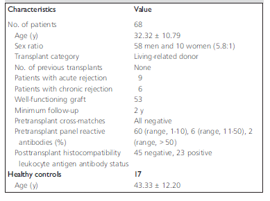
Table 1. Demographic profile of the study subjects.
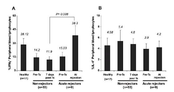
Figure 1, A and B. Percentage of frequencies of T cells expressing (A) interferon-γ or (B) interleukin 4 in renal ransplant recipients and in healthy controls. A quantity of 1-2 × 10⁶ peripheral blood lymphocytes was isolated from renal transplant recipients before transplant (Pre-Tx), 7 days after transplant, and during a rejection episode and also from healthy individuals. Those cells were stimulated for 6 hours with horbol myristate acetate (5 ng/mL) and ionomycin (2 mM) in the presence of monensin (2 µM), and were hen stained to reveal intracellular interferon-γ and interleukin 4 before analysis via flow cytometry as described in the Materials and Methods section.
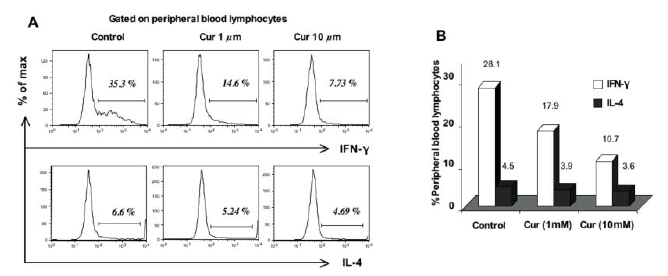
Figure 2, A and B. Dose-dependent effect of curcumin on the frequency of interferon-γ and interleukin 4 expression in peripheral blood lymphocytes from healthy controls. Peripheral blood lymphocytes from healthy controls were pretreated with 1 µM or 10 µM of curcumin for 3 hours and were then stimulated with phorbol myristate acetate and ionomycin. The cells were stained to reveal interferon-γ and interleukin 4 and were analyzed with flow cytometry as described in the Materials and Methods section. (A) Representative flow cytometry histogram plots indicating the reduction of interferon-γ expression and the only marginal effect of curcumin on interleukin 4 expression. (B) Cumulative data on curcumin-treated peripheral blood lymphocytes expressing interferon-γ and interleukin 4 in 10 healthy individuals.
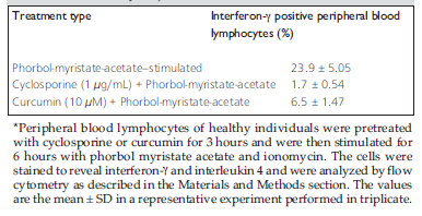
Table 2. Comparative analysis of cyclosporine and curcumin-induced inhibition in the study subjects*.
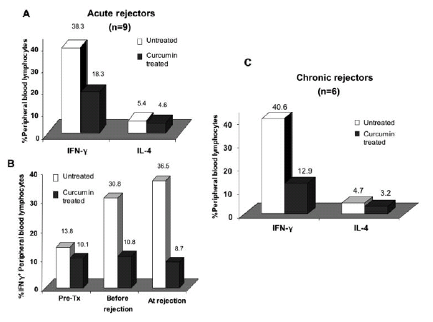
Figure 3, A–C. Effect of curcumin on the frequency of interferon-γ and interleukin 4 expression in the peripheral blood lymphocytes of renal transplant recipients experiencing acute or chronic rejection. Peripheral blood lymphocytes isolated from patients with acute graft rejection were treated with curcumin (10 µM) for 3 hours and were then stimulated for 6 hours with phorbol myristate acetate and ionomycin. The cells were stained and then analyzed, via flow cytometry as described in the Materials and Methods section, for peripheral blood lymphocytes expressing interferon-γ and interleukin 4. (A) Cumulative data on curcumin-treated peripheral blood lymphocytes expressing interferon-γ and interleukin 4 in 9 patients who experienced an acute rejection episode. (B) The effect of curcumin on interferon-γ expression by peripheral blood lymphocytes of renal transplant recipients before transplant and after transplant without and with an acute rejection episode. (C) Peripheral blood lymphocytes from renal transplant recipients expressing rejection episode after 1 year or more were pretreated with 10 µM of curcumin and were then stimulated with phorbol myristate acetate and ionomycin. The cells were stained and were then analyzed, via flow cytometry as described in the Materials and Methods section, to reveal peripheral blood lymphocytes expressing interferon-γ and interleukin 4. The plot shows cumulative data on 6 patients experiencing chronic rejection.
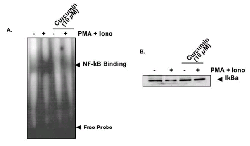
Figure 4, A and B. Effect of curcumin on the induction of nuclear factor kappa beta and I kappa B alpha degradation. Peripheral blood lymphocytes isolated from 5 healthy donors were pretreated with curcumin (10 µM) for 3 hours and were then stimulated for 1 hour with phorbol myristate acetate and ionomycin. Nuclear and cytoplasmic protein extracts from those cells were prepared and tested for nuclear factor kappa beta deoxyribonucleic acid binding activity (A) and the expression of I kappa B alpha (B), respectively, as described in the Materials and Methods section.
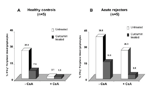
Figure 5, A and B. Synergistic inhibitory effect of curcumin and cyclosporine on peripheral blood lymphocytes from acute renal transplant rejectors. Peripheral blood lymphocytes from (A) healthy controls (n = 5) or (B) acute renal allograft rejectors (n = 5) were treated with cyclosporine (1 µg/mL) in the absence or presence of curcumin (10 µM) for 3 hours before polyclonal stimulation with phorbol myristate acetate and ionomycin. The cells were then stained and were analyzed, with flow cytometry, for peripheral blood lymphocytes expressing interferon-γ and interleukin 4.