Volume: 8 Issue: 2 June 2010
FULL TEXT
Kidney Transplant in Highly Sensitized Patients After Desensitization With Plasmapheresis and Low-dose Intravenous Immunoglobulin
Materials and Methods: Thirty-five highly sensitized patients (HLA class I panel reactive antibody > 50%) received plasmapheresis, plus low-dose intravenous immunoglobulin treatment. In 25 patients (group 1), a positive T- and/or B-cell cytotoxicity crossmatch was rendered negative by plasmapheresis, plus low-dose intravenous immunoglobulin treatment. Two patients did not receive renal transplants owing to persistent positive crossmatch. Eight patients already had a negative crossmatch before desensitization. During the same time, 32 highly sensitized patients (group 2), without desensitization, had a negative crossmatch and received deceased-donor renal transplants.
Results: Group 1 showed a numerically higher rate of acute rejection (32.0% vs
21.9%; P = .6) and antibody-mediated rejection (20.0% vs 9.4%; P = .3), but the
difference was not statistically significant. Four of 5 cases of antibody-mediated
rejection in group 1 had a peak donor specific antibody titer
≥
1:8. Comparable mean serum creatinine levels at 24 months were observed
(group 1: 130 ± 38 µmol/L vs group 2: 123 ± 41 µmol/L; P = .5). No difference in
Kaplan-Meier graft survival was found between group 1 and group 2 after follow-up
of
52 ± 26 months (P = .7).
Conclusions: Desensitization with plasmapheresis, plus low-dose intravenous immunoglobulin enables successful deceased-donor renal transplant in highly sensitized patients with a positive crossmatch. Antibody-mediated rejection occurred predominantly in recipients with donor-specific antibodies of high titers.
Key words : HLA antibodies, Kidney transplantation, Antibody-mediated rejection
A positive crossmatch indicates the presence of donor-specific antibodies to human leucocyte antigen in the serum of a potential recipient, and is associated with a rate of graft loss that exceeds 80% (1, 2). Alloantibodies develop after exposure to foreign human leucocyte antigen molecules, usually through pregnancy, transfusion, and previous organ transplant. Positive lymphocyte crossmatch represents an immunologic barrier to kidney transplant in highly sensitized patients. Recently, 2 regimens have evolved to overcome this barrier: the high-dose intravenous immunoglobulin-based protocol, and plasmapheresis plus low-dose intravenous immunoglobulin protocol (3-5). The former protocol has been used in living-donor and deceased-donor renal transplants (3). However, the latter was mainly reported in living-donor renal transplants because the time to transplant cannot be predicted in deceased transplants (4, 5).
Because there is no national organ allocation organization, it is difficult to find highly matched kidney donors for highly sensitized patients. Highly sensitized patients must remain on the waiting list for extended periods for a suitable crossmatch negative organ. Desensitization provides an alternative approach to this problem. Here, we present our results of successful kidney transplant in highly sensitized patients from deceased-donors after desensitization with plasmapheresis, plus low-dose intravenous immunoglobulin.
Materials and Methods
Patient demographics
Between January 2001 and December 2007, 35 highly sensitized patients (human
leucocyte antigen class I panel reactive antibodies > 50%) waiting for deceased-donor
renal transplants received plasmapheresis, plus low-dose intravenous
immunoglobulin treatment. In 25 patients (group 1), a positive crossmatch
between the donor’s lymphocyte and the recipient’s historic sera was rendered
negative. Two patients did not receive renal transplants owing to persistent
positive crossmatch. Eight patients already had a negative crossmatch before
plasmapheresis, plus low-dose intravenous immunoglobulin treatment, and received
renal transplants. During the same time, 32 highly sensitized patients (group 2)
without desensitization treatment received renal transplants with negative
crossmatch. Mean follow-up of the patients was
52 ± 26 months (range, 6-105 months). Prior to the study, the study protocol,
which conforms with the ethical guidelines of the 1975 Helsinki Declaration, was
approved by our local institutional ethics committee. Written informed consent
was obtained from all of the subjects. The demographics and immunologic factors
of the 2 groups are listed in Table 1.
Cytotoxicity crossmatch
Complement-dependent cytotoxicity crossmatch was performed by a modified
National Institutes of Health complement-dependent cytotoxicity method using
serial doubling dilutions of recipient serum. Magnetically sorted T- and B-lymphocytes
were incubated with sera for 60 minutes, followed by 120 minutes further
incubation with rabbit compliment. The last reaction resulting in greater than
10% cell death above background was considered to be the anti-donor titer. We
did not routinely perform an auto-crossmatch. The contribution of IgM was ruled
out by testing in the presence of dithiothreitol. Anti-human globulin
enhancement was not used. When donors became available, cross-matching was done
on all serum samples collected before and after desensitization treatment, an
acceptable crossmatch was defined as negative current T- and B-cell, complement-dependent
cytotoxicity crossmatch.
Specific analysis of human leucocyte antigen antibodies
All pretransplant sera were screened by enzyme-linked immunosorbent assay (ELISA)
assays (LAT-M, One Lambda, Inc., Canoga Park, CA, USA) to determine the presence
or absence of anti-human leucocyte antigen class I and/or class II antibodies of
the IgG isotype. The presence of donor-specific antibodies was retrospectively
screened by human leucocyte antigen-specific ELISA assays. Identification of
anti-human leucocyte antigen class I and class II antibody specificities were
performed using ELISA kits LAT-1HD and LAT 2–40 (One Lambda). Both ELISA tests
were performed as recommended by the manufacturer.
Desensitization protocol
Before transplant, patients received plasmapheresis 3 times weekly. One plasma
volume exchange with 5% albumin was used, with a membrane apheresis device (Plasmaflux
P2S, Fresenius Medical Care Ltd., Bad Homburg, Germany). Standard intravenous
immunoglobulin (Gamimune N 10%, Bayer Biological, Elkhart, IN, USA) 100 mg/kg
was given after each plasmapheresis treatment to suppress alloantibody rebound.
All patients received tacrolimus (Prograf, Fujisawa Ireland Limited, Killorglin,
Ireland) (0.1 mg/kg/d) and mycophenolate mofetil (Cellcept, Roche, Shanghai,
China) (1.5 g/d) on the day of the first plasmapheresis, plus low-dose
intravenous immunoglobulin session. The dosage of tacrolimus was adjusted to
maintain a target level of
8 to 10 ng/mL. In addition, all recipients in group 1 underwent 1 to 3 sessions
of plasmapheresis, plus low-dose intravenous immunoglobulin after transplant.
Two patients in group 1 with panel reactive antibody > 80% received a single
dose of rituximab (375 mg/m2; Rituxan Genentech, Inc., San Francisco, CA, USA)
on day 1.
Immunosuppressive therapy
All patients received induction therapy with single dose of daclizumab (Zenapax,
Roche, Nutley, NJ)
50 mg immediately before transplant operation. All patients also received
induction therapy with 3 methylprednisolone pulses (3 × 500 mg) and
antithymocyte globulin (ATG, Fresenius Medical Care Ltd., Bad Homburg, Germany)
1.5 mg/kg/day up to 7 days (range, 5-7 days). Subsequent maintenance
immunosuppression consisted of prednisone
20 mg/day with a taper to 10 mg/day by 3 months posttransplant, mycophenolate
mofetil 750 mg twice daily, and tacrolimus to maintain a target level of 8 to 10
ng/mL for the first 3 months, 6 to 8 ng/mL for months 3 to 6, and 5 to 7 ng/mL
after 6 months.
Diagnosis and treatment of acute rejection
Diagnosis of acute rejection was confirmed by kidney biopsy and kidney pathology
was classified using Banff 97 criteria. The diagnostic criteria for antibody-mediated
rejection included clinical evidence of acute graft dysfunction, histologic
evidence of tissue injury, immunopathologic evidence for antibody action (C4d in
peritubular capillaries), and serologic evidence of anti-human leucocyte antigen
antibody at time of biopsy (6). Antibody-mediated rejection was treated with
plasmapheresis, plus low-dose intravenous immunoglobulin every other day for
1 to 5 sessions, plus anti-thymocyte globulin
1.5 mg/kg/day for 3 to 5 days. Patients with acute cellular rejection were
treated by 3 methylprednisolone pulses (3 × 500 mg).
Statistical analyses
Results are expressed as numerical values and percentages for categorical
variables and as mean ± standard deviation for continuous variables. Statistical
analyses were performed with SPSS software for Windows (Statistical Product and
Service Solutions, version 16.0, SPSS Inc, Chicago, IL, USA). Comparisons were
based on the chi-square test for categorical data, the t test for normally
distributed continuous data, and the Mann-Whitney U tests for ranked data. The
Mantel cox log-rank test was used to compare Kaplan-Meier graft survival between
groups. Results were considered significant when P was less than .05.
Results
Complement-dependent cytotoxicity crossmatch results and donor-specific antibody
specificities
In patients of group 1, both T- and B-cell positive crossmatch with historic
sera were found in 21 patients, only B-cell positive crossmatch with historic
sera was found in 4 patients. Both human leucocyte antigen class I and class II
donor-specific antibodies were found in 2 patients, only class I donor-specific
antibodies were present in 17 patients, and only class II donor-specific
antibodies were present in 4 patients. In 2 patients, no definite anti-donor
specificity was identified (Table 2).
Efficacy of the desensitization protocol
A positive crossmatch with historic sera was rendered negative in 92.6% patients
(25/27). In patients with both positive T- and B-cell crossmatch, the highest
titer was adopted for analysis. The majority (68.0%) of successfully
desensitized recipients had a donor-specific antibodies titer less than 1:8.
Panel reactive antibody level of human leucocyte antigen class I antibodies was
decreased from 77% ± 14% to 36% ± 17% (P < .001); panel reactive antibody level
of human leucocyte antigen class II antibodies was decreased from 52% ± 33% to
37% ± 30% (P < .001). The number of total plasmapheresis, plus low-dose
intravenous immunoglobulin sessions, before transplant, was significantly larger
than the number of sessions needed to achieve a negative crossmatch (6.2 ± 2.1
vs 3.6 ± 1.7, P < .001).
Acute rejection episodes
Group 1 showed a higher rate of acute rejection (32.0% vs 21.9%, P = .6) and
antibody-mediated rejection (20.0% vs 9.4%, P = .3), but the difference was not
statistically significant. All antibody-mediated rejection episodes were
reversed by plasmapheresis, plus low-dose intravenous immunoglobulin and anti-thymocyte
globulin treatment. Delayed graft function occurred in 1 patient after antibody-mediated
rejection in group 1. The peak donor-specific antibody titers of 5 recipients
with antibody-mediated rejection in group 1 were 1:4, 1:8, 1:16 (n=2), and 1:128
respectively (Table 2).
Main outcomes
Comparable mean serum creatinine levels at
12 months (group 1: 112 ± 18 µmol/L vs group
2: 107 ± 16 µmol/L, P = .3) and 24 months (group 1: 130 ± 38 µmol/L vs group 2:
123 ± 41 µmol/L, P = .5) were observed. No difference in Kaplan-Meier graft
survival rate (patient deaths included) was found between group 1 and group 2
after follow-up of
52 ± 26 months (P = .7) (Figure 1).
Discussion
Our study indicates that highly sensitized patients with positive lymphocyte complement-dependent cytotoxicity crossmatch, desensitized with plasmapheresis, plus low-dose intravenous immunoglobulin, can achieve results similar to highly sensitized patients with negative crossmatch. We chose the plasmapheresis, plus low-dose intravenous immunoglobulin protocol, according to the early reports of Montgomery and associates and Schweitzer and associates (4, 5). Effect of plasmapheresis is short-lived, and a rebound occurs usually when plasmapheresis is discontinued. Plasmapheresis, in combination with intravenous immunoglobulin, produces durable, donor-specific antibody suppression. Intravenous immunoglobulin is known to activate anti-idiotypic circuits, suppress endogenous antibody secretion, and inhibit complement activity.
But if the transplant does not follow closely on the heels of the preconditioning, donor-specific antibodies may return. Kidney graft may be critical for antibody elimination to occur and be sustained (7). Another explanation for the persistent suppression of donor-specific antibodies is immunosuppressive therapy. Immunosuppressive agents can suppress antibody production in experimental studies and clinical observation (7, 8).
One of the disadvantages of desensitization is the high rate of antibody-mediated rejection. The antibody-mediated rejection rate of our study (20%) is comparable to reported rates of 21% to 37% of plasmapheresis, plus low-dose intravenous immunoglobulin protocols (9, 10). In our study, the number of total plasmapheresis, plus low-dose intravenous immunoglobulin treatments before transplant was significantly larger than the number of treatments for achieving a negative crossmatch, which decreased the donor-specific antibodies to more-acceptable level, and contributed to less antibody-mediated rejection episodes.
We observed that 4 of 5 antibody-mediated rejection cases had a peak donor-specific antibodies titer ≥ 1:8, indicating that donor-specific antibodies of a high titer were not eliminated to optimal level. The association between baseline donor-specific antibody titer and antibody-mediated rejection also was proved by another report (11). Using more-sensitive single antigen Luminex bead assay, donor-specific antibodies levels expressed as standard fluorescence intensity in patients with antibody-mediated rejection were significantly higher than they were in patients without antibody-mediated rejection, patients with donor-specific antibodies greater than 105 standard fluorescence intensity are at higher risk for antibody-mediated rejection (12).
In the current study, we used anti-thymocyte globulin for induction. In sensitized recipients without desensitization, anti-thymocyte globulin induction has been proved to be effective at a lower incidence of biopsy-proven acute rejection episodes, increased 1-year graft survival, and improved graft function (13). But in desensitized patients with donor-specific antibodies, anti-thymocyte globulin induction did not show efficacy in preventing antibody-mediated rejection (9, 10). Akalin and associates (14) showed that the addition of plasmapheresis to high-dosage intravenous immunoglobulin dramatically decreased the incidence of acute rejection from 66% (44% antibody-mediated rejection) to 7% but the crossmatch results of their patients were T-cell crossmatch negative but complement-dependent cytotoxicity B-cell and/or flow cytometry crossmatch positive, which were considered as milieus only with a lower titer of donor-specific antibodies, or nonhuman leucocyte antigen antibodies.
There are some limitations of our study. First, patients who could not receive a renal graft in 4 to 6 weeks had to go back on the waiting list and receive high-dose intravenous immunoglobulin to prevent donor-specific antibody rebound. Second, posttransplant monitoring of the donor-specific antibody levels could not be accomplished because donor material for posttransplant crossmatching was not available. Third, surveillance biopsy was not routinely performed, so the rate of subclinical antibody-mediated rejection was unknown, which may contribute to development of chronic allograft nephropathy (15).
In summary, our study proved the efficacy of a plasmapheresis, plus low-dose intravenous immunoglobulin protocol in highly sensitized patients. Desensitization with plasmapheresis, plus low-dose intravenous immunoglobulin protocols provide an alternative approach for highly sensitized patients waiting for deceased donors. It will be logistic and cost-effective in combination with the acceptable mismatch program (16).
References:
- Patel R, Terasaki PI. Significance of the positive crossmatch test in kidney transplantation. N Engl J Med. 1969;280(14):735-739.
- Williams GM, Hume DM, Hudson RP Jr, Morris PJ, Kano K, Milgrom F. "Hyperacute" renal-homograft rejection in man. N Engl J Med. 1968;279(12):611-618.
- Jordan SC, Vo A, Bunnapradist S, et al. Intravenous immune globulin treatment inhibits crossmatch positivity and allows for successful transplantation of incompatible organs in living-donor and cadaver recipients. Transplantation. 2003;76(4):631-636.
- Montgomery RA, Zachary AA, Racusen LC, et al. Plasmapheresis and intravenous immune globulin provides effective rescue therapy for refractory humoral rejection and allows kidneys to be successfully transplanted into cross-match-positive recipients. Transplantation. 2000;70(6):887-895.
- Schweitzer EJ, Wilson JS, Fernandez-Vina M, et al. A high panel-reactive antibody rescue protocol for cross-match-positive live donor kidney transplants. Transplantation. 2000;70(10):1531-1536.
- Takemoto SK, Zeevi A, Feng S, et al. National conference to assess antibody-mediated rejection in solid organ transplantation. Am J Transplant. 2004;4(7):1033-1041.
- Montgomery RA, Zachary AA. Transplanting patients with a positive donor-specific crossmatch: a single center's perspective. Pediatr Transplant. 2004;8(6):535-542.
- Kozlowski T, Tibell A, Morpurgo E, Sundberg B, Sundin U, Groth C. Suppression of immunoglobulin resynthesis after plasmapheresis: efficacy of various immunosuppressive drugs--a study in rats. Transplant Proc. 1995;27(6):3545-3546.
- Stegall MD, Gloor J, Winters JL, Moore SB, Degoey S. A comparison of plasmapheresis versus high-dose IVIG desensitization in renal allograft recipients with high levels of donor specific alloantibody. Am J Transplant. 2006;6(2):346-351.
- Vo AA, Toyoda M, Peng A, Bunnapradist S, Lukovsky M, Jordan SC. Effect of induction therapy protocols on transplant outcomes in crossmatch positive renal allograft recipients desensitized with IVIG. Am J Transplant. 2006;6(10):2384-2390.
- Gloor JM, DeGoey SR, Pineda AA, et al. Overcoming a positive crossmatch in living-donor kidney transplantation. Am J Transplant. 2003;3(8):1017-1023.
- Reinsmoen NL, Lai CH, Vo A, et al. Acceptable donor-specific antibody levels allowing for successful deceased and living donor kidney transplantation after desensitization therapy. Transplantation. 2008;86(6):820-825.
- Thibaudin D, Alamartine E, de Filippis JP, Diab N, Laurent B, Berthoux F. Advantage of antithymocyte globulin induction in sensitized kidney recipients: a randomized prospective study comparing induction with and without antithymocyte globulin. Nephrol Dial Transplant. 1998;13(3):711-715.
- Akalin E, Dinavahi R, Friedlander R, et al. Addition of plasmapheresis decreases the incidence of acute antibody-mediated rejection in sensitized patients with strong donor-specific antibodies. Clin J Am Soc Nephrol. 2008;3(4):1160-1167.
- Haas M, Montgomery RA, Segev DL, et al. Subclinical acute antibody-mediated rejection in positive crossmatch renal allografts. Am J Transplant. 2007;7(3):576-585.
- Claas FH, Rahmel A, Doxiadis II. Enhanced kidney allocation to highly sensitized patients by the acceptable mismatch program. Transplantation. 2009;88(4):447-452.

Volume : 8
Issue : 2
Pages : 130 - 135
From the 1Department of Organ Transplant, First Affiliated Hospital of Sun
Yat-sen University, Guangzhou; and the 2Department of Organ Transplant, Taiping
People’s Hospital, Dongguan, China
Address reprint requests to: Chang-xi Wang, Department of Organ Transplant,
First Affiliated Hospital of Sun Yat-sen University, 58 Zhongshan 2nd Road,
Guangzhou, 510080, China
Phone: +86 13600450862
Fax: +86 20 87335825
E-mail: wcx6363@163.com
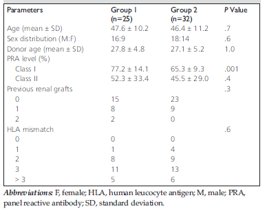
Table 1. Patients demographics and immunologic data.
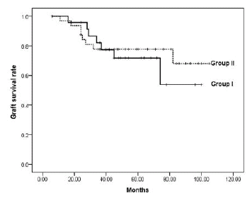
Figure 1. Comparison of group 1 (solid line) and group 2 (dashed line) patients for graft survival rate (patient deaths included). TheMantel-Cox log-rank test was used to compare Kaplan-Meier graft survival between groups (P = .7).
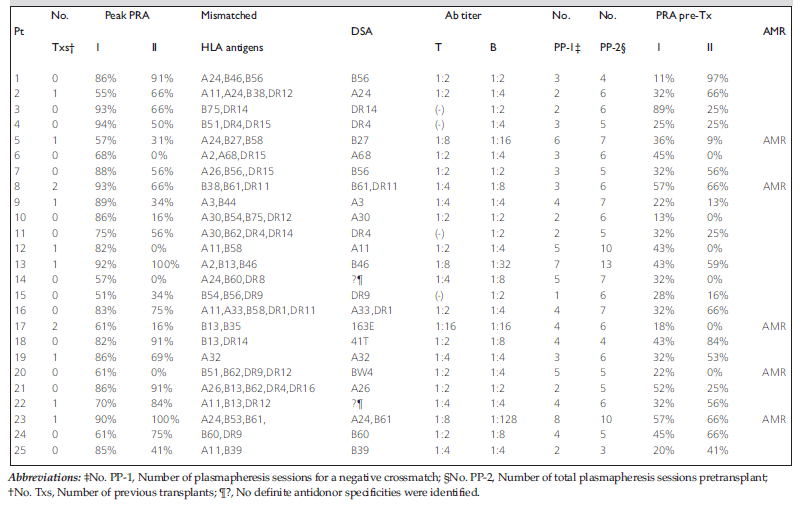
Table 2. Baseline serologic characteristics of desensitized patients.