Volume: 6 Issue: 4 December 2008
FULL TEXT
Objectives: Renal allograft dysfunction can be caused by renal vessel thrombosis, acute tubular necrosis, hyperacute or acute rejection, nephrotoxicity induced by cyclosporine or tacrolimus, thrombotic microangiopathy, or urinary tract obstruction.
Materials and Methods: We describe a renal transplant recipient in whom oliguria developed during the first week after transplant, although his early renal allograft function was good.
Results: A Doppler ultrasonographic study revealed a lack of perfusion in the lower pole of the allograft. A perfusion defect was noted in the lower pole that was supplied by a polar artery, which had been damaged during engraftment. Light microscopy disclosed tubular cell necrosis without evidence of vascular or humoral rejection.
Conclusions: We suggest that toxic molecules such as tumor necrosis factor-alpha released from a segmental infarcted area can induce tubular cell damage and necrosis leading to renal allograft dysfunction.
Key words : Kidney, Infarction, Rejection, Transplant, Tubular necrosis
Renal allograft dysfunction that occurs during the first week after transplant surgery could be due to severe hypovolemia, renal vessel thrombosis, acute tubular necrosis, hyperacute or acute rejection, nephrotoxicity induced by cyclosporine or tacrolimus, thrombotic microangiopathy, or a urinary tract obstruction (1). Segmental renal infarction, which is a poorly characterized complication of renal transplant that often develops during the early postoperative period, is due to the disruption or thrombosis of the renal arterial branches. Segmental renal infarction may produce no symptoms and is associated with allograft dysfunction or rejection (2). In this report, we describe a patient with renal allograft dysfunction caused by segmental infarction. We suggest that necrotic material may have induced tubular cell damage in this individual.
Case Report
A 30-year-old man received a living-unrelated renal transplant after 2 years of end-stage renal disease (ESRD) that previously, had been treated with hemodialysis. The cause of his ESRD was chronic glomerulonephritis. Postsurgical treatment included immunosuppressive therapy with an interleukin-2 receptor blocker (basiliximab), a steroid, mycophenolate mofetil, and cyclosporine. His renal allograft demonstrated good function immediately after surgery and produced more than 10 L urine on the first posttransplant day. On the second posttransplant day, however, his urine output decreased, and his blood pressure increased from 130/80 to 180/120 mm Hg. Atenolol was prescribed to reduce his hypertension. Laboratory analyses revealed the following results: white blood cell count, 11 × 109/L (11 000/µL); platelet count, 256 × 109/L (256 000/µL); hemoglobin, 95 g/L (9.5 g/dL); total bilirubin, 18.8 µmol/L (1.1 mg/dL) (normal range, < 20.5 µmol/L); prothrombin time, 13 sec; and serum creatinine level, 530.4 µmol/L (6 mg/dL). On the fourth posttransplant day, laboratory testing yielded the following values: serum lactic dehydrogenase, 1730 U/L (normal range, 250-500 U/L); alanine aminotransferase, 120 U/L (normal range, 5-40 U/L); and aspartate aminotransferase, 160 U/L (normal range, 5-40 U/L). Ultrasonographic evaluation showed no urinary tract obstruction. Doppler ultrasonography revealed an intact main renal artery and vein, but a lack of perfusion in the lower pole of the allograft (Figure 1). The lower pole was supplied by a polar artery, which has been damaged during engraftment (Figure 2).
On the fifth posttransplant day, the patient’s urine output decreased to less than 0.5 L/d. Acute rejection was diagnosed, and treatment with hemodialysis and antithymocyte globulin was initiated. On the seventh posttransplant day, an ultrasonographically guided renal biopsy specimen was obtained from the upper pole of the allograft. Light microscopy revealed detached, vacuolized tubular cells and necrosis. Neither interstitial cellular infiltration nor tubulitis was noted. The glomeruli and vessels (Figure 3) were normal. The results of immunofluorescent staining to detect peritubular capillary deposits of C4d (as described in reference 3) were negative. Plasmapheresis, which was initiated on the tenth posttransplant day, was continued for 6 days. At the third week after transplant, the patient’s serum creatinine levels remained higher than 884 µmol/L (10 mg/dL), and his urine output was less than 200 mL/d. A second renal biopsy that was performed on the twenty-sixth posttransplant day revealed tubular cells, necrosis, and vacuolization with interstitial fibrosis. At that time, Doppler ultrasonography showed upper-pole perfusion that was within normal limits and the return of weak perfusion in the lower pole of the allograft. On the posttransplant day 32, all immunosuppressive therapy was discontinued, and the patient continued to receive treatment with hemodialysis.
Discussion
In this report, we described a renal transplant recipient whose renal allograft function was good immediately after surgery but worsened shortly thereafter. A lack of perfusion in the lower pole of the allograft indicated an infarction. The results of a renal allograft biopsy suggested acute tubular necrosis. We found no sign of acute humoral rejection or acute cellular rejection. In a few patients with acute humoral rejection, an allograft biopsy showed acute tubular necrosis as the most significant finding (4), but we found no peritubular C4d deposition that would indicate acute humoral rejection. Other histologic findings of acute humoral rejection, such as neutrophil margination within the glomeruli, a fibrin thrombus, or glomerular or arterial fibrinoid necrosis (4) also were absent, as were interstitial mononuclear cell infiltration and tubulitis, which are the classic histologic findings of acute cellular rejection (4).
We suggest that during renal segmental infarction, toxic substances (described below) released from the necrotic area can infiltrate the renal cortex and act as endogenous tubular toxins. The increase in blood pressure that occurred in our patient on the second posttransplant day could be explained by the sudden release of renin from the necrotic and ischemic areas in the allograft. Elevated levels of aspartate aminotransferase, alanine aminotransferase, and serum lactic dehydrogenase suggested an infarction in the allograft (2).
During prolonged and severe renal ischemia, production of tumor necrosis factor alpha (TNF-α) increases (5, 6). TNF-α directly triggers apoptosis of renal tubular cells and indirectly increases renal vasoconstriction and ischemia by increasing the endothelin level (7, 8). TNF-α also has been implicated in the pathogenesis of ischemic and nephrotoxic acute renal failure (7, 8). Necrotic cells release other toxic substances (eg, heat shock proteins HSP70 and HSP60) that can induce future tissue damage via Toll-like receptors (9-11). There are similarities between acute tubular necrosis induced by necrotic debris and acute tubular necrosis associated with gram-negative sepsis. Lipopolysaccharides and other endotoxins can damage renal tubular cells by creating a storm of cytokines and chemokines (12, 13). Toxic substances such as TNF-α released from the infarcted area also can cause renal tubular cell damage and necrosis. In our opinion, renal segmental infarction should be considered in the differential diagnosis of renal allograft dysfunction.
References:
- Halloran PF, Hunsicker LG. Delayed graft function: state of the art, November 10-11, 2000. Summit meeting, Scottsdale, Arizona, USA. Am J Transplant. 2001;1(2):115-120.
- Kanchanabat B, Siddins M, Coates T, et al. Segmental infarction with graft dysfunction: an emerging syndrome in renal transplantation? Nephrol Dial Transplant. 2002;17(1):123-128.
- Regele H, Exner M, Watschinger B, et al. Endothelial C4d deposition is associated with inferior kidney allograft outcome independently of cellular rejection. Nephrol Dial Transplant. 2001;16(10):2058-2066.
- Racusen LC, Haas M. Antibody-mediated rejection in renal allografts: lessons from pathology. Clin J Am Soc Nephrol. 2006;1(3):415-420.
- Kelly KJ. Distant effects of experimental renal ischemia/reperfusion injury. J Am Soc Nephrol. 2003;14(6):1549-1558.
- Tsuruya K, Ninomiya T, Tokumoto M, et al. Direct involvement of the receptor-mediated apoptotic pathways in cisplatin-induced renal tubular cell death. Kidney Int. 2003;63(1):72-82.
- Wolfs TG, Buurman WA, van Schadewijk A, et al. In vivo expression of Toll-like receptor 2 and 4 by renal epithelial cells: IFN-gamma and TNF-alpha mediated up-regulation during inflammation. J Immunol. 2002;168(3):1286-1293.
- Ramesh G, Reeves WB. TNFR2-mediated apoptosis and necrosis in cisplatin-induced acute renal failure. Am J Physiol Renal Physiol. 2003;285(4):F610-F618.
- Li M, Carpio DF, Zheng Y, et al. An essential role of the NF-kappa B/Toll-like receptor pathway in induction of inflammatory and tissue-repair gene expression by necrotic cells. J Immunol. 2001;166(12):7128-7135.
- Leemans JC, Stokman G, Claessen N, et al. Renal-associated TLR2 mediates ischemia/reperfusion injury in the kidney. J Clin Invest. 2005;115(10):2894-2903.
- Wu H, Chen G, Wyburn KR, et al. TLR4 activation mediates kidney ischemia/reperfusion injury. J Clin Invest. 2007;117(10):2847-2859.
- Schor N. Acute renal failure and the sepsis syndrome. Kidney Int. 2002;61(2):764-776.
- Cunningham PN, Wang Y, Guo R, He G, Quigg RJ. Role of Toll-like receptor 4 in endotoxin-induced acute renal failure. J Immunol. 2004;172(4):2629-2635.

Volume : 6
Issue : 4
Pages : 312 - 314
From the 1Department of Nephrology, Tabriz University (Medical Sciences), Tabriz, Iran; the
2Department of Internal Medicine, Shahrkurd University of Medical Sciences, Shahrkurd, Iran; and the
3Tuberculosis and Lung Disease Research Center, Tabriz University (Medical Sciences), Tabriz, Iran
Address reprint requests to: Mohammad Reza Ardalan, MD, Department of Nephrology, Tabriz University (Medical Sciences), Tabriz, Iran
Phone: +98 9141168518
Fax: +411 3344280
E-mail: ardalan34@yahoo.com
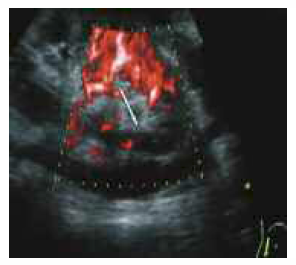
Figure 1. Power Doppler Ultrasound study of the renal allograft revealed lack of perfusion in the lower pole region of the renal allograft.
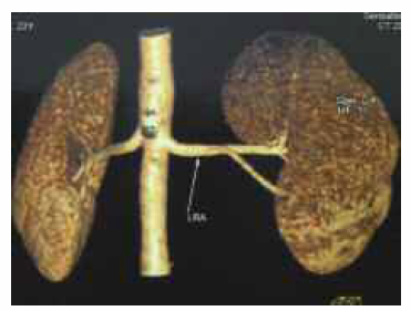
Figure 2. CT angiography of donor kidney. Lower pole of the left kidney was supplied by a polar artery. It was damaged during engraftment.
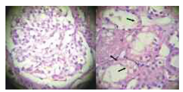
Figure 3. Light microscopic study of the allograft biopsy disclosed tubular cells necrosis, detachment, vacuolization, and coagulation necrosis (black arrows). Interstitial infiltrations and tabulitis were absent. Glomerulus and Vessels were relatively normal. Hematoxylin and Eosin 40×10.