Volume: 6 Issue: 4 December 2008
FULL TEXT
Objectives: Pulmonary complications after liver transplant significantly affect mortality and morbidity; however, their relation has not been clearly established. We sought to determine pulmonary complications during the early and late term after liver transplant and identify risk factors for mortality.
Materials and Methods: At our institution, 130 liver transplant patients (mean age, 40.1 ± 14.6 years; 71.1% male) were retrospectively evaluated, and 114 adult orthotopic liver transplant patients were included. Cause of liver disease, pulmonary function test results, arterial blood gas analyses, surgery duration, length of stay in the intensive care unit and the hospital, pulmonary complications, and mortality causes were noted.
Results: Pulmonary complications were detected in 48 patients (42.1%), pneumonia in 24 patients (21.1%), and pleural effusion in 21 patients (18.4%). Development of pulmonary complications was found to be significantly related to survival (P = .001). Fifty-two patients (45.6%) were smokers, a significant predictor of pulmonary complications (P = .03). There was no relation between pulmonary function test results and orthodeoxia and pulmonary complications and mortality. Early and late survival rates were significantly lower in patients in whom a microorganism was isolated on deep tracheal aspirate culture, while early survival was significantly reduced in the presence of a pleural effusion (P < .005).
Conclusions: Pulmonary complications after liver transplant are common. Care must be taken to determine preoperative risk factors, and patients should be observed closely for development of respiratory complications after liver transplant.
Key words : Liver transplant, Pulmonary complications, Mortality
Liver transplant is a successful treatment for patients with acute liver failure and end-stage liver cirrhosis, but the procedure carries with it risks for morbidity and mortality. Postoperative pulmonary complications contribute to the morbidity and mortality in liver transplant recipients; however, the risk factors related with these complications have not been completely defined. This study seeks to assess the risk factors for short- and long-term pulmonary complications as well as the rate of mortality related to pulmonary complications in adult liver transplant recipients and to determine the variables associated with pulmonary complications and mortality.
Materials and Methods
From January 1999 to December 2007, a total of 230 liver transplants were done at the Baskent University Hospital in Ankara, Turkey. Patients younger than 16 years (n=97) and those who underwent re-transplant (liver) (n=3) were excluded. The records of 130 adult liver transplant recipients were retrospectively evaluated. Patients in whom a heterotopic adult liver transplant had been done (n=12), those who had undergone a retransplant (n=3), and those whose data were not available (n=1) were excluded. Thus, 114 adult orthotopic liver transplant patients were included. The surgical technique for orthotopic liver transplant has been described (1, 2). Intravenous methylprednisolone (10 mg/kg), administered on the day of the transplant, was tapered rapidly over the next 7 to 10 days. Maintenance prednisone (0.25 mg/kg/d) was continued for 3 months when steroid therapy was stopped. Tacrolimus or cyclosporine was used for maintenance therapy. Maintenance dosages of tacrolimus were adjusted to maintain a level of 10 to 12 ng/mL during the first 2 months and 5 to 7 ng/mL thereafter. For cyclosporine maintenance, dosages to achieve trough levels of 150 to 250 ng/mL were prescribed in the first 2 months, and after that, 100 to 125 ng/mL were prescribed. All patients received sulfamethoxazole/ trimethoprim for prophylaxis against Pneumocystis carinii (Pneumocystis jiroveci) for 6 months, fluconazole for fungal infection for 6 months, acyclovir for antiviral prophylaxis for 3 months, and postoperative antibiotic treatment included cefotaxime and ampicillin for 3 days.
Pretransplant clinical status, in terms of severity of hepatic dysfunction, was determined using the Child-Pugh classification (3). The smoking status of each patient was recorded. Medical records, chest roentgenograms, pulmonary function test results, arterial blood gas analyses, duration of the operation, time to extubation, length of stay in the intensive care unit of all transplant recipients were recorded. Arterial blood gas analyses were done with the subjects breathing room air in an upright position at rest and lying down. Orthodeoxia was defined as decrease in the PaO2 value of more than 10 mm Hg from the lying position to sitting upright.
Short (< 90 days) and long-term (> 90 days) postoperative mortality and pulmonary complications were noted. Pleural effusion, pneumonia, acute respiratory failure, pulmonary edema, alveolar hemorrhage, and acute respiratory distress syndrome were considered pulmonary complications. These data were obtained by physical examination, chest radiograph, thoracic computed tomography, or bronchoscopic findings. Chest radiographs of the patients were reviewed preoperatively and within 10 days postoperatively. Pneumonia was defined as the presence of pulmonary infiltrate, fever, leukocytosis, and new onset of respiratory symptoms (cough, sputum, and dyspnea). Culturing of pulmonary secretions (sputum analysis, deep tracheal aspirate specimens, bronchoscopic evaluation including bronchoalveolar lavage) and blood samples were done for all patients with pneumonia. Acute respiratory distress syndrome was defined by bilateral infiltrates on chest radiograph, pulmonary artery wedge pressure less than 18 mm Hg, and a PaO2/FiO2 ratio less than 200. Atelectasis was defined if the pulmonary infiltrate regressed within 48 hours in the absence of an infiltrate cause. Pulmonary function tests were obtained as part of routine preoperative evaluation and on postoperative follow-up (approximately 9 months or 1 year after the transplant) in the sitting position. The percentage of forced vital capacity (FVC%), percentage of forced expiratory flow in 1 second (FEV1%), FEV1/FVC ratio, predicted forced midexpiratory flow rate (FEF25%-75%), total lung capacity, and residual volume were obtained. Single-breath carbon monoxide diffusing capacity was measured in each patient after the pulmonary function tests had been assessed. Diffusion lung capacity adjusted for hemoglobin concentration (DLAdj) and the other variables were measured with a clinical spirometer (SensorMedics Vmax spectra 229, Bilthoven, The Netherlands).
The study protocol complies with the Helsinki Declaration of 1975, and the research protocol was approved by the institutional review board.
Statistical Analyses
Statistical analyses were done with SPSS software (Statistical Product and Services Solutions, version 9.0, SPSS Inc, Chicago, IL, USA). Continuous variables are expressed as means ± standard deviation. Continuous variables were compared using the
t test, and the chi-square test was used to compare the qualitative variables. All
P values are 2-sided, and values for P less than .05 were considered statistically significant.
Results
The mean age of the study population was 40.1 ± 14.6 years, and 81 patients (71.1%) were male. Seventy-six transplants (66.7%) were from a living donor, and 38 (33.3%) were from a deceased donor. The mean surgery duration was 10.9 ± 1.6 hours; the mean time to extubation was 1.1 ± 1.0 days, mean length of stay in the intensive care unit was 3.4 ± 3.1 days, and total mean length of stay in the hospital was 38.0 ± 42.6 days. Smoking history was noted in 52 patients (45.6%). Supplementary oxygen was given to all patients while in the intensive care unit. The primary diagnosis in liver disease, Child-Pugh class, smoking history, and preoperative laboratory values of the study population are shown in Table 1.
The results of preoperative chest radiographs were normal in 71 patients (62.3%). In 43 patients (37.7%) in whom the chest radiograph findings were not normal, right diaphragmatic elevation was the most frequent disorder (n=29 patients; 25.4%). Pleural effusion was noted as the most common disorder on early postoperative chest radiographs, detected in 37 patients (32.5%) of which 22 (19.3%) were right sided. Mean serum albumin levels were similar in patients with and without pleural effusion (2.94 ± 0.51 vs 3.15 ± 0.61) (P = .2). A thoracentesis was done to 8 patients preoperatively (7%), with a removal of a mean of 1566 mL (200-2500 mL) pleural fluid. A pleural catheter was placed in a patient in whom a total of 2500 mL drainage was observed. Biochemical analysis of the fluid was transudative in 7 and exudative in 1 patient. No microorganism was identified on culture of the thoracentesis fluid. No complication developed after thoracentesis. Postoperatively, thoracentesis was done to 37 patients (32.5%), of these, the pleural fluid was exudative in 16 (43.2%), and a microorganism was identified on culture in 7 (18.9%). Compared with those with negative results on a thoracentesis culture, early mortality was greater in patients in whom the results of a thoracentesis culture were positive (10.0 vs 42.9, respectively; P = .03).
Pulmonary function tests were done in 98 patients (86%) preoperatively. The results were normal in 61 patients (62.2%); a restrictive pattern was detected in 25 (25.5%), and an obstructive pattern was noted in 12 patients (12.2%). Of the 85 patients to whom a diffusion test was given, diffusion capacity was decreased in 41 (48.2%). Control pulmonary function tests were done to 28 of 85 patients (32.9%); the results were normal in 22 (78.6%), obstructive in 3 (10.7%), and restrictive in 3 patients (10.7%). Diffusion capacity was evaluated in 18 patients (21.2%) postoperatively; in 11 patients (61.1%), it was decreased. There was no significant difference between preoperative and postoperative DLAdj values (79.0 ± 14.1 and 77.9 ± 10.0; P = .7). The decrease in preoperative diffusion capacity was not predictive of the development of postoperative pulmonary complications and mortality (P > .05).
Arterial blood gas analyses were obtained in 91 patients, hypoxia was detected in 32 (35.2%) (pO2 < 80 mm Hg). No relation was noted between preoperative hypoxia and the rates of mortality and pulmonary complications. With regard to preoperative values, after the operation, the decrease in pH and the increase in pCO2 and pO2 values were significant (P < .05), while change in HCO3 and oxygen saturation were not significant (P > .05). Respiratory alkalosis associated with severe liver disease resolved after successful transplant. Orthodeoxia was detected in 14 patients (16.1%). Orthodeoxia was higher in patients having restrictive pulmonary function test results and in those with abnormal results on a chest radiograph. There was no significant relation between orthodeoxia and pulmonary complication and early and late mortality (P > .05). The preoperative and postoperative chest radiograph findings, pulmonary function tests parameters, and arterial blood gas analyses are summarized in Table 2.
Postoperatively, 20 patients (17.5%) died of various causes in the early period (< 90 days after surgery) and 9 patients (7.9%) died in the late period (> 90 days after surgery). The mean duration of survival in those with early mortality was 23 days (range, 0-80 days); the mean duration of survival in those with late mortality was 16.2 months (range, 3.5-34 months). There were no significant differences between mortality and age, Child-Pugh score, donor characteristics, history of smoking, and results of pulmonary function tests (obstructive or restrictive pattern) (P > .05 for all). Table 3 shows the causes of early and late mortality.
Time to extubation and duration of stay in the intensive care unit were found to be significant predictors of early mortality (P < .05 for all). Positive deep tracheal aspirate culture and development of postoperative pulmonary complications also were associated with higher early mortality (P < .05). On the other hand, time to extubation, duration of stay in hospital, development of pulmonary complications, and isolation of a microorganism on deep tracheal aspirate culture were found to be variables associated with late-term mortality (P < .05 for all). Postoperative pulmonary complications developed in 48 patients (42.1%) (Table 4).
Smoking, duration of stay in the hospital, and positive deep tracheal aspirate culture were the factors that significantly affected the development of pulmonary complications (P < .05 for all). The mortality rate was higher in patients who developed a pulmonary complication (P = .001). In 19 patients, pneumonia was diagnosed with isolation of the related microorganism (in deep tracheal aspirate culture in 13, in sputum culture in 3, and in bronchoalveolar lavage culture in 3 patients). The rate of bacterial pneumonia was 73.6%; the rate of fungal pneumonia was 26% of our orthotopic liver transplant patients. One patient’s bronchoalveolar lavage culture was positive for cytomegalovirus. Of the rest, pneumonia was diagnosed with clinical, laboratory, and radiological findings in 5 patients who had positive blood cultures. Table 5 presents the list of microorganisms that caused pneumonia in our patient group.
Discussion
Hepatosplenomegaly, ascites, and hypoalbuminemia are among the major findings in chronic liver disease. Elevation of the diaphragm, pleural effusion, and atelectasis due to hypoalbuminemia and ascites are seen preoperatively in these patients during assessment for liver transplant. Pleural effusion and atelectasis are the most common pulmonary complications after liver transplant. Pleural effusion is usually located in the right side and is self-limiting; it regresses over the time postoperatively. Hypoalbuminemia, surgical manipulation, long surgery time, high fluid inputs during surgery, inadequate deep inspiration owing to a wide incision, and postoperative pain are thought to lead to early postoperative pleural effusion and atelectasis (4). Golferi and associates reported that pleural effusion and atelectasis, attributed by the authors to direct surgical manipulation, are the most frequent respiratory complications after orthotopic liver transplant (5). In our study, pleural effusion was detected in 19 patients (16.7%), and atelectasis was detected in 18 patients (15.8%) preoperatively, the corresponding numbers postoperatively were 37 (32.5%) and 24 (21%). We suggest that in addition to surgical manipulation, long surgical time and hypoalbuminemia can also be attributed to pleural effusion and atelectasis. The numbers of early and late deaths, as well as the rate of pulmonary complications were higher in patients who had pleural effusion postoperatively than in patients who had no effusion. As the severity of liver disease (as assessed by the Child-Pugh score) increased, the frequency of postoperative pleural effusion increased as well. According to Child-Pugh score, pleural effusion was detected in 59.5%, 37.8%, and 2.7% of patients with Child-Pugh class C, B, and A respectively. Early survival was shorter in patients with postoperative pleural effusion and a positive thoracentesis culture. Patients with chronic, advanced liver disease frequently show abnormalities in numerous organs. Survival shortens substantially after severe hepatic failure, hepatic coma, and development of pulmonary complications such as pleural effusion and atelectasis (6). For this reason, to decrease rates of mortality and postoperative complications, liver transplant should be done before multisystem involvement and the development of severe organ dysfunction.
Pulmonary function testing is a noninvasive examination that should be used preoperatively to identify which patients might develop postoperative complications. In chronic liver disease, depending upon ascites, pleural effusion, interstitial edema, and the patient’s smoking habit, a restrictive or an obstructive pattern, as well as a decrease in diffusion capacity, can be seen on pulmonary function tests. Ascites and/or pleural effusion, massive hepatomegaly, and basal atelectasis are easily recognizable factors that may lead to restricted lung function and impaired oxygenation. Liver-induced changes in the pulmonary vascular bed, interstitial edema, and/or ascites, as a result of alveolar ventilation to pulmonary capillary blood flow imbalance, have been reported as reasons for decreases seen in the diffusion capacity (7). Impaired DLAdj was the most common abnormal finding among the lung function test results in patients with end-stage liver disease (8). Hourani and coworkers (9) evaluated 116 patients with chronic liver disease. In most instances, different types of respiratory function disorders were found. A decrease in diffusion capacity was the most common, and obstructive pulmonary function was the least common disorder on pulmonary function tests. Krowka and associates found that the effectiveness of pulmonary gas exchange, as measured by diffusing capacity of the lung for carbon monoxide (DLCO), was the most frequent pulmonary abnormality before transplant. The mean DLCO for the patients with the most-severe liver disease improved significantly in the posttransplant period (10). In our study, we detected a decrease in diffusion capacity followed mostly by a restrictive pattern; the mean DLAdj before and after transplant was abnormal, although the change in postoperative DLAdj was not statistically significant. The authors of another study reported an association between preoperative DLCO values and posttransplant pulmonary complications such as acute respiratory distress syndrome, nonspecific pneumonitis, presence of hemothorax, and pleural effusion requiring insertion of a chest tube (10). However, in our study, we found no relation between the decrease in DLAdj and the development of pulmonary complications or severity of liver disease. This finding might be related to 2 factors: First, it might be the result of an inadequate number of our patients having pulmonary function tests postoperatively; second, the persistence of low DLCO values after liver transplant would point to an underlying pulmonary vascular derangement (it has been reported that 15 months or more are required for a hepatopulmonary syndrome to recover fully after liver transplant) (11). In our patients, a restrictive pattern was noted as the second most common pulmonary function test abnormality. As the severity of liver disease (as defined by Child-Pugh score) increased, the frequency of a restrictive pattern in pulmonary function tests and rates of pleural effusion and atelectasis increased (P = .004). In our study, no relation was found between pulmonary function test results, mortality, and development of pulmonary complications.
Pulmonary gas exchange abnormalities are common in patients with advanced liver disease. Ascites and/or pleural effusion, massive hepatomegaly, and basal atelectasis lead to impaired oxygenation. The main mechanisms involved in the development of arterial hypoxemia are ventilation-perfusion mismatch, diffusion limitation, and development of intrapulmonary shunts (12). Because of abnormal pulmonary gas exchange and intrapulmonary vascular dilatation, patients with advanced liver disease usually hyperventilate, resulting in hypocapnia and respiratory alkalosis. Respiratory alkalosis associated with severe liver disease resolves after successful transplant. In our study, with regard to preoperative values, the decrease in pH and the increase in pCO2 and pO2 were significant (P < .005) after transplant. There were improvements in respiratory alkalosis, oxygenation, and hypocapnia that were thought to be the result of regression of ascites and hepatomegaly and recovery of normal pulmonary gas exchange. Our results suggest that hypoxia, which is determined preoperatively because of the liver disease, should not be considered the distinctive factor in determining pulmonary complications and mortality rate. In chronic liver disease, orthodeoxia develops depending on intrapulmonary shunts; the rate of shunts increases in vertical position and hypoxemia becomes evident. In our study, the results of chest radiographs of our patients with orthodeoxia were abnormal, and pulmonary function test results of these patients displayed a restrictive pattern. No correlation was found between orthodeoxia and postoperative pulmonary complications. Therefore, orthodeoxia should not be accepted as a distinctive parameter in patients being evaluated for a liver transplant.
After liver transplant, survival rate has increased up to 80% in 5 years (13). Despite advances in surgical technique and immunosuppression, infectious complications are still frequently seen, and they constitute the most significant causes of death. The most frequently diagnosed infections are abdominal infections, bacteremia, and pneumonia (14). Severe pneumonia in adult orthotopic liver transplant patients is a dangerous condition with significant morbidity and mortality. Factors increasing the tendency to infections are the long hospitalization and intensive care stay owing to the chronic diseases of patients having flora originating from nosocomial agents, extensive surgical procedures, the length of mechanical ventilation period during the operation, implementation of invasive procedures (eg, central catheterization), and high dosages of immunosuppressive drugs administered during the early postoperative period. Lung infections are frequently seen after solid organ transplant, reported in 15% to 52% of patients; death is reported in 36% to 60% of patients (5). In the present study, lung infections were noted in 24 patients (21.1%), which resulted in 45.8% (11 out of 24 cases with pneumonia) of the deaths. Pneumonia was the cause of sepsis in 11 patients who died. Our results in this regard are similar to those in the literature. Bacterial agents were the most common cause of pneumonia, followed by fungi. In an attempt to reduce fungal infections, as in other centers, we include fluconazole treatment as a prophylactic measure during the first 6 months after transplant.
Factors like the patients’ Child-Pugh score, the duration of surgery, mechanical ventilation (extended intubation), administration of a high volume of fluid during surgery, and implementation of immunosuppressive treatments after the operation are responsible for the high rate of postoperative respiratory complications. In our study, postoperative pulmonary complications developed in 48 patients (42.1%), and there was a significant relation between pulmonary complications and patient’s smoking habits, duration of hospitalization, and positive culture of deep tracheal aspirate. To predict postoperative pulmonary complications, chest physicians routinely ask patients during the preoperative evaluation about a history of smoking. In the current study, 45.6% of the patients had a history of smoking, and development of postoperative pulmonary complications was significantly higher in these patients. A breakdown of mucocutaneous defensive barriers is seen in patients with a history of smoking requiring a long period of intubation after liver transplant. Consecutive hospitalizations render patients to a greater exposure to nosocomial agents and a decrease in their immune functioning. For these reasons, patients are advised to quit smoking at least 8 weeks before the operation. In addition, in patients whose hospitalization and intensive care period is long, deep tracheal aspirate cultures should be taken during the early postoperative period, and in case of a positive culture, antimicrobial therapy should be instituted to prevent death. In patients with a high risk of pulmonary complications, early diagnostic and therapeutic management is essential for increasing survival rates.
Pulmonary complications, particularly pleural effusion and atelectasis, have a significant effect on mortality and morbidity of patients following orthotopic liver transplant. In addition, by prolonging the length of stay in the hospital and in the intensive care unit as well as the duration of intubation, higher hospital costs result. Patients at risk of pulmonary complications such as smokers, those with prolonged duration of hospitalization, and positive results on deep tracheal aspirate culture should be followed closely after surgery. Invasive and noninvasive diagnostic methods should be used early; pulmonary function test results and arterial blood gas results should not be the sole preoperative criteria for determining risk for undergoing liver transplant. To decrease pulmonary complications after surgery, liver transplant should be planned before multisystem involvement is seen.
References:
- Karakayali H, Boyvat F, Coskun M, et al. Venous complications after orthotopic liver transplantation. Transplant Proc. 2006;38(2):604-606.
- Haberal M, Sevmis S, Karakayali H, et al. A novel technique for hepatic arterial reconstruction in living-donor liver transplant. Exp Clin Transplant. 2007;5(1):585-589.
- Pugh RN, Murray-Lyon IM, Dawson JL, Pietroni MC, Williams R. Transection of the oesophagus for bleeding oesophageal varices. Br J Surg. 1973;60(8):646-649.
- Durán FG, Piqueras B, Romero M, et al. Pulmonary complications following orthotopic liver transplant. Transpl Int. 1998;11(1):255-259.
- Golfieri R, Giampalma E, Morselli Labate AM, et al. Pulmonary complications of liver transplantation: radiological appearance and statistical evaluation of risk factors in 300 cases. Eur Radiol. 2000;10(7):1169-1183.
- Kim WR, Wiesner RH, Therneau TM, et al. Optimal timing of liver transplantation for primary biliary cirrhosis. Hepatology. 1998;28(1):33-38.
- Martínez-Palli G, Gómez FP, Barberà JA, et al. Sustained low diffusing capacity in hepatopulmonary syndrome after liver transplantation. World J Gastroenterol. 2006;12(36):5878-5883.
- Arnow PM, Zachary KC, Thistlethwaite JR, Thompson KD, Bova JL, Newell KA. Pathogenesis of early operative site infections after orthotopic liver transplantation. Transplantation. 1998;65(11): 1500-1503.
- Hourani JM, Bellamy PE, Tashkin DP, Batra P, Simmons MS. Pulmonary dysfunction in advanced liver disease: frequent occurrence of an abnormal diffusing capacity. Am J Med. 1991;90(6):693-700.
- Krowka MJ, Dickson ER, Wiesner RH, Krom RA, Atkinson B, Cortese DA. A prospective study of pulmonary function and gas exchange following liver transplantation. Chest. 1992;102(4):1161-1166.
- Castro M, Krowka MJ. Hepatopulmonary syndrome. A pulmonary vascular complication of liver disease. Clin Chest Med. 1996;17(1):35-48.
- Krowka MJ, Cortese DA. Hepatopulmonary syndrome. Current concepts in diagnostic and therapeutic considerations. Chest. 1994;105(5):1528-1537.
- Gustafsson BI, Friman S, Mjornstedt L, Olausson M, Backman L. Liver transplantation for polycystic liver disease--indications and outcome. Transplant Proc. 2003;35(2):813-814.
- Souza MV, Barth AL, Alvares-da-Silva MR, Machado AR. Infections after liver transplantation in adults: data from a university hospital in southern Brazil (1996-2000). Arq Gastroenterol. 2007;44(2):128-132.

Volume : 6
Issue : 4
Pages : 264 - 270
From the Department of 1Pulmonary Medicine and 2General Surgery and Transplantation, Baskent University Faculty of Medicine
Address reprint requests to: Serife Savas Bozbas, MD, Baskent University Hospital, F. Cakmak Cad, Bahcelievler, 06490 Ankara, Turkey
Phone: +90 505 6690610
Fax: +90 312 2152631
E-mail: serifesb@gmail.com
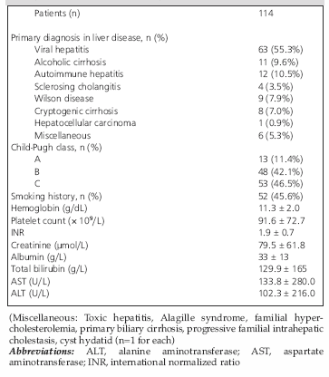
Table 1. Primary diagnosis in liver disease, Child-Pugh class, smoking history, and preoperative laboratory values for the 114 patients
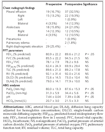
Table 2. Preoperative and postoperative chest radiograph findings, PFT parameters, and ABG analyses
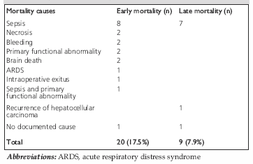
Table 3. Early and late mortality causes
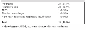
Table 4. Postoperative pulmonary complications
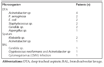
Table 5. Microorganisms obtained from DTA, sputum, and BAL cultures in adult liver transplanted patients