Volume: 6 Issue: 4 December 2008
FULL TEXT
Objectives: Uncontrolled release of cytokines has been linked to graft dysfunction or rejection and contributes to an increase in mortality and morbidity. We argue that temporary vascular clamping of the hepatic pedicle during major hepatic surgery is a potential stimulus for an excessive release of cytokines and the activity of neutrophils.
Materials and Methods: Thirty patients underwent partial liver resection or transplant. Samples were drawn preoperatively, immediately before portal vein clamping, at the early reperfusion period, and on days 1, 3, 5, and 7 after the operation. Central venous plasma concentrations of IL-6, IL-8, and TNF-α were compared to portal venous plasma. The influence of neutrophils on metabolic activity was measured by flow cytometry.
Results: In both patient groups, no significant differences in cytokine concentrations between central and portal venous plasma were found. However, significant differences of neutrophils activity were observed in patients undergoing partial liver resection compared to patients after transplant.
Conclusion: Portal vein stasis induced by clamping the hepatic pedicle has no influence on the local release of IL-6, IL-8, and TNF-α. However, preoperatively increased plasma levels of TNF-α play a decisive role in the metabolic activity of neutrophils in patients with final-stage liver disease.
Key words : IL-6, IL-8, liver resection, liver transplant, respiratory burst, TNF-alpha
Introduction
Major hepatic surgery like partial liver resection or liver transplant is performed under temporary vascular clampage of the hepatic pedicle to prevent massive bleeding. As a result of portal vein occlusion, ischemia followed by reperfusion results in a systemic inflammatory response that is related to the release of various mediators and changes in the metabolic activity. The mechanisms leading to ischemia reperfusion injury are various and are triggered among others by the activity of polymorphonuclear neutrophils and a release of cytokines.
Cytokines are involved in regulating inflammatory and immune responses at the sites of injury or infection (1, 2). They are produced by different types of immune cells and play a pivotal role in changes that occur in the immune, metabolic, and endocrine functions during the intra- and postoperative period (3).
The respiratory burst is one of the major functions by which polymorphonuclear neutrophils protect the human body against bacterial and fungal infections in the early postoperative stage (4). Inhibition of superoxide production by polymorphonuclear neutrophils contributes to various adverse effects. On the other hand, oxygen radicals show damaging effects on cell function and may lead to organ rejection (5). The evidence today indicates that there is a complex balance between pro- and anti-inflammatory cytokines and superoxide production by polymorphonuclear neutrophils after tissue manipulation or surgical treatment. An imbalance is linked to situations like organ dysfunction or failure, and graft damage or rejection (6, 7). The postoperative levels of cytokines correlate to the severity of surgery and the occurrence of complications. These complications, like coagulation disorders, hemodynamic instability or allograft rejection, increase the mortality and morbidity rate (8, 9). The coherences described above are well-accepted phenomena in patients undergoing major hepatic surgery. However, the importance of preoperative cytokine levels and the interaction between cytokine release and superoxide production by polymorphonuclear neutrophils as well as the predictive values for determining organ or graft dysfunction in humans are controversially discussed. In this clinical trial, we attempted to determine whether temporary vascular clamping of the hepatic pedicle has a crucial influence on the local release of cytokines and the activity of neutrophils. Excessive intraoperative production of TNF-α, IL-6, and IL-8 may amplify reperfusion injury of the graft. In addition, we attempted to determine whether already increased cytokine levels at the preoperative stage play a decisive role for the activity of neutrophils after liver resection or orthotopic liver transplant, aggravating organ dysfunction or graft rejection.
Materials and Methods
Patients
The study was approved by the local ethics committee. Informed consent was obtained from every patient. In total 30 patients were recruited for this study. In the liver resection group 15 patients (13 men, 2 women, mean age, 57 ± 14.0 years) underwent partial liver resection for primary or secondary malignancies. A “Pringle” maneuver was carried out for the time of parenchyma dissection (10). Portal clamping time was 0.27 ± 0.12 hours. In the liver transplant group, 15 patients (12 men, 3 women, mean age, 45 ± 14.2 years) had undergone orthotopic full-sized liver transplant for final-stage liver disease (medical urgency criteria T2/T3). The grafts for liver transplant had a cold ischemic time of 9.6 ± 2.0 hours and an anhepatic time of 0.49 ± 0.11 hours. All grafts were preserved with histidine tryptophan ketoglutarate (HTK) preservation solution (Dr. Köhler Chemie, Alsbach-Hähnlein, Germany). Exclusion criteria were systemic inflammatory response syndrome (SIRS) and sepsis, tumor infiltration of adjacent organs, and immunodeficiency. Patients’ data are summarized in Table 1.
Surgical and anaesthesia procedures
Liver resection and transplant were carried out according to a standard operating technique after laparotomy. Orthotopic full-sized liver transplant was performed without the use of femoral-subclavian bypass and without splenectomy under temporary clamping of the hepatic pedicle according to Pringle to prevent massive bleeding.
General anaesthesia was induced with thiopental 3 to 5 mg/kg-1 per bodyweight and fentanyl 1.5 to 2.5 µg/kg-1 per bodyweight. Oral intubation was carried out after application of atracurium 0.5 mg/kg-1 per bodyweight. All patients were normoventilated with an air oxygen mixture of 50%, the end-expiratory carbon dioxide was maintained between 4.5% and 5%. During reperfusion, patients were ventilated with 100% oxygen. Patients were anaesthetized with continuous sevoflurane inhalation (end-expiratory concentration 1.5-2.5 vol %), supplemented by bolus application of fentanyl 2 to 3 µg/kg/h and of atracurium 0.2 to 0.3 mg/kg/h. Arterial and central venous lines were used to monitor cardiovascular stability. Postoperatively, all patients received a patient-controlled analgesia with piritramid. Heparin therapy was initiated at the end of surgery and was administered for 2 to 4 days before switching to low-molecular weight heparin.
The immunosuppression protocol consisted of a bolus of methylprednisolone intraoperatively, which was gradually reduced over the next postoperative days. Furthermore, an interleukin 2 (IL-2) receptor antibody (Basiliximab; Novartis, Basel, Switzerland) was given 6 hours postoperatively, and on postoperative day 4; and cyclosporin was administered intravenously, starting on postoperative day 1. In cases of hepatitis B or C, only 1 single dose of corticosteroids was given intraoperatively, and mycophenolate mofetil (MMF; Roche, Grenzach-Wyhlen, Germany) was added on postoperative day 4.
Samples
Heparinized peripheral blood samples were taken through a central venous line inserted into the superior vena cava before surgery
(T1), immediately before portal vein clamping (T2), at the beginning of reperfusion
(T3), 30 minutes after reperfusion (T4) and then on days 1
(T5), 3 (T6), 5 (T7), and 7 (T8) after the operation. Additionally, heparinized portal venous blood samples were taken by direct venepuncture immediately before portal vein clamping
(T2), at the beginning of reperfusion (T3) and 30 minutes after reperfusion
(T4).
Cytokine assays
Blood samples (5 mL each) for cytokine analysis were collected in tubes (Monovette, Sarstedt, Nümbrecht, Germany) containing ethylene diamine tetraacetic acid (EDTA). Cytokine levels were determined by a commercial enzyme-linked immunosorbent assay kit according to the manufacturer’s instructions.
Respiratory burst
To measure respiratory burst, blood samples (7.5 mL each) were collected in lithium heparin-coated disposable blood-sampling tubes (Monovette, Sarstedt, Nümbrecht, Germany) and processed immediately by using the protocol of Rothe and associates. The blood was layered on equal Ficoll quantity (Ficoll-Hypaque, density
1.077g/dL-1, Biochrom, Berlin, Germany). The nucleated blood cells of the supernatant were harvested without centrifugation. Phosphate buffered saline (PBS, Dulbecco’s without
Ca2+ and MgCl2, GIBCO BRL, Eggenstein, Germany) was portioned (1 mL each) in cups (Eppendorf) and heated to 37°C. Thirty microliters of the supernatant, containing 5 ×
105 cells/mL-1 on average, were incubated with 1 × 10⁻³ mol DHR (MoBiTec, Göttingen, Germany) at 37°C for 5 minutes. Respiratory burst activity was induced either by adding 20 µL of Escherichia coli (1 x
109 mL-1, E. coli, HB 101), 1 × 10⁻⁷ N-formyl-methionyl-leucyl-phenylalanine (0.01 mM, 10 µL fMLP, Sigma, Deisenhofen, Germany) or the combination of 10 ng recombinant tumor necrosis factor alpha (1 µg/mL, 10 µL, rh TNF-α, Sigma, Deisenhofen, Germany) and 1 ×
10-7 N-formyl-methionyl-leucyl-phenylalanine (0.01 mM, 10 µL fMLP, Sigma, Deisenhofen, Germany). After 20 minutes incubation at 37°C, the reaction was terminated by transferring the samples onto ice. To determine viability, dead cells were counterstained by adding 1 mM propidium iodide (PI, Serva, Heidelberg, Germany). The samples were stored on ice and immediately subjected to flow cytometry. Negative controls were performed without stimulation to detect possible preactivation of neutrophils. The samples were analyzed in a flow cytometer equipped with an argon ion laser, adjusted to a wavelength of 488 nm (Epics, Beckman-Coulter, Krefeld, Germany). Fifteen thousand events were measured for each sample. The rhodamine emission was filtered and measured with a green photomultiplier (FL1; 515 - 545 nm). The propidium iodide (PI) emission was measured with a photomultiplier in the red fluorescence channel (FL 3; 650 nm). Sideward scatter (SSC) and forward scatter (FSC) were assessed in linear mode and FL 1 and FL 3 in logarithmic mode without compensation. The photomultiplier voltage and gains of FSC, SSC, FL1, and FL3 were adjusted for each negative control and remained constant. Data files were stored in a list mode and analyzed in dot plots using a computer software package (EXPO 2.0; Beckman-Coulter).
Calculation and statistics
All statistical data showed a Gaussian distribution (Kolmogorov-Smirnov test). A univariate analysis of variance (ANOVA) was used to identify effects at various points of time, followed post hoc by the Bonferroni or Games-Howell test if Levene’s test for homogeneity of variance failed (SPSS/PS V 15.0 software package, SPSS, Munich, Germany). The t test was used to compare portal and systemic blood, furthermore to compare intergroup data. Results were expressed as mean values ± the standard error of the mean (SEM). A
P value of less than .05 was considered to be statistically significant.
Results
Interleukin-6
IL-6 (Table 2) plasma concentration was increased slightly before portal vein clamping in patients who underwent partial liver resection
(T2 = 62.4 ± 23.4) or liver transplant (T2 = 64.4 ± 20.4). The IL-6 levels showed a marked increase before reperfusion in both groups
(T3 = 179.2 ± 41.0 vs 227.3 ± 54.6), reaching a maximum 30 minutes after reperfusion in liver transplant recipients
(T4 = 280.2 ± 70.4). The IL-6 central venous concentrations showed no significant differences in comparison to the portal venous blood concentrations at patients undergoing partial liver resection
(T2 = 62.4 ± 23.4 vs 84.9 ± 35.5; T3 = 179.2 ± 41.0 vs 261.9 ± 83.2) or in the liver transplant recipients
(T2 = 64.4 ± 20.4 vs 65.6 ± 24.2; T3 = 227.3 ± 54.6 vs 275.6 ± 16.3;
T4 = 280.2 ± 70.4 vs 265.0 ± 71.3). On the first to third postoperative day, the IL-6 concentrations showed a small downward drop in both groups
(T5 = 112.2 ± 42.9 vs 37.1 ± 11.9). From the fifth postoperative day onwards, IL-6 was increased again and remained above the preoperative values in both groups
(T7 = 259.3 ± 112.9 vs 124.9 ± 57.7).
Interleukin-8
IL-8 (Table 3) increased progressively until the fifth postoperative day in the liver resection group and remained above the preoperative levels
(T1 = 29.4 ± 5.8; T7 = 53.8 ± 13.5). However, the levels were significantly lower following hilus occlusion
(P = .056) and reperfusion (P = .025) in this group compared to liver transplant recipients.
In liver transplant recipients, the IL-8 concentration was increased before portal vein clamping (T2 = 68.0 ± 16.1) reaching a maximum concentration in the portal venous blood 30 minutes after reperfusion (T4 = 81.6 ± 14.1). The postoperative plasma levels were reduced and remained below preoperative levels during the entire postoperative period (T8 = 35.9 ± 6.7). The IL-8 central venous plasma concentrations showed no significant differences compared to portal venous blood concentrations in patients that underwent partial liver resection (T2 = 33.8 ± 8.1 vs 32.2 ± 6.2) or orthotopic full-sized liver transplant (T2 = 68.0 ± 16.1 vs 56.4 ± 15.5).
TNF-α
TNF-α (Table 4) first increased on the third postoperative day after liver resection and remained above the preoperative concentrations until day 5 (T1 = 11.6 ± 4.3;
T7 = 26.1 ± 9.5). Enhanced values of TNF-α were found preoperatively in liver transplant recipients
(T1 = 27.4 ± 8.7), decreasing to lower levels before portal vein clamping
(T2 = 22.4 ± 5.6 vs 16.6 ± 5.6). The central venous plasma concentration of TNF-α reached a maximum before reperfusion
(T3 = 54.5 ± 18.4) and showed a significant difference compared to portal venous blood concentration
(T3 = 8.4 ± 3.3) in liver transplant recipients. Immediately after reperfusion, central venous and portal venous blood concentrations increased to nearly the same levels
(T4 = 42.1 ± 13.5 vs 44.1 ± 15.9) and remained above the preoperative concentrations during the entire postoperative period
(T8 = 39.8 ± 10.8). Liver transplant recipients showed significantly higher TNF-α levels
(P = .034) following hilus occlusion until the early reperfusion period
(T5, P = .010) in comparison to patients for liver resection.
Respiratory burst
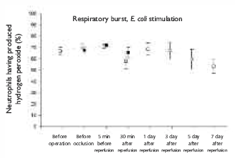
After stimulation with E. coli, an increased respiratory burst was observed in both groups without any significant differences at any time. Overall, the hydrogen peroxide production was significantly higher in both groups after
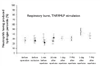
E. coli stimulation compared to stimulation with TNF/fMLP. However, after stimulation with TNF/fMLP a significantly increased hydrogen peroxide production of neutrophils was already found preoperatively in patients for liver resection, compared to patients for liver transplant
(P = .037).
Discussion
The present clinical trial demonstrates the local inflammatory cytokine response of IL-6, IL-8, and TNF-α in the portal vein compared to the central venous plasma response in patients who underwent partial liver resection or orthotopic liver transplant at different points in time. In addition, the influence of the observed cytokines on the activity of polymorphonuclear neutrophils was demonstrated.
IL-6, IL-8, TNF-α
The preoperative central venous plasma concentration of IL-6 in patients for partial liver resection was 8.7 ± 4.7 pg/mL, and in patients for orthotopic full-sized liver transplants it was 24.8 ± 9.8 pg/mL. The venous plasma concentration increased before the beginning of the anhepatic period, showed a temporary minor decrease during the early postoperative period and remained above the preoperative central venous plasma concentration in both groups until day 5. The corresponding concentrations of IL-6 in the portal venous stasis blood, taken from the portal vein, showed a similar distribution. Interestingly, no significant differences between venous and portal concentrations of IL-6 were found through the entire clamping time and the early reperfusion period. These findings may reflect no excessive splanchnic production of IL-6 during the anhepatic period induced by temporary vascular clampage of the hepatic pedicle. These results suggest that the ischemia of the gut, a possible source of IL-6, did not play a pivotal role in major hepatic surgery. In addition, the observed temporary minor decrease of IL-6 in the early reperfusion period and on day 1 to 3 may indicate a hepatic trapping of IL-6.
On the one hand, our findings of IL-6 plasma concentrations in patients that underwent partial liver resection or orthotopic liver transplant are in agreement to prior studies (11, 12). Otherwise, prior studies described an increased splanchnic production of IL-6 during the anhepatic period so that our findings seem to be contrary in this point (4). IL-8 concentrations increased steadily during surgery in patients undergoing partial liver resection or liver transplant, with nonsignificant differences in plasma concentrations between portal venous blood compared to central venous blood. IL-8 remained above the preoperative levels in patients that underwent partial liver resection. However, a massive drop below preoperative levels was observed in patients after orthotopic liver transplant during the entire postoperative period. The relevance of this distribution appears to be unclear. Whereas the preoperative increase of IL-8 blood levels in the orthotopic full-sized liver transplant group could be caused by primarily increased TNF-α concentration, TNF-α is known to induce production of IL-8, a chemo-attractant for neutrophils (13). In our investigation enhanced values of TNF-α were found in liver transplant recipients preoperatively. It is possible that this condition is caused by final-stage liver disease.
Respiratory burst
Interestingly, we found a significantly reduced capacity of polymorphonuclear cells (PMN) to produce reactive oxygen species after TNF/fMLP stimulation in liver transplant recipients compared to patients for partial liver resection. This suppression of the PMN function lasted for the early postoperative period and then increased on day 5 and 7. Ex vivo stimulation with E. coli induced a strong amount of released active oxygen compounds in both patients groups at any time (Figure 1, 2, 3 and 4). The first phenomenon should be assumed to be a result of permanent stimulation of neutrophils by inflammatory factors. Prior studies observed high concentrations of soluble adhesive molecules VCAM-1, ICAM-1, and E-selectin, which can account for marked vascular endothelium damage in liver cirrhosis. Endothelial cell failure or stimulation can occur due to high levels of proinflammatory cytokines (14). A persistent stimulation of neutrophils by inflammatory factors (eg, TNF-α) can lead to exhaustion of their function (15, 16).
Further studies confirmed that proinflammatory factor elimination from peripheral blood is impaired in patients with liver cirrhosis, which facilitates persistent stimulation of peripheral blood neutrophils (17). Our orthotopic full-sized liver transplant patients who also had final-stage liver disease, showed reactions similar to patients with liver cirrhosis as described above. It is possible that the polymorphonuclear neutrophils in our orthotopic full-sized liver transplant group also were under permanent stimulation because of impaired elimination of circulating proinflammatory factors. Finally, this permanent stimulation leads to the same exhaustion, similar to patients with liver cirrhosis. On the other hand, the higher release of oxygen metabolites observed after E. coli stimulation seems to be incomprehensible at first. However, this phenomenon can be explained by different stimulation mechanisms for polymorphonuclear neutrophils. Neutrophils are cells with a large phagocytizing activity, which play an important role in the immunologic processes of the organism. After migration to the inflammatory site induced by chemotactic factors and cytokines, the polymorphonuclear neutrophils can be stimulated by microorganisms (eg, E. coli). This stimulation leads to an elevation of active oxygen metabolites responsible for killing microorganisms (18).
Overall, polymorphonuclear neutrophils are an important host immunity barrier and they protect the human body in the early stages of bacterial and fungal infections. Many factors, for example, immunosuppressive treatment after organ transplant, may influence the PMN functions. The suppressed oxidative burst observed in our investigation cannot be explained by this immunosuppressive therapy alone. Rather our results suggest that an increased preoperative concentration of cytokines with a persistent stimulation of leukocytes leads to the exhaustion of polymorphonuclear neutrophils. Among other known factors, this depressed oxidative burst after persistent stimulation plays another decisive role for an impaired immune function, which may contribute to an exacerbation of bacterial and fungal infections after organ transplant.
References:
- Toledo-Pereyra LH, Suzuki S. Neutrophils, cytokines, and adhesion molecules in hepatic ischemia and reperfusion injury. J Am Coll Surg. 1994;179(6):758-762.
- Pesonen EJ, Höckerstedt K, Mäkisalo H, et al. Transhepatic neutrophil and monocyte activation during clinical liver transplantation. Transplantation. 2000;69(7):1458-1464.
- Chuang D, Paddison JS, Booth RJ, Hill AG. Differential production of cytokines following colorectal surgery. ANZ J Surg. 2006;76(9):821-824.
- Stevenson GW, Hall SC, Rudnick S, Seleny FL, Stevenson HC. The effect of anesthetic agents on the human immune response. Anesthesiology. 1990;72(3):542-552.
- Kurokawa T, Nonami T, Kobayashi H, et al. Inhibition by cyclosporine of the production of superoxide radicals. N Engl J Med. 1992;326(12):840.
- Fernandes H, Koneru B, Fernandes N, et al. Investigation of promoter polymorphisms in the tumor necrosis factor-alpha and interleukin-10 genes in liver transplant patients. Transplantation. 2002;73(12):1886-1891.
- Arranz J, Soriano A, Garcia I, et al. Effect of proinflammatory cytokines (IL-6, TNF-alpha, IL-1beta) on hemodynamic performance during orthotopic liver transplantation. Transplant Proc. 2003;35(5):1884-1887.
- Miki C, McMaster P, Mayer AD, Iriyama K, Suzuki H, Buckels JA. Factors predicting perioperative cytokine response in patients undergoing liver transplantation. Crit Care Med. 2000;28(2):351-354.
- Bishop GA, Rokahr KL, Napoli J, McCaughan GW. Intragraft cytokine mRNA levels in human liver allograft rejection analysed by reverse transcription and semiquantitative polymerase chain reaction amplification. Transpl Immunol. 1993;1(4):253-261.
- Pringle JH. V. Notes on the arrest of hepatic hemorrhage due to trauma. Ann Surg. 1908;48(4):541-549.
- Kimura F, Miyazaki M, Suwa T, et al. Evaluation of total hepatic vascular exclusion and Pringle maneuver in liver resection. Hepatogastroenterology. 2002;49(43):225-230.
- Santiago F, Bueno P, Olmedo C, et al. Time course of intraoperative cytokine levels in liver transplant recipients. Transplant Proc. 2006;38(8):2492-2494.
- Zhao X, Koshiba T, Fujimoto Y, et al. Proinflammatory and antiinflammatory cytokine production during ischemia-reperfusion injury in a case of identical twin living donor liver transplantation using no immunosuppression. Transplant Proc. 2005;37(1):392-394.
- Panasiuk A, Wysocka J, Maciorkowska E, et al. Phagocytic and oxidative burst activity of neutrophils in the end stage of liver cirrhosis. World J Gastroenterol. 2005;11(48):7661-7665.
- Fiuza C, Salcedo M, Clemente G, Tellado JM. In vivo neutrophil dysfunction in cirrhotic patients with advanced liver disease. J Infect Dis. 2000;182(2):526-533.
- Rajkovic IA, Williams R. Abnormalities of neutrophil phagocytosis, intracellular killing and metabolic activity in alcoholic cirrhosis and hepatitis. Hepatology. 1986;6(2):252-262.
- Saitoh O, Sugi K, Lojima K, et al. Increased prevalence of intestinal inflammation in patients with liver cirrhosis. World J Gastroenterol. 1999;5(5):391-396.
- Aratani Y, Kura F, Watanabe H, et al. In vivo role of myeloperoxidase for the host defense. Jpn J Infect Dis. 2004;57(5):S15.

Volume : 6
Issue : 4
Pages : 254 - 260
From the 1Department of Anaesthesiology and the 2Department of Visceral and Transplant Surgery, Hannover Medical School, 30625 Hannover, Germany
Address reprint requests to: Arthur Studzinski, MD, Hannover Medical School, Department of Anaesthesiology, Carl-Neuberg-Str. 1, 30625 Hannover, Germany
Phone: ++49-511-532 3689
Fax: +49-511-532 3642
E-mail: studzinski.arthur@mh-hannover.de
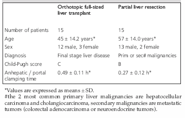
Table 1. Demographic and clinical data of patients for orthotopic full-sized liver transplant and partial liver resection.
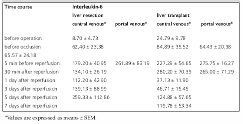
Table 2. Central and portal venous plasma concentration of Interleukin-6 [pg/mL].
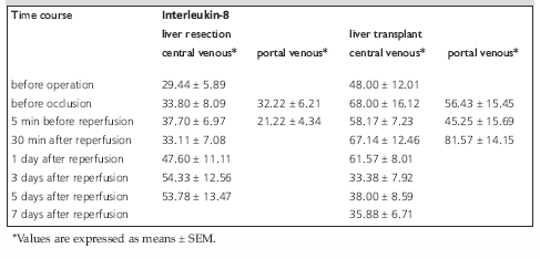
Table 3. Central and portal venous plasma concentration of Interleukin-8 [pg/mL].
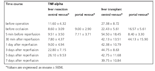
Table 4. Central and portal venous plasma concentration of TNF-alpha [pg/mL].
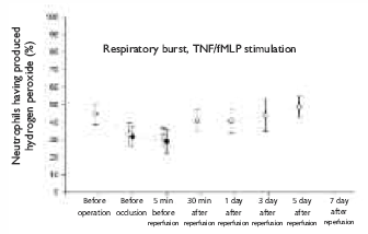
Figure 1. Percentage of respiratory burst after stimulationwith
TNF/fMLP
in patients after partial liver resection. ο = central venous concentration;
• = portal venous concentration. Values are expressed as mean ± SEM.
 = central venous concentration;
 = portal venous concentration. Values are expressed as mean ± SEM.
" style="margin-bottom: 10px; cursor: pointer">
= portal venous concentration. Values are expressed as mean ± SEM.
" style="margin-bottom: 10px; cursor: pointer">
Figure 2. Percentage of respiratory burst after stimulationwith
TNF/fMLP
in patients after liver transplant.  = central venous concentration;
= portal venous concentration. Values are expressed as mean ± SEM.
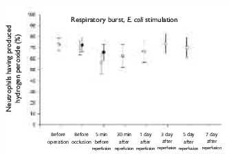
Figure 3. Percentage of respiratory burst after stimulation with E.
coli in
patients after partial liver resection. ο = central venous concentration;
• = portal venous concentration. Values are expressed as mean ± SEM.
 = central venous concentration;
= portal venous concentration. Values are expressed as mean ± SEM.
= central venous concentration;
= portal venous concentration. Values are expressed as mean ± SEM." style="margin-bottom: 10px; cursor: pointer">
Figure 4. Percentage of respiratory burst after stimulation with E.
coli in
patients after liver transplant.
= central venous concentration;
= portal venous concentration. Values are expressed as mean ± SEM.