Volume: 3 Issue: 2 December 2005
FULL TEXT
Posttransplant lymphoproliferative disorders are severe complications that arise after solid organ transplantation, which are often related to Epstein-Barr virus. Reports are anecdotal, and a standardized therapy does not exist. We report a case of a 36-year-old man who developed posttransplant lymphoproliferative disorder of the oropharynx 1 year after receiving a heart transplant. A short review of the literature is presented, after which a new therapeutic approach that combines antiviral therapy, monoclonal antibodies, and a sirolimus-based maintenance immunosuppression regimen with reduced target trough levels of tacrolimus is introduced. The patient achieved complete remission and was free from recurrence 18 months after the therapy was initiated.
Key words : Oropharynx, immunosuppression, posttransplant lymphoproliferative disorders, rituximab, antibodies
Posttransplant lymphoproliferative disorder (PTLD) is a severe complication after solid-organ transplantation [1], with the highest incidence in the first 2 years after transplantation [2]. PTLD is, in fact, a group of heterogeneous lymphoid proliferations, usually of B-cell origin, and in fewer cases, of T-cell origin [3]. In most cases, PTLD can be related to infection with Epstein-Barr virus (EBV) and is diagnosed predominantly in immunosuppressed patients [4]. The incidence of PTLD among transplanted patients ranges from 1%-20% in different centers [5]. Potential risk factors for development of PTLD are pediatric age, allograft type, EBV seronegativity at the time of transplantation, and cytokine gene polymorphisms [2]. Treatment of PTLD remains controversial, and there is no common gold standard for successful PTLD treatment.
Herein, we report a case of successful PTLD treatment following heart transplantation and introduce a therapeutic regimen based on rituximab and cidofovir in combination with a sirolimus-based immunosuppression.
Case Report
A 36-year-old EBV-seronegative man underwent heart transplantation in June 2002. He had dilative cardiomyopathy (New York Heart Association Classification III-IV) originating from biopsy-proven viral myocarditis in 2001.
The postoperative immunosuppression regimen consisted of sirolimus, tacrolimus, and prednisone, which is one of our standard immunosuppression protocols [6]. Target trough levels for both sirolimus and tacrolimus were 6-8 ng/mL. There were no episodes of elevated sirolimus or tacrolimus trough levels. The prednisone dosage was 7.5 mg/day and was stopped 6 months after transplantation. Acute rejection episodes were determined by endomyocardial biopsies, cytoimmunologic monitoring, electrocardiogram, and echocardiography. The patient had no rejection episodes on follow-up. His postoperative course was uneventful, and he was discharged from the hospital on the 17th postoperative day.
In February 2003, he underwent bilateral tonsillectomy at another hospital for abscessing tonsillitis. In August 2003, he was again admitted to another hospital for fever, pain in the oropharynx, and mucous expectorations. He was treated with ciprofloxacin and imipenem IV for 12 days with no improvement of symptoms. The patient was transferred to our center for further diagnostics and treatment.
On admission, a panendoscopic examination revealed a tumor in the lateral oropharyngeal wall with fibrinous exudation. A biopsy of the tumor was obtained. Histopathologic examination revealed lymphoepithelial tissue infiltrated by polymorphic blastoid cells of B-cell origin accompanied by a T-cell population, which led to the diagnosis of polymorphic PTLD. Immunohistochemistry proved CD20-positivity.
Serum EBV polymerase chain reaction (PCR) was 32500 gEq/mL, and it was 70000 gEq/mL in saliva. Figure 1 shows a computed tomography (CT) scan of the tumor before treatment. Sequential chemotherapy with the anti-CD20 antibody, rituximab, and cyclophosphamide, adriamycin, vincristine, and prednisone (CHOP) + granulocyte colony-stimulating factor (G-CSF) chemotherapy was planned.
The patient received 4 cycles of the anti-CD20 monoclonal antibody, rituximab (375 mg/m2 IV once per week for 4 weeks), and the antiviral substance, cidofovir (5 mg/kg IV once per week for 4 weeks). CHOP chemotherapy was scheduled for the seventh week after the start of treatment in case of tumor progression. As reduction of immunosuppression is one of the basic principles of PTLD therapy, we reduced the target trough levels of tacrolimus to 3-5 ng/mL in an sirolimus-based immunosuppressive protocol.
After the first 2 cycles of rituximab/cidofovir, serum EBV-PCR revealed 1900 gEq/mL. One week after the fourth cycle, 450 gEq/mL was detected. The saliva samples were free from EBV in all further examinations. Four weeks after the treatment was initiated, a computed tomography (CT) scan revealed complete regression of the mass (Figure 2). The formerly planned CHOP chemotherapy was not given. The patient was discharged and received regular follow-up examinations.
Eighteen months after treatment, CT scans of the cranium, neck, thorax, and abdomen revealed no signs of PTLD recurrence. However, the serum EBV-PCR result was 130000 gEq/mL, and EBV persistence also was detected in peripheral lymphocytes with 150 gEq/mL in 20000 cells. After 4 weeks’ treatment with valganciclovir, serum PCR revealed 2400 gEq/mL. Further EBV screenings are scheduled for this patient.
Discussion
The patient presented with enlargement of the tonsils. Unfortunately, no material was subjected to histologic examination. It might be important to suspect tonsil enlargement in a transplant recipient as a possible manifestation of PTLD, particularly if the recipient were EBV negative at the time of the transplant.
Our patient was treated successfully with rituximab, cidofovir, and sirolimus achieving a complete remission of PTLD for 18 months. In contrast, EBV-PCR remained positive in the lymphocytes and serum at 18 months after the initial therapy. The elevated serologic values of EBV could be related to the loss of B cells due to the rituximab therapy. Under cidofovir therapy, the patient had low levels of serum EBV-PCR. However, EBV-PCR increased steadily during the next 6 months after the cessation of cidofovir. A seroconversion did not occur, and the patient remained EBV-antibody negative, which might be related to the absence of B cells.
Reports dealing with PTLD treatment after thoracic organ transplantation are anecdotal, and a standard therapy for PTLD does not exist [7, 8, 9]. We decided to combine antiviral treatment with monoclonal antibodies and rapamycin-based immunosuppression to achieve complete remission.
A combined approach with rituximab, cidofovir, and sirolimus seemed the best therapeutic option, because several authors had reported promising results of antiviral therapy in polyclonal PTLD [10]. Experiences with monoclonal antibodies in the treatment of PTLD were first reported by Fischer and coworkers [11, 12]. Oertel and coworkers and Zilz and colleagues have reported the treatment of PTLD with rituximab alone [13, 14]. In addition, rapamycin inhibits the interleukin-10 signal transduction pathway and the growth of EBV B-cell lymphomas [15]. Recent reports offer some evidence for the beneficial effects of a combined rituximab and sirolimus therapeutic approach for PTLD [16]. CHOP chemotherapy also has demonstrated acceptable results in the treatment of PTLD [17]; however, CHOP chemotherapy was not performed in our case owing to anticipated adverse effects and the patient’s prompt response to our therapeutic approach.
After the patient had been diagnosed with PTLD, we lowered the target trough levels of tacrolimus to 3-5 ng/mL because reduction of immunosuppression is a further goal of PTLD treatment. Sirolimus was adjusted slightly to 5-8 ng/mL because it possesses antiviral and antiproliferative abilities [18].
Treatment of PTLD to date has not been standardized. It is not known which interventions contributed to the therapeutic success in our patient. Lowering immunosuppression seems to be inevitable. For EBV-related PTLD, antiviral therapy is a therapeutic option. Monoclonal antibodies add a further therapeutic option to PTLD treatment. Antiviral therapy can be combined with rituximab in EBV-related PTLD. A sirolimus-based immunosuppression regimen might guarantee a longer freedom from recurrence. Chemotherapy (eg, CHOP) is not the first choice in PTLD patients at our center but should be considered if remission is not achieved with a combination of antiviral therapy, monoclonal antibodies, and sirolimus-based immunosuppression. Additional trials utilizing this form of therapy should be undertaken.
References:
- Reams BD, McAdams HP, Howell DN, Steele MP, Davis RD, Palmer SM. Posttransplant lymphoproliferative disorder: incidence, presentation, and response to treatment in lung transplant recipients. Chest 2003; 124: 1242-1249
- Nalesnik MA. Clinicopathologic characteristics of post-transplant lymphoproliferative disorders. Recent Results Cancer Res 2002; 159: 9-18
- Haldas J, Wang W, Lazarchick J. Post-transplant lymphoproliferative disorders: T-cell lymphoma following cardiac transplant. Leuk Lymphoma 2002; 43(2): 447-450
- Franco A, Jimenz L, Aranda I, Alvarez L, Gonzalez M, Rocamora N, et al. Diffuse lymphoproliferative disease after renal transplantation and its relation with Epstein-Barr virus. Experience at one center. Nefrologia 2002; 22(5): 463-469
- Merlino C, Giacchino F, Bergallo M, Bonello F, Bollero C, Segoloni GP, et al. Epstein Barr viral load monitoring in mononuclear lymphocytes and serum of renal transplant recipients using a quantitative PCR protocol. G Ital Nefrol 2003; 20(2): 170-175
- Mueller M, Meiser B, Groetzner J, Kaczmarek I, Reichart B. Sirolimus in combination with tacrolimus for primary immunosuppression after heart transplantation. J Heart Lung Transplant 2004; 23: S46
- Herman J, Vandenberghe P, van den Heuvel I, Van Cleemput J, Winnepenninckx V, Van Damme-Lombaerts R. Successful treatment with rituximab of lymphoproliferative disorder in a child after cardiac transplantation. J Heart Lung Transplant 2002; 21(12): 1304-1309
- Legere BM, Saad CP, Mehta AC. Endobronchial post-transplant lymphoproliferative disorder and its management with photodynamic therapy: a case report. J Heart Lung Transplant 2003; 22(4): 474-477
- Gao SZ, Chaparro SV, Perlroth M, Montoya JG, Miller JL, DiMiceli S, et al. Post-transplantation lymphoproliferative disease in heart and heart-lung transplant recipients: 30-year experience at Stanford University. J Heart Lung Transplant 2003; 22(5): 505-514
- Hanto DW, Gajl-Peczalska KJ, Frizzera G, Arthur DC, Balfour HH Jr, McClain K, et al. Epstein-Barr virus (EBV) induced polyclonal and monoclonal B-cell lymphoproliferative diseases occurring after renal transplantation. Clinical, pathologic, and virologic findings and implications for therapy. Ann Surg 1983; 198(3): 356-369
- Fischer A, Blanche S, Le Bidois J, Bordigoni P, Garnier JL, Niaudet P, et al. Anti-B-cell monoclonal antibodies in the treatment of severe B-cell lymphoproliferative syndrome following bone marrow and organ transplantation. N Engl J Med 1991; 324(21): 1451-1456
- Stephan JL, Le Deist F, Blanche S, Le Bidois J, Peuchmaur M, Lellouch-Tubiana A, et al. Treatment of central nervous system B lymphoproliferative syndrome by local infusion of a B cell-specific monoclonal antibody. Transplantation 1992; 54(2): 246-249
- Oertel SH, Anagnostopoulos I, Bechstein WO, Liehr H, Riess HB. Treatment of posttransplant lymphoproliferative disorder with the anti-CD20 monoclonal antibody rituximab alone in an adult after liver transplantation: a new drug in therapy of patients with posttransplant lymphoproliferative disorder after solid organ transplantation? Transplantation 2000; 69(3): 430-432
- Zilz ND, Olson LJ, McGregor CG. Treatment of post-transplant lymphoproliferative disorder with monoclonal CD20 antibody (rituximab) after heart transplantation. J Heart Lung Transplant 2001; 20(7): 770-772
- Nepomuceno RR, Balatoni CE, Natkunam Y, Snow AL, Krams SM, Martinez OM. Rapamycin inhibits the interleukin 10 signal transduction pathway and the growth of Epstein Barr virus B-cell lymphomas. Cancer Res 2003; 63(15): 4472-4480
- Garcia VD, Bonamigo Filho JL, Neumann J, Fogliatto L, Geiger AM, Garcia CD, et al. Rituximab in association with rapamycin for post-transplant lymphoproliferative disease treatment. Transpl Int 2003; 16(3): 202-206. Epub 2003 Feb 13
- Garrett TJ, Chadburn A, Barr ML, Drusin RE, Chen JM, Schulman LL, et al. Posttransplantation lymphoproliferative disorders treated with cyclophosphamide-doxorubicin-vincristine-prednisone chemotherapy. Cancer 1993; 72(9): 2782-2785
- Sehgal SN. Sirolimus: its discovery, biological properties, and mechanism of action. Transplant Proc 2003; 35(3 Suppl): 7S-14S

Volume : 3
Issue : 2
Pages : 381 - 384
Department of Cardiac Surgery, University Hospital Grosshadern, Ludwig-Maximilians-University, Marchioninistrasse 15, 81377 Munich, Germany
Address reprint requests to: Andres Beiras-Fernandez, MD, Herzchirurgische Poliklinik, Marchioninistr. 15, 81377 Muenchen, Germany
Phone: 00 49 89 70 952 436
Fax: 00 49 89 70 958 873
E-mail: Andres.Beiras@med.uni-muenchen.de
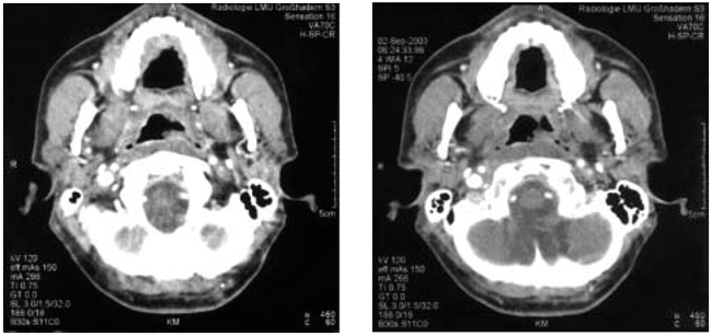
Figure 1. (A, B) A contrast-enhanced CT scan revealing prominent lymphoid tissue in the nasopharynx. Adjacent to the left tube, an exophytic mass is obvious.
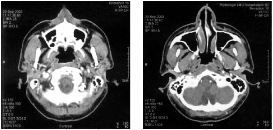
Figure 2. (A, B) Four weeks after the beginning of the treatment, CT scans revealed a complete regression of the mass.