Volume: 24 Issue: 5 May 2026
FULL TEXT
Successful Anesthetic Management of Deceased Donor Liver Transplant in a Patient With Neonatal Sclerosing Cholangitis As a Result of DCDC2 (doublecortin domain-containing protein 2) Mutation: A Case Report
Neonatal sclerosing cholangitis, which develops due to mutations in doublecortin domain-containing protein 2, is a rare and serious autosomal recessive inherited end-stage liver disease and frequently requires liver transplant due to decompensated biliary cirrhosis in childhood. Here, we present the anesthetic management of a deceased donor liver transplant in an 8-year-old female patient with hepatopulmonary syndrome and pulmonary arteriovenous fistula due to neonatal sclerosing cholangitis. Liver transplant is the only treatment option for hepatopulmonary syndrome and pulmonary arteriovenous fistula due to neonatal sclerosing cholangitis, but liver transplant has high mortality and morbidity. In patients with neonatal sclerosing cholangitis, detailed evaluations of preoper-ative conditions and underlying pathophysiological factors are crucial. Anesthesia management requires a multidisciplinary approach, comprehensive periopera-tive assessment, and close intraoperative monitoring to achieve successful outcomes and improve prognosis, thereby reducing mortality rates.
Key words : Anesthesia management, Hepatopulmonary syndrome, Pulmonary arteriovenous fistula
Introduction
Doublecortin domain-containing protein 2 (DCDC2) gene mutations can cause neonatal sclerosing cho-langitis (NSC), a rare and serious progressive biliary fibrosis that eventually progresses to end-stage liver disease (ESLD), necessitating liver transplant (LT) in childhood.1 The vital clinical findings in NSC include hyperbilirubinemia, jaundice, hepatosplenomegaly, cholestasis, pale stools, and elevated serum gamma-glutamyl transferase activity.1,2 Extrahepatic findings, involving nephronophthisis, renal cysts, vascular malformations, neurological manifestations, and sensorineural hearing loss, have been described in previous case series.1-3 In NSC, the bile duct is patent but strictured, and there is no biliary atresia. Children with the DCDC2 variant experience rapidly progres-sive portal hypertension and variceal bleeding within the first 5 years of life. Varicose vein eradication is often difficult in these patients, with only 27.3% eradicated, and recurrent bleeding rates are high. Response to endotherapy is the sole determinant of long-term liver survival, and LT is the only treatment option for those who do not respond.4
Hepatopulmonary syndrome (HPS) is a serious complication of ESLD that progresses gradually, and the mortality rate of HPS can be up to 48%. Charac-teristic findings of HPS include hypoxemia, increased alveolar-arterial oxygen gradient at rest in a sitting position breathing room air due to intrapulmonary vascular dilatation (IPVD), and hepatic cirrhosis.5,6 Although previously contraindicated in patients with HPS due to ESLD because of potential perioperative complications, LT has recently become the gold standard treatment for these patients. Furthermore, the risk of developing arteriovenous fistula (AVF) in patients with HPS is high, which may contribute to the development of hypoxemia; therefore, factors that trigger hypoxemia should be avoided as much as possible.6
We report the successful anesthetic management of a deceased donor LT in a patient with ESLD, HPS, and pulmonary AVF due to a DCDC2 mutation. Verbal and written informed consent was obtained from the patient’s parents for publication of our case.
Case Report
An 8-year-old female patient (height 125 cm, weight 26 kg, body mass index 16.6 kg/m2) with HPS and pulmonary AVF diagnosis, who developed NSC due to a DCDC2 mutation, was admitted to the intensive care unit for LT. The patient was tested by the pediatric metabolism department after birth at aged 3 years. The patient developed portal hypertension, hypersplenism, jaundice, ascites, esophageal varices, tubulopathy, and metabolic acidosis due to cirrhosis. The patient underwent band ligation twice because of esophageal varices. The patient presented with dyspnea, orthodeoxia, platypnea, cyanosis, and weakness.
Transthoracic echocardiography revealed micro-bubbles in the left atrium, but no pulmonary hyper-tension was detected. Lung perfusion scintigraphy showed a 20% intrapulmonary right-to-left shunt, and pulmonary arteriography demonstrated diffuse IPVD and pulmonary AVF. The liver computed tomography angiography revealed a diffuse hepatoportal shunt and splenomegaly in irregular areas of the liver parenchyma (Figure 1).
During follow-up, the patient was scheduled for living donor LT due to reduced arterial oxygen saturation (83%), dyspnea, orthodeoxia, platypnea, increasing cyanosis, and hypoxemia. On room air in the supine position, venous oxygen pressure was 59.5 mm Hg and O2 saturation was 85%; in the sitting position, venous oxygen pressure was 45.7 mm Hg and O2 saturation was 68.9%. Preoperative hemog-lobin (11.3 g/dL), hematocrit (33.9%), platelets (66 × 103/mL), alanine aminotransferase level (126 IU/L), aspartate transaminase level (107 IU/L), gamma-glutamyl transferase (107 IU/L), total bilirubin (1.9 mg/dL), direct bilirubin (1.2 mg/dL), albumin (2.7 g/dL), and international normalized ratio (1.1) were normal.
The patient’s Child-Pugh-Turcotte score was B, and the Pediatric End-Stage Liver Disease score was 6.4. The patient’s physical classification, as deter-mined by the American Society of Anesthesiologists, was class III, and the Mallampati score was II.
The deceased donor was a 13-year-old male patient (weight 50 kg, height 168 cm); cause of death was subarachnoid hemorrhage due to a traffic accident. Standard monitoring tests, including elec-trocardiogram, peripheral oxygen saturation (SpO2), noninvasive blood pressure, capnography (end-tidal carbon dioxide), temperature, and bispectral index, were performed. After 3 minutes of preoxygenation with 80% O2, anesthesia was induced with lidocaine, propofol, fentanyl, and rocuronium. Intubation was performed without complications using a size 6.0 cuffed endotracheal tube, and the patient was connected to a ventilator. The mean arterial pressure was targeted as >60 mm Hg throughout the surgery, and norepinephrine infusion (0.02-0.5 mg/kg/min) was initiated for hypotension.
A 1050-g liver tissue was extracted from the patient (Figure 2), and a 1000-g deceased donor liver was implanted (Figure 3). After the dissection phase, anastomosis of the great vessels was completed during the anhepatic phase, which lasted about 68 minutes. The clamp on the portal vein was removed, and reperfusion was started. The patient’s blood pressure dropped to 60/32 mm Hg 2 minutes after reperfusion, and norepinephrine infusion dose was raised; ephedrine and calcium chloride were administered twice.
Doppler ultrasonography performed at the end of the surgery revealed normal perfusion in the portal vein, hepatic artery, and hepatic venous system (Figures 4, 5, and 6). Postoperatively, the patient was intubated and ventilated, with fraction of inspired oxygen (FiO2) at 40% and SpO2 between 85% and 92%. On day 2, SpO2 ranged from 89% to 94%.
With resolution of hypoxemia, the patient was extubated, and SpO2 remained normal on room air. The patient’s hemodynamic state, condition, and consciousness were stable, and blood gas and lactate values, as well as liver transaminases, returned to reference values within a few days. Immunosup-pressive medications, steroids, and other necessary treatments were administered. The patient was transferred to the hospital ward on day 4 of follow-up and discharged 14 days after surgery without any postoperative complications.
Discussion
To our knowledge, we have described for the first time in the literature the anesthetic management of LT in a patient who developed NSC due to a DCDC2 mutation. Our patient had many clinical and physical disorders as a result of NSC and because of HPS and pulmonary AVF, which made LT quite challenging for us in terms of both anesthesia and surgery.
Although any NSC arising from a patient with a DCDC2 mutation is complex, we recommend that the anesthesiologist’s plans of anesthesia management during LT primarily consider the disease’s pathophys-iology and associated comorbidities. The mortality rate as a result of HPS is quite high in these children without LT, with pulmonary and intracranial hemorrhage, portal vein thrombosis, infection, sepsis, and multiorgan failure being the most significant causes of death.5 The mortality rate is 30% in LT candidates with severe preoperative hypoxemia (partial arterial oxygen pressure <50 mm Hg). Even after successful LT, these patients may require prolonged mechanical ventilation.5,6 Hypoxemia in patients with HPS who have intrapulmonary right-to-left shunts and pulmonary AVFs can worsen the clinical picture during surgery. Therefore, to safely anesthetize patients with HPS, whether with or without a pulmonary AVF, situations that could worsen hypoxemia and increase pulmonary vaso-dilation should be avoided as much as possible.6
Our patient had platypnea, orthodeoxia, and dyspnea before surgery. In the supine position, venous oxygen pressure was 59.5 mm Hg and SpO2 was 85%; in the sitting position, venous oxygen pressure was 45.7 mm Hg and SpO2 was 68.9%. We successfully performed LT in our patient (8-year-old girl with HPS due to NSC caused by the DCDC2 mutation and pulmonary AVF) who had room air SpO2 of 84% to 88% and intrapulmonary right-to-left shunt of 20%.
Postreperfusion syndrome can trigger and worsen hypoxia by increasing the shunt fraction in patients with pulmonary AVF and HPS.6 In our case, after the development of postreperfusion syndrome, short-term hypoxemia occurred intraoperatively due to HPS and pulmonary AVF. Our anesthesia plan aimed to minimize factors that could trigger hypoxemia and hypercarbia. We attempted to treat this by increasing inspiratory pauses and mechanical ventilator pressures. Patients with HPS and pulmonary AVF are at rise risk for paradoxical embolism. We were careful to prevent air from entering the surgical field during the operation and to prevent air bubbles from forming during fluid or drug administration.5-7 We attempted to keep pulmonary end-expiratory pressure at 4 to 6 mm Hg. Because our patient had HPS and AVF, we used pulse contour cardiac output to closely monitor for potential hemodynamic and oxygenation difficulties.
Lee and colleagues8 reported a 9-year-old girl with HPS and diffuse pulmonary AVF due to ESLD caused by biliary atresia who underwent living donor LT. The investigators used total intravenous anesthesia as an anesthetic management method to prevent further shunting and hypoxemia and successfully performed LT. We successfully performed deceased donor LT in our 8-year-old female (Child-Pugh-Turcotte class B, Pediatric End-Stage Liver Disease score 6.0) with HPS, pulmonary AVF, a 20% intra-pulmonary right-to-left shunt, and diffuse IPVD due to the DCDC2 mutation. Our anesthesia plan aimed to minimize factors that could trigger hypoxemia, hypercarbia, and shunting.
Postoperative hypoxemia and intrapulmonary shunt recovery time are related to the severity of preoperative hypoxemia. In most cases, HPS-related hypoxemia resolves within 1 year after LT.6 After transplant, our patient was intubated and ventilated, with fraction of inspired oxygen of 40% and SpO2 between 85% and 92%. On day 2, SpO2 ranged from 89% to 94%. With hypoxemia resolved, the patient was extubated after weaning. After 2 days of 2 L/min mask oxygen supplementation, SpO2 remained normal on room air.
Conclusions
Our patient had multiple clinical abnormalities (HPS and pulmonary AVF due to NSC resulting from the DCDC2 mutation), making LT quite challenging from both the anesthesia and surgical perspectives. Liver transplant performed with successful anesthesia and surgical management can lead to improved oxygenation, shunt regression, and improved outcomes, improving prognosis, reducing mortality rates, and enhancing quality of life. Anesthesiologists and surgeons should be knowledgeable about comprehensive preoperative evaluation, underlying pathophysiology, and close intraoperative moni-toring in patients with HPS and pulmonary AVF due to NSC from the DCDC2 mutation.
References:
- Chen HL, Wu SH, Hsu SH, Liou BY, Chen HL, Chang MH. Jaundice revisited: recent advances in the diagnosis and treatment of inherited cholestatic liver diseases. J Biomed Sci. 2018;25(1):75. doi:10.1186/s12929-018-0475-8
CrossRef - PubMed - Grammatikopoulos T, Sambrotta M, Strautnieks S, et al. Mutations in DCDC2 (doublecortin domain containing protein 2) in neonatal sclerosing cholangitis. J Hepatol. 2016;65(6):1179-1187. doi:10.1016/j.jhep. 2016.07.017
CrossRef - PubMed - Kaur P, Lal BB, Janakiraman D, et al. Portal hypertension in doublecortin domain-containing protein 2 (DCDC2)-related neonatal sclerosing cholangitis. J Pediatr Gastroenterol Nutr. 2025;80(1):189-196. doi:10.1002/jpn3.12414
CrossRef - PubMed - Syryn H, Hoorens A, Grammatikopoulos T, et al. Two cases of DCDC2-related neonatal sclerosing cholangitis with developmental delay and literature review. Clin Genet. 2021;100(4):447-452. doi:10.1111/cge.14012
CrossRef - PubMed - Krowka MJ, Fallon MB, Kawut SM, et al. International Liver Transplant Society practice guidelines: diagnosis and management of hepatopulmonary syndrome and portopulmonary hypertension. Transplantation. 2016;100(7):1440-1452.
CrossRef - PubMed - Younis I, Sarwar S, Butt Z, Tanveer S, Qaadir A, Jadoon NA. Clinical characteristics, predictors, and survival among patients with hepatopulmonary syndrome. Ann Hepatol. 2015;14(3):354-360.
CrossRef - PubMed - Lumb AB, Slinger P. Hypoxic pulmonary vasoconstriction: physiology and anaesthetic implications. Anesthesiology. 2015;122(4):932-946. doi:10.1097/ALN.0000000000000569
CrossRef - PubMed - Lee HJ, Lee JM, Jung CW, Lee J. Anesthetic management of a pediatric patient with pulmonary arteriovenous fistula undergoing liver transplantation: a case report. Pediatr Transplant. 2016;20(5):711-716. doi:10.1111/petr.12716
CrossRef - PubMed

Volume : 24
Issue : 5
Pages : 432 - 436
DOI : 10.6002/ect.2026.0008
From the 1Department of Anesthesiology and Intensive Care Unit, and the 2Department of General Surgery, Baskent University, Faculty of Medicine, Ankara, Turkey
Acknowledgements: The authors have not received any funding or grants in support of the presented research or for the preparation of this work and have no declarations of potential conflicts of interest.
Author contributions: All authors contributed to the design and writing of the case report and read and approved the final version. N. Çekmen drafted and critically reviewed the article and provided constructive suggestions.
Corresponding author: Denada Haka, Baskent University, Faculty of Medicine, Department of Anesthesiology and Intensive Care Unit, Ankara, Turkey
Phone: +90 312 203 68 68-4867
E-mail: denadahaka97@gmail.com
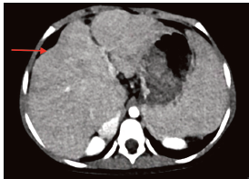
Figure 1.Computed Tomography Angiography of Patient’s Liver, With Diffuse Hepatoportal Shunt and Splenomegaly Found in the Liver Parenchyma
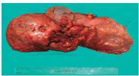
Figure 2.Liver Tissue (1050 g) Removed From the Patient
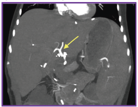
Figure 3.Liver From Donor (1000 g)
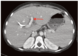
Figure 4.Normal Perfusion in the Portal Vein Shown in Doppler Ultrasonography Performed After Liver Transplant
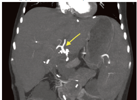
Figure 5.Assessment of Hepatic Arterial Perfusion With Doppler Ultrasonography Performed After Liver Transplant
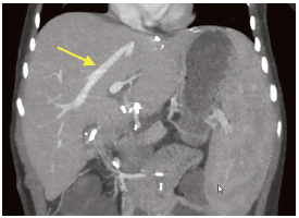
Figure 6.Normal Perfusion in the Hepatic Vein Shown in Doppler Ultrasonography Performed After Liver Transplant