Volume: 17 Issue: 1 January 2019 - Supplement - 1
FULL TEXT
Chronic kidney disease is defined as irreversible and progressive damage of the kidney. Chronicity is defined by the presence of renal dysfunction for at least 3 months, and renal dysfunction is defined through combinations of investigation (abnormal radiologic findings, abnormal urine or abnormal biochemistry reflecting renal dysfunction) and/or documentation of glomerular filtration rate below 60 mL/min/1.73 m2. The case patient was a girl of 11 years of age, with diagnosis of chronic renal disease, of unknown cause, under renal replacement therapy with peritoneal dialysis, with progressive deterioration of general status and decrease of functional capacity and tolerance to physical activity, presence of fatigue, pulmonary congestion, retention of liquids, and edema in lower extremities, even with adjustment of medical treatment. Transplant was performed from a related living donor, without incidents or complications. The results of pediatric kidney transplant are excellent, offering a high quality of life for recipients; many patients return to school. It is considered the criterion standard for the treatment of pediatric end-stage renal disease with excellent allograft function and subsequent resolution of systolic dysfunction.
Key words : Chronic kidney disease, Hypertension, Hypertrophy, Pediatric renal transplant
Introduction
Chronic kidney disease is defined as irreversible and progressive damage of the kidney. Chronicity is defined by the presence of renal dysfunction for at least 3 months, and renal dysfunction is defined through combinations of investigation (abnormal radiologic findings, abnormal urine, or abnormal biochemistry reflecting renal dysfunction) and/or documentation of glomerular filtration rate below 60 mL/min/1.73 m2.1,2 The most common diagnosis for pediatric renal transplantation is aplastic, hypoplastic, or dysplastic kidneys (15.8%).3
Hypertension and left ventricular hypertrophy are common and should be expected in children who are on dialysis. Remodeling of the myocardium results in structural changes of the geometry of the heart (concentric and eccentric hypertrophy). Concentric hypertrophy is seen in response to increased afterload, and eccentric hypertrophy develops in response to increased preload. Early markers of cardiomyopathy may be diagnosed by echocardiography. From one-third to one-half of deaths in children with chronic renal failure have been attributed to cardiac causes.4,5
Treatment requires a multidisciplinary team for the care of these often challenging patients. Multidisciplinary teams, consisting of surgeons, anesthesiologists, nephrologists, cardiologists, operating room staff, and pediatric intensivists, among many others, are all important for ensuring good outcomes in this population.6,7
The importance of adequately assessing the preoperative patient will reduce surgical morbidity and mortality, due to the identification of risk factors that can be modified and eliminated before the event.
Case Report
The patient was an 11-year-old girl, with diagnosis of chronic renal disease of unknown cause, who was being treated with renal replacement therapy and peritoneal dialysis. She had previous hospitalizations because of events of peritonitis and dyscontrol of blood hypertension. For these reasons, her treatment was changed to hemodialysis and adjustments were made to treatment with antagonist agents, including calcium channel blockers and angiotensin-converting enzyme inhibitors. Six months after initial presentation, she had progressive deterioration of general status, including decreased functional capacity and decreased tolerance to physical activity, presence of fatigue, pulmonary congestion, retention of liquids, and edema in lower extremities (Figure 1).
Complementary laboratory studies reported anemia with 8.9 g of hemoglobin, hematocrit level of 27%, reticulocyte level of 1.2%, platelet count of 179 × 103/L, white blood cell count of 6 × 10⁹/L, albumin level of 3.8 g/dL, and total protein of 60 g/L. Hydroelectrolytic alterations showed potassium level of 7.4 mmol/L, sodium level of 127 mmol/L, chloride level of 95 mmol/L, total calcium of 7.8 g/dL, glucose level of 104 mg/dL, creatinine level of 12.3 mg/dL, and serum urea nitrogen level of 171 mg/dL. Coagulation times were within normal limits. Echocardiogram data indicated concentric hypertrophy of the left ventricle, ejection fraction of 25%, shortening fraction of 16%, dilatation of left cavities with condition of mild mitral insufficiency, and trivalved aortic valve without data of insufficiency or stenosis.
The transplant procedure was conducted with a related living donor (the patient’s father). The kidney allograft was placed in the extraperitoneal iliac fossa via a curvilinear incision along the lateral margin of the rectus muscle, approximately 8 to 10 inches from just above the pubic bone to just above the umbilicus.
A left kidney was inserted into the right iliac fossa while the patient was under general balanced anesthesia. Two arteries were anastomosed, with the smallest artery polar to the internal iliac artery and the main artery to the external iliac artery. A single vein was anastomosed to the right external iliac vein. Once the vessels were anastomosed, the ureter was implanted with the use of the Lich-Gregoir technique (Figure 2).
For perioperative management, we applied a central venous catheter (7F), a catheter in the right radial artery, and support with levosimendan 0.1 μg/kg/min, norepinephrine 0.06 μg/kg/min, and dopamine 3 μg/kg/min, all in continuous intravenous infusion. Therapy for the period of reperfusion was necessary to optimize the state of volemia with crystalloids and colloids. Central venous pressure was maintained at 12 mm Hg, and mean arterial pressure was maintained above 65 mm Hg. In addition to the use of mannitol at 0.25 mg/kg bolus and intravenous furosemide at 1.5 mg/kg, we administered methylprednisolone 10 mg/kg and diphenhydramine 0.5 mg/kg.
There were no notable incidents or complications. Surgical bleeding was 100 mL; therefore, there was no need for blood products. The patient met the parameters for early extubation and transfer to the intensive care unit for continuous monitoring, showing good treatment response and amount of uresis.
Conclusions
Results after pediatric kidney transplant procedures are excellent, offering a high quality of life for recipients; many patients may return to their normal affairs (such as attending school). Transplant is considered the criterion standard for the treatment of pediatric patients with end-stage renal disease, resulting in excellent allograft function and subsequent resolution of systolic dysfunction. The treatment of patients with heart failure and chronic kidney disease is unclear, as there is very little solid evidence to support any recommendation.9,10 Guidelines for the management of these patients should be based on hemodynamic and metabolic behaviors that they present.
References:
- Wasson NR, Deer JD, Suresh S. Anesthetic management of pediatric liver and kidney transplantation. Anesthesiol Clin. 2017;35(3):421-438.
CrossRef - PubMed - North American Pediatric Renal Trials and Collaborative Studies Web site. NAPRTCS 2014 Annual Transplant Report. https://web.emmes.com/study/ped/annlrept/annualrept2014.pdf.
- United States Renal Data System. 2017 USRDS Annual Data Report: Epidemiology of Kidney Disease in the United States. Bethesda, MD: National Institutes of Health, National Institute of Diabetes and Digestive and Kidney Diseases; 2017. https://www.usrds.org/2017/view/Default.aspx
CrossRef - PubMed - Gargiulo R, Suhail F, Lerma EV. Cardiovascular disease and chronic kidney disease. Dis Mon. 2015;61(9):403-413.
CrossRef - PubMed - Mitsnefes MM. Cardiovascular disease in children with chronic kidney disease. J Am Soc Nephrol. 2012;23(4):578-585.
CrossRef - PubMed - Santiago MJ, López-Herce J, Urbano J, et al. Complications of continuous renal replacement therapy in critically ill children: a prospective observational evaluation study. Crit Care. 2009;13(6):R184.
CrossRef - PubMed - El-Husseini AA, Sheashaa HA, Hassan NA, et al. Echocardiographic changes and risk factors for left ventricular hypertrophy in children and adolescents after renal transplantation. Pediatr Transplant. 2004;8(3):249-254.
CrossRef - PubMed - Matteucci MC, Chinali M, Rinelli G, et al. ESCAPE Trial Group. Change in cardiac geometry and function in CKD children during strict BP control: a randomized study. Clin J Am Soc Nephrol. 2013;8(2):203-210.
CrossRef - PubMed - Wong H, Mylrea K, Feber J, Drukker A, Filler G. Prevalence of complications in children with chronic kidney disease according to KDOQI. Kidney Int. 2006;70(3):585-590.
CrossRef - PubMed - Tuegel C, Bansal N. Heart failure in patients with kidney disease. Heart. 2017;103(23):1848-1853.
CrossRef - PubMed
Volume : 17
Issue : 1
Pages : 153 - 155
DOI : 10.6002/ect.MESOT2018.P23
From the Departments of 1Transplantation and 2Anesthesiology, Servicios de Salud
Del Estado de Queretaro; and the 3Postgraduate Medical Research Universidad
Autónoma de Queretaro, Hospital General de Querétaro, Querétaro, México
Acknowledgements: The authors have no sources of funding for this study and have
no conflicts of interest to declare.
Corresponding author: Juan Carlos Delgado Márquez or Juan Manuel Sandoval
Cuellar, Cardiac Surgery and Transplant programme. Av. 5 de Febrero num 101,
Col. Los Virreyes, CP 76170. Santiago de Querétaro, Mexico
Phone: +52 01 442 216 0039 / +442 467 56 13
E-mail: cjuanc@hotmail.com /
juanmasa215@hotmail.com
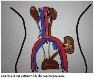
Figure 1. Kidney Transplant in Children
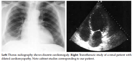
Figure 2. Imaging Studies