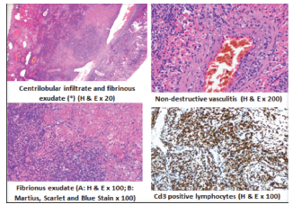
Figure 2. Microscopic Examination of Lung Biopsy With Fibrinous and Organizing Pneumonia and Lymphocytic Interstitial Pneumonia (hematoxylin and eosin scarlet and blue stains)