Volume: 24 Issue: 8 August 2026
FULL TEXT
Kidney Autotransplant With Ex Vivo Repair for Bilateral Renal Artery Aneurysm: Case Report
Renal artery aneurysm is a rare vascular condition that could remain silent or could lead to potential life-threatening consequences, especially when there are multiple lesions. Endovascular treatment and open in situ surgical reconstruction are often considered first-line options, although not always feasible or durable. We report the case of a successful kidney autotransplant in a 56-year-old female patient who had bilateral multiple renal artery aneurysms with calcified walls and parietal thrombotic apposition. Given the history of hypertension and the risks related to aneurysm rupture, the patient underwent lapa-roscopic nephrectomy followed by ex situ repair and heterotopic graft reimplant. We also compared our approach with the literature to report on the current evidence and the recommended management for renal artery aneurysms.
Key words : Ex situ artery repair, Hypertension, Laparoscopic nephrectomy, Renal artery aneurysm, Renal autotransplantation
Introduction
Renal artery aneurysm (RAA) is an uncommon vas-cular lesion, with an incidence <0.1%,1 predo-minantly in female individuals in their sixth decade of life; presentations often receive physicians’ attention when patients present during other consultations, for example, following imaging studies. Although most RAAs remain silent for years, the potential consequences could be fatal, for example, in the case of rupture after excessive enlargement (>2 cm), distal embolization, progressive perfusion loss of the kidney parenchyma, or, more frequently, refractory renovascular hypertension.
When RAA is multiple or bilateral, cases become challenging to wait and watch in a conservative manner, rather than intervening. Typically, in most cases, when there is a single saccular aneurysm arising from the main renal artery, endovascular treatment represents the first-line intervention.2 However, this modality heavily relies on a cooperative anatomy, that is, with a neck that allows secure exclusion of the sac, a position that does not jeopardize essential segmental branches, and a configuration that avoids leaving areas of the kidney without blood supply. If the aneurysm lies at a bifurcation or involves several branches, then the limitations of endovascular treatment become more evident. Any attempt to place a stent-graft or coil creates a risk for arterial flow disruption and potential infarction. For these reasons, a substantial proportion of RAAs remain outside the scope of endovascular treatment.
Open in situ reconstruction is the next step in the therapeutic decision ladder; however, especially for deep hilar aneurysms, open in situ reconstruction is far from straightforward. Access to the lesion may be limited, and, even with a skilled vascular team, the dissection in the confined space of the hilum can require extended surgical time. The kidney must tolerate a period of warm ischemia while repairs are attempted, and small branches can be inadvertently damaged or left inadequately reconstructed. When both kidneys are affected, the consequences of even a partial loss of function become particularly relevant.
Kidney autotransplant for renal artery disease was first performed by Ota and colleagues in 1967.3 Since then, given the above-mentioned limitations, a limited number of more complex renal lesions have been treated similarly.
Here, we report a case with multiple bilateral RAAs and complex hilar anatomy. The combination of laparoscopic nephrectomy and ex vivo repair allowed for a controlled reconstruction with minimal warm ischemia time and, ultimately, a successful reimplant of the graft.
Case Report
A 56-year-old female patient, affected by hypertension, dyslipidemia, umbilical hernia, and previous thyroidectomy, was referred after incidental findings were shown at computed tomography angiography scan of multiple RAAs involving the kidneys bilaterally (Figure 1).
Imaging showed 2 sacs measuring 27 × 16 mm and 11 × 11 mm close to the hilum in the right renal artery; on the left renal artery, an aneurysmatic dilation was also present measuring 26 × 18 mm. Both vessels were characterized by calcified walls and parietal thrombotic apposition.
The main concerns were the cumulative risk of rupture and the possibility of progressive loss of renal perfused parenchyma. At the moment of presentation, renal function was preserved (creatinine level of 0.58 mg/dL); however, in consideration of the patient’s comorbidities and of the challenges of an endovascular or in situ treatment, a multidisciplinary consensus posed indication to bilateral nephrectomy and reimplant of the left kidney.
The operation combined a transperitoneal bila-teral laparoscopic nephrectomy (Figure 2) with rapid cooling and perfusion of the left organ, ex vivo resection of the aneurysmal segments, and bench reconstruction of the hilar artery (Figure 3).
The left kidney was then heterotopically reim-planted into the right iliac fossa with end-to-side vascular anastomoses to the external iliac vessels and uretero-bladder anastomosis over a double-J stent. The kidney had a single anatomy of 1 artery, 1 vein, and 1 ureter. Postoperative course was uneventful, and Doppler imaging confirmed immediate per-fusion and stable graft function at 8 months of follow-up.
Discussion
Ex vivo repair of RAA with kidney autotransplant represents the preference solution in patients with anatomically complex or bilateral RAA when opti-mal exposure to allow a refined repair could not be guaranteed without a period of warm ischemia at the renal parenchyma. In contrast, during back-table repair, performed under hypothermic temperature, the ischemic damage is better tolerated by the organ, and thereby the limited exposure obtainable with in situ surgery is overcome.4
Bench surgery, a term attributed to Guerriero and colleagues,5 refers to reconstructive surgery per-formed on diseased kidneys that are kept viable through perfusion outside the body. The bench surgery approach, which was originally intended for the preservation of deceased donor kidneys and subsequent transplant, has also been adopted in complex urologic operative procedures allowing autotransplant.6
A general recommendation to intervene on an asymptomatic RAA is considered for a vessel diameter >2 cm; however, even at the smaller calibers, complications associated with rupture and/or syste-mic comorbidities have been described, thus indi-cating the necessity for repair. If an emergency occurs, then open nephrectomy is often the only possible option.
In elective settings, minimally invasive surgery in the form of laparoscopic or robotic nephrectomy is instead increasingly chosen, followed by back-table reconstruction of the renal artery. In this regard, various techniques have been proposed, including direct repair, autologous saphenous vein or bovine pericardium patch, or the use of internal iliac artery.7
Most series report only sporadic cases,8 with limited literature on larger series,9,10 mostly due to technical complexity and low frequency of ex situ repair for RAAs. Thus, it is always recommendable to perform such procedures in specialized transplant centers. We also recommend a multidisciplinary team involvement of skilled transplant and vascular surgeons, cardiologists, and nephrologists.
In conclusion, our present case aligns with the existing evidence regarding refractory hypertension treatment in female patients in the sixth decade of life and affected by complex multiple RAAs, for which kidney autotransplant represents the only safe, efficient, and durable treatment modality.
References:
- Coleman DM, Stanley JC. Renal artery aneurysms. J Vasc Surg. 2015;62(3):779-785. doi:10.1016/j.jvs.2015.05.034.
CrossRef - PubMed - Zhang Z, Yang M, Song L, Tong X, Zou Y. Endovascular treatment of renal artery aneurysms and renal arteriovenous fistulas. J Vasc Surg. 2013;57(3):765-770. doi:10.1016/j.jvs.2012.09.042
CrossRef - PubMed - Ota K, Mori S, Awane Y, Ueno A. Ex situ repair of renal artery for renovascular hypertension. Arch Surg. 1967;94(3):370-373. doi:10.1001/archsurg.1967.01330090064016
CrossRef - PubMed - Henke PK, Cardneau JD, Welling TH 3rd, et al. Renal artery aneurysms: a 35-year clinical experience with 252 aneurysms in 168 patients. Ann Surg. 2001;234(4):454-462. doi:10.1097/00000658-200110000-00005
CrossRef - PubMed - Guerriero WG, Scott R, Joyce L. Development of extracorporeal renal perfusion as an adjunct for bench renal surgery. J Urol. 1972;107(1):4-8. doi:10.1016/s0022-5347(17)60930-3
CrossRef - PubMed - Putnam CW, Halgrimson CG, Stables DP, et al. Ex vivo renal perfusion and autotransplantation in treatment of calculous disease or abdominal aortic aneurysm. Urology. 1975;05(3):337-342. doi:10.1016/0090-4295(75)90149-1
CrossRef - PubMed - Forgács B, Augustine T. Renal autotransplant in patients with complex hilar renal artery aneurysms. Exp Clin Transplant. 2013;11(5):450-453. doi:10.6002/ect.2012.0230
CrossRef - PubMed - Sevmis S, Karakayali H, Boyvat F, et al. Renal autotransplantation for complex renal arterial disease: a case report. Exp Clin Transplant. 2006;4(2):559-561.
CrossRef - PubMed - Contarini E, Takagi K, Kimenai H, et al. Kidney autotransplantation for renal artery aneurysm: case series and a systematic review. Ann Vasc Surg. 2021;77:349.e5-349.e18. doi:10.1016/j.avsg.2021.05.039
CrossRef - PubMed - Fang Y, Kimenai H, de Bruin RWF, et al. Treatment of renal artery aneurysm by ex situ repair and autotransplantation: a nationwide cohort study. Eur Surg Res. 2024;65(1):123-129. doi:10.1159/000541814
CrossRef - PubMed

Volume : 24
Issue : 8
Pages : 659 - 661
DOI : 10.6002/ect.2026.0121
From the 1Department of Surgery, Sapienza University, Rome; the 2Bind Department, Palermo University, Palermo; the 3General and Emergency Surgery Unit, National Relevance and High Specialization Hospital (ARNAS), Ospedale Civico Di Cristina Benfratelli, Palermo; and the 4School of Medicine and Surgery, Palermo University, Palermo, Italy
Acknowledgements: The authors have not received any funding or grants in support of the presented research or for the preparation of this work and have no declarations of potential conflicts of interest.
*Maria Irene Bellini, Giovanni Tomasello, and Giacomo Urso contributed equally to this work.
Corresponding author: Maria Irene Bellini, Department of Surgery, Sapienza University, Viale Regina Elena 324, 00161, Rome, Italy
E-mail: mariairene.bellini@uniroma1.it
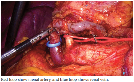
Figure 2. Isolation of Renal Hilum During Laparoscopic Nephrectomy
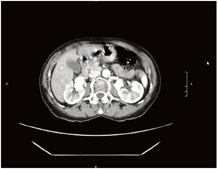
Figure 1. Preoperative Computed Tomography Scan Findings of Bilateral Renal Aneurysms
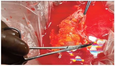
Figure 3. Back-Table Repair of the Renal Artery Aneurysm