Volume: 22 Issue: 5 May 2024
FULL TEXT
We present a challenging clinical case of a 68-year-old female kidney transplant recipient who had a complicated posttransplant course marked by borderline T-cell-mediated rejection and BK virus nephropathy. The treatment for borderline rejection with steroids resulted in overimmunosuppression, and the patient acquired cytomegalovirus infection manifesting as colitis and SARS-CoV-2 infection. This progressed rapidly to collapsing glomerulopathy and allograft failure. This study also highlights the challenges in surveillance with donor-derived cell-free DNA in the setting of allograft injury by multiple viral infections.
Key words : COVID-19, Severe acute respiratory syndrome coronavirus 2, T-cell-mediated rejection
Case Report
We present a challenging clinical case of a 68-year-old female kidney transplant recipient who received a kidney from a 30-year-old deceased donor with a Kidney Donor Profile Index of 22% and one haplotype A, B, and DR antigen match. The donor was Cytomegalovirus (CMV) positive, whereas the recipient was CMV negative. Induction immunosuppression included basiliximab and methylprednisolone because of the advanced age of the recipient. Maintenance immunosuppression included tacrolimus with goal 12-hour trough levels of 8 to 10 ng/dL, mycophenolate mofetil (MMF) dose of 1000 mg by mouth (PO) twice daily, and prednisone 5 mg PO daily. Standard CMV prophylaxis with valgan-ciclovir for donor-positive and recipient-negative status was administered for a period of 6 months. The recipient’s posttransplant course was complicated by sigmoid colon perforation on postoperative day 4, requiring an exploratory laparotomy with sigmoid colectomy and a colostomy. The patient had slow graft function and achieved a baseline serum creatinine of 1.45 mg/dL at 3 weeks posttransplant. Tacrolimus levels remained therapeutic at goal. She was noted to have BK viremia with BK virus level in serum as measured by quantitative polymerase chain reaction (qPCR) of 539 copies/mL at 2 months after transplant. She also had donor-derived cell free DMA (dd-cf-DNA) level of 0.29%.
The patient progressed to BK virus nephropathy (BKVN) with BK viremia of 10 527 copies/mL by qPCR at 3 months after transplant with dd-cf-DNA level of 0.21%. As a result, MMF dose was reduced to 750 mg PO twice daily. Serum creatinine increased from baseline to 1.7 mg/dL.
At 6 months posttransplant, the patient under-went colostomy reversal but developed acute kidney injury (AKI) following the procedure, with serum creatinine level of 2.24 mg/dL. The patient showed persistent BKVN with BK virus level of 16 094 copies/mL by qPCR. Because of ongoing BKVN, MMF dose was further reduced to 500 mg PO twice daily. At 7 months posttransplant, BK virus qPCR decreased to 3394 copies/mL; however, serum creatinine level increased to 2.78 mg/dL and CMV qPCR of <34.5 IU/L was also noted. The dd-cf-DNA level was 0.25%.
Because of worsening AKI, a renal allograft biopsy was requested. The biopsy showed a T-cell-rich lymphocytic infiltrate in the interstitium involving <10% along with mild tubulitis and negative SV40 staining (Figure 1A). The findings were reported as borderline changes, suspicious for T-cell-mediated rejection (TCMR). The patient was treated with 1000 mg of methylprednisolone intravenously in 2 divided doses. One week after treatment completion, the patient presented with SARS-CoV-2 infection, and BK qPCR increased to 9808 copies/mL. The patient was treated for SARS-CoV-2 with intravenous remdesivir, and MMF was discontinued. Five days after treatment completion for SARS-CoV-2 infection, MMF was restarted at a dose of 250 mg PO twice daily.
Three weeks after treatment for borderline TCMR, the patient was noted to have CMV qPCR of 1 160 000 IU/L, BK qPCR of 13 016 copies/mL, serum creatinine of 2.98 mg/dL, and immune cell function assay level of 60. The patient developed CMV colitis with diarrhea. Tacrolimus levels remained therapeutic in the range of 8 to 10 ng/dL throughout this time. As a result of concomitant CMV disease and BKVN, MMF and prednisone were both stopped, Tacrolimus dose was adjusted to 5 to 7 ng/dL, and treatment was initiated for CMV disease with intravenous ganciclovir with dose adjusted to renal function. Five days after treatment initiation with ganciclovir, CMV qPCR decreased to 96 400 copies/mL, but AKI worsened, with the patient becoming severely oliguric and serum creatinine level increasing to 4.59 mg/dL. An increase of dd-cf-DNA was noted at 4.8%, which was a substantial increase from the baseline level of 0.2%. Because of worsening AKI, ganciclovir was changed to maribavir, tacrolimus was discontinued, and the patient was started on belatacept at 5 mg/kg monthly. At 8 months posttransplant, a second renal allograft biopsy was requested.
The second biopsy showed a T-cell-rich infiltrate with focal mild endothelitis and all glomeruli demonstrated evidence of collapsing glomerulopathy. SV40 immunostain was positive for BK virus, but CMV immunostain was negative. Compared with the first biopsy, there was increased global glomerulosclerosis (6/50), associated with increase in degree of interstitial fibrosis from <10% to 30% to 40%, (Figure 1c). Endothelitis was also noted on few arteries, but CD138 immunostain confirmed absence of plasma cell infiltrates. Donor-specific antibodies remained negative, and there was no C4d staining in peritubular capillaries. Biopsy did not meet criteria for cellular or antibody-mediated rejection.
One week after the second biopsy, the patient progressed to anuria and developed pulmonary edema for which temporary hemodialysis was started. Two weeks after initiation of treatment for CMV disease, CMV qPCR decreased to 313 IU/L and BK qPCR decreased to 1795 copies/ mL; however, serum creatinine level remained elevated at 4.35 mg/dL. Over the next month, the patient remained anuric and became dialysis dependent. Renal allograft failure was attributed to collapsing glomerulopathy and concomitant BK virus-associated nephropathy.
Discussion
We presented an unusual situation in a kidney transplant recipient with 3 serious viral infections (BKVN, CMV colitis, and SARS-CoV-2 infection) at the same time. The immune cell function assay of 60 did show that the patient was overimmunosuppressed1,2 despite appropriate reduction in immunosuppression to manage ongoing viral infections. Collapsing glomerulopathy is usually associated with HIV infection3 but has also been described with CMV nephropathy4,5 and SARS-CoV-2 nephropathy.6 We were not able to demonstrate CMV on pathology, but that could be related to treatment with ganciclovir and, by the time biopsy was performed, CMV titer by qPCR had decreased to 313 IU/L. Both CMV and SARS-CoV-27,8 infections are known risk factors for collapsing glomerulopathy in patients of African American ancestry, who harbor ApoL1 risk mutations. Although our recipient was White, the donor was of African American ancestry. The absence of collapsing features during the first biopsy and widespread collapsing features after CMV and SARS-CoV-2 infections support the hypothesis that collapsing glomerulopathy was likely related to viral infections. T-cell-rich tubulointerstitial infiltrate observed on pathology could be attributed to viral activation of the immune system. One haplotype A, B, and DR antigen match with the donor, advanced age, severe immunosuppression as indicated by an immune cell function assay level of 60, and 3 concomitant viral infections in the setting of thera-peutic immunosuppression render the possibility of TCMR less likely, although it cannot be completely excluded.
This case also highlighted the potential pitfalls of currently available biomarkers for surveillance of allograft rejection. We used dd-cf-DNA surveillance, which is a marker of injury and has shown good correlation with diagnosis of rejection when values are >1.0%. Although the test has a very high negative predictive value (90%), the positive predictive value is only in the range of 30% to 50%.9 The patient’s value was 0.2% when the biopsy was read as borderline TCMR. However, it is possible that the biopsy changes interpreted as borderline were related to BKVN. Because we consider allograft biopsy as the gold standard for diagnosing rejection, we treated this with methylprednisolone despite a low level of dd-cf-DNA. We believe that the treatment for borderline TCMR tipped the delicate balance of immunosup-pression and the patient became so immunosup-pressed that she acquired several viral infections concomitantly. A dd-cf DNA value of 4.8% prompted the second biopsy; however, allograft biopsy could not conclusively determine whether this was indeed an allograft rejection. Thus, dd-cf-DNA elevations should be interpreted with caution. We believe that the renal allograft failure was secondary to collapsing glomerulopathy, with most likely association being CMV, although SARS-CoV-2 infection could also have contributed.
Conclusions
Kidney transplant recipients of advanced age are at a higher risk of overimmunosuppression and opportunistic infections. Monitoring immunosup-pression with immune cell function assays in this patient population can be an inexpensive and useful modality to prevent complications. Treatment for borderline TCMR in kidney transplant should be reconsidered in patients who are severely immuno-suppressed, and optimization of immunosuppres-sion instead of treatment with methylprednisolone should be considered. A high value of dd-cf-DNA does not necessarily indicate allograft rejection but can represent allograft injury from multiple viruses.
References:
- Batal I, Zeevi A, Heider A, et al. Measurements of global cell-mediated immunity in renal transplant recipients with BK virus reactivation. Am J Clin Pathol. 2008;129(4):587-591. doi:10.1309/23YGPB1E758ECCFP
CrossRef - PubMed - Zeevi A, Lunz J. Cylex ImmuKnow cell function assay. Methods Mol Biol. 2013;1034:343-351. doi:10.1007/978-1-62703-493-7_21
CrossRef - PubMed - Chandra P, Kopp JB. Viruses and collapsing glomerulopathy: a brief critical review. Clin Kidney J. 2013;6(1):1-5. doi:10.1093/ckj/sft002
CrossRef - PubMed - Grover V, Gaiki MR, DeVita MV, Schwimmer JA. Cytomegalovirus-induced collapsing focal segmental glomerulosclerosis. Clin Kidney J. 2013;6(1):71-73. doi:10.1093/ckj/sfs097
CrossRef - PubMed - Tomlinson L, Boriskin Y, McPhee I, Holwill S, Rice P. Acute cytomegalovirus infection complicated by collapsing glomerulopathy. Nephrol Dial Transplant. 2003;18(1):187-189. doi: 10.1093/ndt/18.1.187
CrossRef - PubMed - Hassler L, Reyes F, Sparks MA, Welling P, Batlle D. Evidence for and against direct kidney infection by SARS-CoV-2 in patients with COVID-19. Clin J Am Soc Nephrol. 2021;16(11):1755-1765. doi:10.2215/CJN.04560421
CrossRef - PubMed - Chang JH, Husain SA, Santoriello D, et al. Donor’s APOL1 risk genotype and “second hits” associated with de novo collapsing glomerulopathy in deceased donor kidney transplant recipients: a report of 5 cases. Am J Kidney Dis. 2019;73(1):134-139. doi:10.1053/j.ajkd.2018.05.008
CrossRef - PubMed - Wu H, Larsen CP, Hernandez-Arroyo CF, et al. AKI and collapsing glomerulopathy associated with COVID-19 and APOL 1 high-risk genotype. J Am Soc Nephrol. 2020;31(8):1688-1695. doi:10.1681/ASN.2020050558
CrossRef - PubMed - Oellerich M, Sherwood K, Keown P, et al. Liquid biopsies: donor-derived cell-free DNA for the detection of kidney allograft injury. Nat Rev Nephrol. 2021;17(9):591-603. doi:10.1038/s41581-021-00428-0
CrossRef - PubMed

Volume : 22
Issue : 5
Pages : 392 - 395
DOI : 10.6002/ect.2024.0029
From the Division of 1Transplant Surgery, West Virginia University Hospitals; 2Transplant Alliance, West Virginia University Hospitals; the Department of 3Transplant Infectious Disease, West Virginia University Hospitals; and the Department of 4Pathology, West Virginia University Hospitals, Morgantown, West Virginia, USA
Acknowledgements: The authors have not received any funding or grants in support of the presented research or for the preparation of this work and have no declarations of potential conflicts of interest.
Corresponding author: Rajeev Sharma, 1 Medical Center Drive, PO Box 9238, HSS Suite 700, Morgantown, WV 26505, USA
Phone: +1 304 293 0283
E-mail: Rajeev.Sharma@hsc.wvu.edu
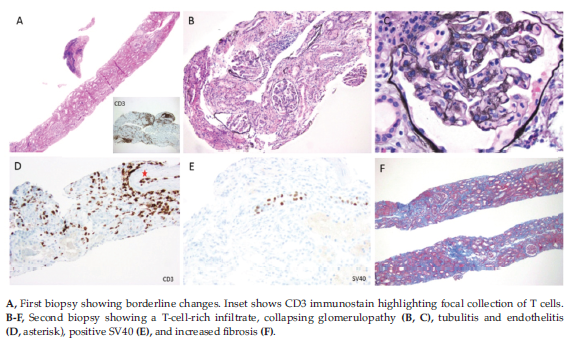
Figure 1. Biopsy Findings