Volume: 22 Issue: 5 May 2024
FULL TEXT
Factors Associated With Recurrence of Underlying Diseases Among Liver Transplant Recipients: A Single-Center Study
Objectives: The recurrence of underlying diseases remains a major cause of graft failure after liver transplant. This study aimed to identify factors associated with the recurrence of underlying diseases and investigate the incidence of these factors and recurrence at the main liver transplant center in Iran.
Materials and Methods: We included adult liver transplant recipients followed at Shiraz Transplant Center between 2011 and 2018 with a confirmed diagnosis of recurrence of underlying disease in our study. We reviewed medical records and extracted data on demographic characteristics, clinical and paraclinical features, medication use, and current status. We used a systematic random sampling method to select a control group of 95 transplant recipients who did not have recurrence. Of 3022 total transplant recipients, 76 recipients experienced a recurrence of their underlying disease.
Results: Model for End-Stage Liver Disease score, underlying disease, recipient blood group, donor sex, donor blood group, and rejection frequency were significantly different between study groups with and without recurrence of underlying diseases. Liver transplant recipients with recurrence had lower mean Model for End-Stage Liver Disease score. Recipients with recurrence also had higher rate of drug consumption (eg, prednisolone, tacrolimus, mycophenolate mofetil, sirolimus). Regression analysis showed that donor sex and rejection frequency had an effect on disease recurrence. Death occurred more frequently in liver transplant recipients with recurrence than in the control group (39.5% vs 26.3%), but the difference was not significant.
Conclusions: Donor sex and acute rejection frequency are independent factors predictive of the recurrence of underlying disease. Modifying risk factors can help minimize the recurrence of underlying diseases after liver transplant.
Key words : Graft failure, Primary disease, Risk factors
Introduction
Liver transplant (LT) is the only effective treatment for end-stage liver disease. However, LT has many challenges, including a limited donor pool, lifetime immune system suppression, and opportunistic infections. Moreover, long-term consequences such as chronic biliary complications, the development of new malignancies, and recurrence of the underlying disease (RUD) are still major obstacles in post-LT surveillance.1-3
In LT recipients, RUD can be classified into 4 main groups: autoimmune, malignant, viral diseases, and lifestyle risk behaviors. Autoimmune diseases such as primary biliary cirrhosis (PBC), autoimmune hepatitis (AIH), and primary sclerosing cholangitis (PSC) are the leading causes of RUD among LT recipients. Nonalcoholic fatty liver disease, nonalcoholic steatohepatitis, and alcoholic liver disease can be considered lifestyle-related diseases that are usually less recurrent because of modifications to lifestyle after LT. However, factors such as age, donor steatosis, metabolic syndrome, and immunosuppression have been found to play a role in recurrence of nonalcoholic steatohepatitis and nonalcoholic fatty liver disease.2,4
Given their viral etiologies, hepatitis C virus (HCV) and hepatitis B virus (HBV) may recur after LT. Recently, the incidence of recurrence of HBV infection after LT has decreased because of the use of hepatitis B immunoglobulin and oral antivirals. Combinations of influencing factors pertaining to the virus, donor, recipient, operation, and immunosup-pression are associated with HCV-related fibrosis after LT.5,6
The rate of recurrence of hepatocellular carcinoma (HCC) varies from 16% to 54%. The risk of recurrence depends on a number of factors, such as size, histological differentiation, vascular invasion, and tumor marker level.7,8 Variations in diagnostic criteria and approaches are additional reasons for differences in recurrence rates. Both LT recipients and physicians should consider the possibility of recurrent disease after transplant; thus identification of the risk factors leading to RUD should be a priority.9 In the present study, we aimed to investigate the incidence of RUD and its associated risk factors in LT recipients at Abu-Ali Sina Hospital, Shiraz Transplant Center, Shiraz, Iran, over a 7-year period.
Materials and Methods
Study population and design
Adult LT recipients followed at Shiraz Transplant Center, Shiraz, Iran, from 2011 to 2018 with a confirmed diagnosis of RUD were included in the study. Transplant committee specialists diagnosed RUD, who considered laboratory, pathology, and radiological findings. Medical records and the Abu-Ali Sina hospital database were reviewed, and data were extracted on demographic, clinical, and transplant characteristics, donor characteristics, results of histopathologic examinations, laboratory data (before LT and at the time of recurrence), medications used, possible mortality, and retransplant. For a control group, we selected 95 LT recipients who did not have recurrence by using a systematic random sampling method.
Ethical considerations
This study complied with the Declaration of Helsinki and was approved by the research ethics committee of Shiraz University of Medical Sciences, Shiraz, Iran. Given the retrospective nature of the study, the Institutional Review Board of Shiraz University of Medical Sciences provided an informed consent waiver (IR.SUMS.MED.REC.1400.002).
Statistical analyses
For continuous and categorical variables, we presented data as the mean ± SD or number (%). To check normal distribution, we used the Shapiro-Wilk test. We used the t-test and the chi-square test to compare the 2 groups. The Kaplan-Meier method with the log-rank test was used for survival analyses. To minimize the effects of confounding factors, we entered information on Model for End-Stage Liver Disease (MELD) score, underlying diseases, recipient blood group, donor sex (female), donor blood group, acute rejection frequency, presence of inflammatory bowel disease (IBD), and use of prednisolone, tacrolimus, mycophenolate mofetil (MMF), and sirolimus into Cox proportional hazard regression models, and RUD was considered an event. We analyzed data with SPSS software (version 16.0, SPSS Inc). We considered significance at P = .05 in all tests.
Results
During the study period (2011-2018), 3022 patients underwent LT at our center. The follow-up period ranged from 4 to 10 years, with a median of 7 years. Of 20709 person-years, 76 RUD occurred, which yielded an estimated RUD incidence of 3.66 per 100 person-years. Mean age (± SD) in LT recipients with recurrence was 42.46 ± 12.24 years. No significant differences were shown between the RUD and control groups in terms of age (P = .548), sex (P = .775), donor age (P = .267), gender concordance (P = .485), cold ischemia time (P = .383), warm ischemia time (P = .478), retransplant (P = .477), or hepatic artery thrombosis (P = .746). On the other hand, significant relationships were found for some other variables. Mean MELD score was significantly lower in the RUD group (P = .028); in terms of underlying disease, PSC, AIH + PBC/overlap, and viral/HCC were significantly more prevalent in the RUD group (P < .001). Moreover, the distribution of blood groups differed significantly in both recipients and donors (P = .011 and P = .044). Those with RUD received more organs from female donors (P = .041) and experienced ≥1 acute rejection episode. The detailed baseline demographic and clinical characteristics of both groups are summarized in (Table 1).
Regarding the etiology of the RUD, the most recurrent etiology was viral and HCC, with an incidence rate of 46.7 per 100 person-years, followed by AIH + PBC/overlap (44.1) and PSC (27.8). The final incidence rates of each etiology are shown in (Table 2).
The most frequent comorbid condition in the group with RUD was IBD (P = .031), and the RUD group used significantly more prednisolone (P < .001), tacrolimus (P = .001), MMF (P = .018), and sirolimus (P < .001) in the post-LT course.
(Table 3) lists comparisons in baseline and most recent laboratory data and their changes between the 2 groups. The most recent aspartate transaminase (AST) value and its change, as well as the baseline and most recent values (along with their changes), of alanine transaminase (ALT) and alkaline phosphatase (ALP) were all significantly higher in the group with RUD versus the group without RUD (P =.925; P < .001, and P = .008 for AST; P = .045, P < .001, and P < .001 for ALT; P = .044, P < .001, and P = .049 for ALP). In addition, baseline platelet count was significantly higher in the RUD group, with a significantly smaller change compared with the control group (P = .001 and P = .023). Complete findings for laboratory data and medication use are listed in (Table 3).
Although the mortality rate was higher in the RUD group (39.5% vs 26.3%), the relationship was not significant (P = .067). No significant difference in survival was shown between the 2 groups (log-rank P = .110; (Figure 1)), and median time to RUD was 2.21 years after LT (Figure 2).
Among the significant relationships in the bivariate analyses that were entered into the multivariate Cox regression model, donor sex and acute rejection frequency had an effect on RUD. Significant variables in the Cox regression model are summarized in (Table 4).
Discussion
Our study focused on RUD among LT recipients followed at the main LT center in Iran, where overall estimated incidence of RUD was 3.66 per 100 person-years. The incidence appeared to vary depending on the underlying etiology of liver cirrhosis, and RUD was more common among LT recipients with viral or autoimmune liver disease than with other RUDs. With an overlap was one of the most frequent causes of RUD among all LT recipients at our center. Autoimmune liver diseases were reported to recur after LT, with incidence rates ranging from 10% to 50%.1 The reported incidence of recurrent PSC ranged from 8.6% to 37%, whereas rate of PBC recurrence rate was 29.4% and rate of AIH recurrence ranged from 17% to 42%.3,10,11
Along with autoimmune causes, recurrence of viral liver disease or HCC, with an incidence rate of 46.7 per 100 person-years, was at the top of the list in our study patients. A systematic review reported a mean HCC recurrence rate of 16% among all patients who had LTs for treatment of HCC.7 The recurrence of HBV and HCV has radically diminished after the development of new antiviral agents.12,13 In our study, the incidence rates were mostly consistent with those published previously, although the lower rates reported in some earlier research may be because of differences in the model of presentation or diagnostic tools, diagnostic approaches, or follow-up times.
Univariate analysis of demographic characteristics showed that only donor sex (more men in the RUD group) differed significantly between groups. However, no association was seen in our regression analyses. Lim and Lake reported that recipient age had an effect on recurrent PSC and that donor age had effect on recurrent PBC.10 Controversy exists on the effect of recipient age on recurrent PBC3; however, Li and Lake found that neither recipient age nor donor age had an effect on recurrent PBC. In another study, recipient sex and age, along with donor sex, were not related to recurrent AIH, although recipient age and sex mismatches were associated with recurrent AIH.14 In a previous study of HCC recurrence, donor age of >60 years was associated with an increased post-LT recurrence rate.15 In the present study, we used a holistic approach by investigating all types of RUDs together. Our findings disclosed no statistical association with donor age; however, female patients were more prevalent in the RUD group (36.9% vs 22.1%; hazard ratio = 2.15). This result may reflect differences in the prevalence of female versus male patients in different underlying liver diseases.
Among the clinical variables in our participants, MELD score, acute rejection frequency, donor and recipient blood group, IBD, and immunosuppressive medication use, underlying disease, donor sex were significantly different between groups in univariate analyses. In the Cox regression model, we found that IBD, acute rejection, and donor sex remained significant.
Of note was that the presence of any IBD was a significant risk factor for recurrence in the present study, which is probably because of the large number of LT recipients with recurrent PSC. This finding, however, is inconsistent with the results of other studies.16,17
Lim and Lake reviewed the risk factors for recurrent disease after LT and found that risk factors for recurrent PSC included a history of recurrent acute cellular rejection and transplant for cholan-giocarcinoma, whereas warm ischemia time was a risk factor for recurrent PBC.10 Acute rejection was an independent risk factor for RUD in the present study.
Interestingly, mean MELD score at the time of LT was significantly lower (by nearly 2 points) in the RUD group. This finding contrasts with previous studies, which linked high MELD scores with recurrent PSC.17-19 Another study of HCV recurrence reported a somewhat similar finding, in which patients with high MELD scores tended not to develop more severe or earlier HCV recurrence compared with patients with low MELD scores.20 Our result may reflect differences in the distribution of underlying diseases in the control and RUD groups.
The distribution of ABO blood types in our recipients and donors differed significantly between the RUD group and the control group. Among both recipients and donors, blood type O was more common in the RUD group. Mohkam and colleagues recently reported that blood type A was associated with higher HCC recurrence rates compared with other blood types.21 Our findings, along with those of Mohkam and colleagues, highlight the need for future studies to focus on immune markers as well as blood type and their roles in RUD after LT.
Regarding immunosuppression regimens, use of prednisolone, tacrolimus, sirolimus, and MMF differed between LT recipients with and without RUD. In this connection, Krishnamoorthy and colleagues proposed low-dose corticosteroids along with maintenance immunosuppression to prevent recurrent AIH and improve survival.11 On the other hand, the usual clinical practice at Shiraz Organ Transplant Center is to use tacrolimus along with low-dose prednisolone as immunosuppression for autoimmune liver disease after LT.
Although treatment with either azathioprine or MMF as an add-on to calcineurin inhibitors and corticosteroids was found to protect against recurrent AIH,22 controversy remains concerning the effects of tacrolimus versus cyclosporin because of an association with an increased risk of recurrent PBC and recurrent AIH.3,10,17
Corticosteroids stimulated HBV replication after LT and were linked with recurrent HBV.6 The recurrence rate of HCC after LT was also associated with higher plasma levels of both tacrolimus and cyclosporin.23 However, across different studies, results were inconsistent, possibly because of different points of view and different RUDs across studies. Although we did not analyze the relationship between medications and each etiology, our findings from the univariate analysis seem consistent with those of previous studies on the potential role of tacrolimus in RUD. We did not find any relationship between cyclosporin and RUD. This may be attributed to the low number of patients who received cyclosporin in our study.
Hepatic transaminases, ALP, and platelet count varied significantly between the RUD and control groups. Generally, higher serum concentrations at both baseline and at most recent follow-up, along with different patterns of increase or decrease during follow-up, were detected in the RUD group for AST, ALT, and ALP, and all relationships were significant except for baseline AST. Our findings for changing patterns and the most recent laboratory data were clear; hence, patients are routinely followed up with these tests in the post-LT clinic. The elevated baseline values of ALT and ALP merit additional and more detailed study, given that high baseline laboratory values may indicate more advanced disease. Pre-LT ALP level has been reported as an important independent predictor of HCC recurrence, with even greater predictive value than α-fetoprotein.24 Moreover, earlier HCV-associated HCC recurrence was linked to high ALT levels.25 On the other hand, Molmenti and colleagues found no significant predictive value for pre-LT ALT, AST, platelet, or ALP levels in recurrence of AIH, although they noted a significant increase after LT in ALT, AST, and ALP levels.26
Regarding platelet count, we found a significantly higher pre-LT platelet count in the RUD group versus the control group, although the most recent counts at follow-up did not differ significantly between groups. Changes in platelet count were also shown to be significant. This is consistent with findings from Han and colleagues, who found higher pre-LT platelet counts to correlate with an increased HCC recurrence rate after living donor LT.27
Many studies have reported that living donor LT can increase the mortality rate in patients with AIH, and mortality rates of about 0.9% were reported for patients with recurrent AIH. Moreover, recurrence of PBC was also increased in LT patients whose graft was from a living donor.28 All patients in our study received grafts from deceased donors. Although more deaths occurred in the RUD group, the difference compared with the control group was not significant. As shown in (Figure 1), in the first 4 years, survival decreased rapidly in both groups, albeit more notably in the RUD group; during the second 6 years, survival continued to decrease more gradually. As shown in (Figure 2), the median time to recurrence was 2.21 years after LT.
Early studies reported times to recurrence of AIH of 4.6, 2.6, and 1 year and mean time to recurrence of PSC of 8.6 (range, 0.4-17 y).10,11,14,17 Another report showed median time to recurrence of HCC of 6.1 years.29 After LT, the time to HBV recurrence has been shown to range from 15 to 2615 days.6,12 The mean time to recurrence of PBC was found to be 3 to 5.5 years post-LT. Other studies noted that tacrolimus, compared with cyclosporin, was associated with a shorter time to PBC recurrence.30,31
Initially, our LT recipients survived with RUD, although survival gradually decreased up to the year 11 after their surgery. This is consistent with findings published by Ueda and colleagues, who reported a rapid reduction in survival in the first 5 years after LT among patients with PSC, followed thereafter by a further gradual reduction. Thus, early RUD may indicate a worse prognosis.32
To the best of our knowledge, this is the first study to investigate risk factors, their incidence rates, and their influence on RUD across different etiologies of liver disease. The findings reported here, together with previous studies, can help clinicians achieve a holistic view of the role of these risk factors in the recurrence of specific conditions. Among the strengths of this study, which enhance the reliability our findings, are use of a control group, the large population, and the long follow-up time (average 7 years). However, it should be noted that data were missing for some variables; moreover, follow-up data were incomplete for some patients, and, in some cases, the diagnosis of the underlying disease was tentative. These limitations were due mainly to the retrospective nature of the study.
Conclusions
This study investigated the risk factors associated with the RUD in LT recipients. Donor sex, IBD, and acute rejection frequency were independent factors predictive of recurrence. Modifying risk factors can help minimize the incidence of RUD after LT.
References:
- Faisal N, Renner EL. Recurrence of autoimmune liver diseases after liver transplantation. World J Hepatol. 2015;7(29):2896-2905. doi:10.4254/wjh.v7.i29.2896
CrossRef - PubMed - Neuberger J. An update on liver transplantation: A critical review. J Autoimmun. 2016;66:51-59. doi:10.1016/j.jaut.2015.08.021
CrossRef - PubMed - Li X, Peng J, Ouyang R, Yang Y, Yu C, Lin H. Risk factors for recurrent primary biliary cirrhosis after liver transplantation: a systematic review and meta-analysis. Dig Liver Dis. 2021;53(3):309-317. doi:10.1016/j.dld.2020.12.005
CrossRef - PubMed - Dooghaie Moghadam A, Eslami P, Razavi-Khorasani N, et al. Recurrence of fatty liver disease following liver transplantation for NAFLD-related cirrhosis: current status and challenges. Caspian J Intern Med. 2020;11(4):346-354. doi:10.22088/cjim.11.4.346
CrossRef - PubMed - Joshi D, Pinzani M, Carey I, Agarwal K. Recurrent HCV after liver transplantation-mechanisms, assessment and therapy. Nat Rev Gastroenterol Hepatol. 2014;11(12):710-721. doi:10.1038/nrgastro.2014.114
CrossRef - PubMed - Nasir M, Wu GY. Prevention of HBV recurrence after liver transplant: A review. J Clin Transl Hepatol. 2020;8(2):150-160. doi:10.14218/JCTH.2020.00003
CrossRef - PubMed - de'Angelis N, Landi F, Carra MC, Azoulay D. Managements of recurrent hepatocellular carcinoma after liver transplantation: A systematic review. World J Gastroenterol. 2015;21(39):11185-11198. doi:10.3748/wjg.v21.i39.11185
CrossRef - PubMed - Burra P, Zanetto A, Germani G. Liver transplantation for alcoholic liver disease and hepatocellular carcinoma. Cancers (Basel). 2018;10(2):46. doi:10.3390/cancers10020046
CrossRef - PubMed - Li KK, Neuberger J. Recurrent nonviral liver disease following liver transplantation. Expert Rev Gastroenterol Hepatol. 2009;3(3):257-268. doi:10.1586/egh.09.16
CrossRef - PubMed - Lim N, Lake J. Recurrent disease after liver transplantation. Curr Hepatol Rep. 2020;19(1):54-62. doi:10.1007/s11901-020-00507-z
CrossRef - PubMed - Krishnamoorthy TL, Miezynska-Kurtycz J, Hodson J, et al. Longterm corticosteroid use after liver transplantation for autoimmune hepatitis is safe and associated with a lower incidence of recurrent disease. Liver Transpl. 2016;22(1):34-41. doi:10.1002/lt.24323
CrossRef - PubMed - Orfanidou A, Papatheodoridis GV, Cholongitas E. Antiviral prophylaxis against hepatitis B recurrence after liver transplantation: current concepts. Liver Int. 2021;41(7):1448-1461. doi:10.1111/liv.14860
CrossRef - PubMed - Durand F. How to improve long-term outcome after liver transplantation? Liver Int. 2018;38(Suppl 1):134-138. doi:10.1111/liv.13651
CrossRef - PubMed - Montano-Loza AJ, Ronca V, Ebadi M, et al. Risk factors and outcomes associated with recurrent autoimmune hepatitis following liver transplantation. J Hepatol. 2022;77(1):84-97. doi:10.1016/j.jhep.2022.01.022
CrossRef - PubMed - Orci LA, Berney T, Majno PE, et al. Donor characteristics and risk of hepatocellular carcinoma recurrence after liver transplantation. Br J Surg. 2015;102(10):1250-1257. doi:10.1002/bjs.9868
CrossRef - PubMed - Akamatsu N, Hasegawa K, Egawa H, et al. Donor age (>/=45 years) and reduced immunosuppression are associated with the recurrent primary sclerosing cholangitis after liver transplantation - a multicenter retrospective study. Transpl Int. 2021;34(5):916-929. doi:10.1111/tri.13852
CrossRef - PubMed - Lindstrom L, Jorgensen KK, Boberg KM, et al. Risk factors and prognosis for recurrent primary sclerosing cholangitis after liver transplantation: a Nordic Multicentre Study. Scand J Gastroenterol. 2018;53(3):297-304. doi:10.1080/00365521.2017.1421705
CrossRef - PubMed - Steenstraten IC, Sebib Korkmaz K, Trivedi PJ, et al. Systematic review with meta-analysis: risk factors for recurrent primary sclerosing cholangitis after liver transplantation. Aliment Pharmacol Ther. 2019;49(6):636-643. doi:10.1111/apt.15148
CrossRef - PubMed - Ravikumar R, Tsochatzis E, Jose S, et al. Risk factors for recurrent primary sclerosing cholangitis after liver transplantation. J Hepatol. 2015;63(5):1139-1146. doi:10.1016/j.jhep.2015.07.005
CrossRef - PubMed - Onaca NN, Levy MF, Netto GJ, et al. Pretransplant MELD score as a predictor of outcome after liver transplantation for chronic hepatitis C. Am J Transplant. 2003;3(5):626-630.
CrossRef - PubMed - Mohkam K, Abdallah R, N'Kontchou G, et al. Influence of the ABO blood group system on hepatocellular carcinoma recurrence after liver transplantation. Transplantation. 2022;106(7):1411-1420. doi:10.1097/TP.0000000000004004
CrossRef - PubMed - Heinemann M, Adam R, Berenguer M, et al. Longterm survival after liver transplantation for autoimmune hepatitis: results from the European Liver Transplant registry. Liver Transpl. 2020;26(7):866-877. doi:10.1002/lt.25739
CrossRef - PubMed - Welker MW, Bechstein WO, Zeuzem S, Trojan J. Recurrent hepatocellular carcinoma after liver transplantation - an emerging clinical challenge. Transpl Int. 2013;26(2):109-118. doi:10.1111/j.1432-2277.2012.01562.x
CrossRef - PubMed - Yu M-C, Chan K-M, Lee C-F, et al. Alkaline phosphatase: does it have a role in predicting hepatocellular carcinoma recurrence? Journal of Gastrointestinal Surgery. 2011;15(8):1440-1449.
CrossRef - PubMed - Tarao K, Rino Y, Takemiya S, et al. Close association between high serum ALT and more rapid recurrence of hepatocellular carcinoma in hepatectomized patients with HCV-associated liver cirrhosis and hepatocellular carcinoma. Intervirology. 2000;43(1):20-26. doi:10.1159/000025019
CrossRef - PubMed - Molmenti EP, Netto GJ, Murray NG, et al. Incidence and recurrence of autoimmune/alloimmune hepatitis in liver transplant recipients. Liver Transpl. 2002;8(6):519-526. doi:10.1053/jlts.2002.32981
CrossRef - PubMed - Han S, Lee S, Yang JD, et al. Risk of posttransplant hepatocellular carcinoma recurrence is greater in recipients with higher platelet counts in living donor liver transplantation. Liver Transpl. 2018;24(1):44-55. doi:10.1002/lt.24961
CrossRef - PubMed - Cholongitas E, Burroughs AK. Recurrence of autoimmune liver diseases after liver transplantation: clinical aspects. Auto Immun Highlights. 2012;3(3):113-118. doi:10.1007/s13317-012-0040-5
CrossRef - PubMed - Wong TH, Ho CM, Hsu HH, et al. Delayed hepatocellular carcinoma recurrence after liver transplantation: comprehensive clinical characterization of case series. J Hepatocell Carcinoma. 2022;9:1081-1091. doi:10.2147/JHC.S383474
CrossRef - PubMed - Kogiso T, Egawa H, Teramukai S, et al. Risk factors for recurrence of primary biliary cholangitis after liver transplantation in female patients: a Japanese multicenter retrospective study. Hepatol Commun. 2017;1(5):394-405. doi:10.1002/hep4.1037
CrossRef - PubMed - Mijic M, Saric I, Delija B, et al. Pretransplant evaluation and liver transplantation outcome in PBC patients. Can J Gastroenterol Hepatol. 2022;2022:7831165. doi:10.1155/2022/7831165
CrossRef - PubMed - Ueda Y, Kaido T, Okajima H, et al. Long-term prognosis and recurrence of primary sclerosing cholangitis after liver transplantation: a single-center experience. Transplant Direct. 2017;3(12):e334. doi:10.1097/TXD.0000000000000751
CrossRef - PubMed

Volume : 22
Issue : 5
Pages : 366 - 372
DOI : 10.6002/ect.2023.0097
From the 1Abu Ali Sina Organ Transplant Center, Shiraz University of Medical Sciences, Shiraz, Iran; the 2Shiraz Transplant Research Center, Shiraz University of Medical Sciences, Shiraz, Iran; the 3Student Research Committee, Shiraz University of Medical Sciences, Shiraz, Iran; and the 4Cardiovascular Research Centre, Shiraz University of Medical Sciences, Shiraz,Iran
Acknowledgements: The authors have not received any funding or grants in support of the presented research or for the preparation of this work and have no declarations of potential conflicts of interest. We thank K. Shashok (AuthorAID in the Eastern Mediterranean) for improving the use of English in the manuscript.
Corresponding author: Nasrin Motazedian, Shiraz Transplant Research Center, Shiraz University of Medical Sciences, Shiraz, Iran
E-mail: motazediann@yahoo.com
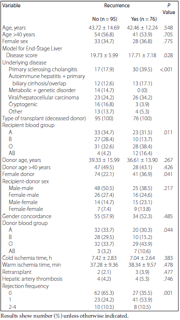
Table 1.Comparison of Demographic and Baseline Clinical Data in Liver Transplant Recipients With and Without Disease Recurrence
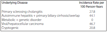
Table 2.Incidence Rate of Recurrence According to Underlying Disease
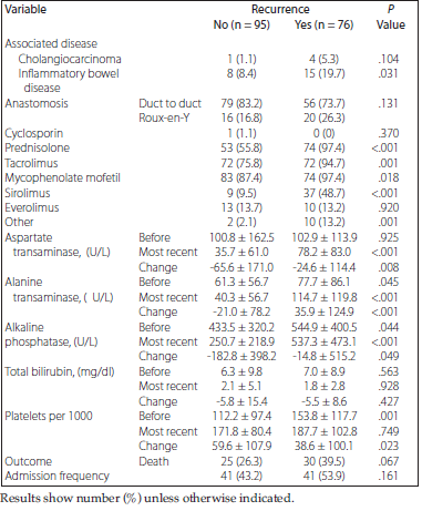
Table 3.Comparison of Associated Diseases, Drugs, and Laboratory Data in Liver Transplant Recipients With and Without Disease Recurrence
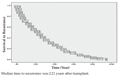
Figure 2.Survival to Recurrence in Patients with Disease Recurrence
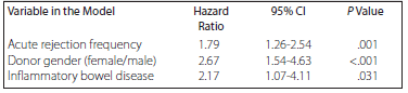
Table 4.Results of Cox Proportional Hazard Model Regression for Likelihood of Recurrence