Volume: 16 Issue: 4 August 2018
FULL TEXT
Objectives: Immunoisolation of pancreatic islets of Langerhans performed by the encapsulation process may be a method to avoid immunosuppressive therapy after transplant. The main problem related to islet transplant is shortage of human pancreata. Resolution of this obstacle may be cryopreservation of encapsulated islets, which enables collection of sufficient numbers of isolated islets required for transplant and long-term storage. Here, we assessed the ability of encapsulated islets to function after long-term banking at low temperature.
Materials and Methods: Islets of Langerhans isolated from rat, pig, and human pancreata were encapsulated within alginate-poly-L-lysine-alginate microcapsules. Cryopreservation was carried out using a controlled method of freezing (Kriomedpol freezer; Kriomedpol, Warsaw, Poland), and samples were stored in liquid nitrogen. After 10 years, the samples were thawed with the rapid method (with 0.75 M of sucrose) and then cultured.
Results: We observed that microcapsules containing islets maintained their shape and integrity after thawing. During culture, free islets were defragmented into single cells, whereas encapsulated islets were still round in shape and compact. After 1, 4, and 7 days of culture of encapsulated islets, the use of 3-(4,5-dimethylthiazol-2-yl)-2,5-diphenyltetrazolium bromide tests showed increased mitochondrial activity. After they were thawed, the insulin secretion capacity was comparable with that obtained with fresh islets.
Conclusions: Cryopreservation and storage of free and microencapsulated islets were possible for about 10 years, although only encapsulated islets retained viability and secretory properties.
Key words : Immunoisolation, Islet transplantation, Microencapsulation
Introduction
Transplant of isolated islets of Langerhans persists as an efficient method for treatment of patients with type 1 diabetes. Effective treatment demands trans-plant of 8000 to 10 000 islet equivalents per kilogram of patient body weight.1 The main problem related to islet transplant is shortage of human pancreata. Solutions for deficiency of organs from deceased donors may be cryopreservation of isolated islets, which involves tissue collection and long-term storage. Cryostorage allows for the accumulation of sufficient amounts of donor tissue and shipping to other institutions.2 Moreover, cryopreservation can decrease immunogenicity of islet allograft by reducing major histocompatibility complex class I antigen expression3 or by depleting “passenger” lymphoid cells.4
The major obstacle associated with cryopreser-vation of pancreatic islets is loss of function of the frozen/thawed islets compared with freshly isolated ones.2 This disadvantage might be overcome by islet microencapsulation in a semipermeable membrane. Application of effective methods for immuno-protection to avoid the shortcomings related to long-term banking of islets is important for islet transplant therapy.
An approach in terms of allogenic islet shortage is application of pig islets for clinical transplant. Human and porcine insulin is structurally similar, and there is an almost unlimited availability of pig donors, providing promise for this treatment in patients with type 1 diabetes. However, there are a number of limitations resulting from xenotransplant procedures. Rapid loss of transplanted islets during the instant blood-mediated inflammatory reaction5 and T-cell-mediated graft rejection6 have both been reported. Regarding the possibility of reduced pancreatic islet immunogenicity, the collection of sufficient numbers of encapsulated islets followed by cryopreservation and long-term banking was tested, showing that pig islet cells remained functional.7
The aim of our study was to assess the influence of long-term storage at low temperature on functional capabilities of encapsulated islets derived from rat, porcine, and human pancreata.
Materials and Methods
Human pancreatic islet isolation, purification, and viability process assessment
Human pancreata were harvested from brain-dead multiorgan donors. Donors were
selected based on the Edmonton protocol for a clinical grade pancreas. All
pancreata were procured using a standardized surgical technique to minimize warm
ischemia time and were flushed with University of Wisconsin (UW) solution for
pancreas perfusion, administered intra-ductally (approximately 1 mL/g pancreas
weight). After the surrounding tissue was removed, organs were perfused with
cold collagenase NB with neutral protease enzyme solution (Serva Electophoresis
GmbH, Heidelberg, Germany) through a catheter placed in the pancreatic duct. The
distended pancreas was subsequently cut into 10 pieces and placed in a Ricordi
chamber and shaken gently at 37°C degree. The digested tissue was collected and
washed with fresh medium (Mediatech solution; Mediatech Inc., Manassas, VA, USA)
to remove the enzyme and then suspended in UW solution (ViaSpan, DuPont, Boston,
MA, USA) before purification. Islets were isolated accordingly to the Ricordi
method.8 After pancreas digestion, the suspended tissue was purified by
continuous density gradient using standard purification method (COBE 2991 cell
processor with Ficoll density gradient solution; COBE, Lakewood, CO, USA). Islet
yield (IE/g), purity, and recovery rate after purification were evaluated in
vitro. Islet viability was assessed using propidium iodide staining to visualize
the distinction between the living islet cells and dead cells (apoptotic,
degranulated); > 95% of cells were viable. The viable islets were identified by
dithizone staining (3 mg/mL; Sigma, St. Louis, MO, USA). The endotoxin
concentration was under detectable levels, and no bacteria or fungus growth was
observed in the islet culture.
This study and the islet isolation protocol including clinical protocol were approved by our bioethics committee (182/2002) at Warsaw Medical University (Warsaw, Poland).
Isolation of pig islets
Porcine pancreata, as the waste material from market-weight pigs (sows weighing
over 150 kg) were processed. The pancreata were dissected after pig
exsanguination, hot water bathing (64°C), and shaving. Pancreata were
subsequently flushed intraductally with cold collagenase P solution (1500-2000
U/mL in Hanks balanced salt solution; Roche Diagnostics GmbH, Penzberg, Germany)
sup-plemented with 10% porcine serum. Instantly after distention (1-2 mL/g of
pancreas), pancreata were dissected and transported to the laboratory in UW
preservation solution (ViaSpan, DuPont, Boston, MA, USA). In the laboratory,
islets were isolated using the semiautomated Ricordi method9 with minimal
shaking at 37°C. The progress of digestion was monitored under a microscope by
analyses of the collected suspension samples every 2 minutes and staining with
dithizone (Sigma). The digestion was stopped when more than 50% of free islets
were detected in the visual area. Islet purification was performed with the use
of Histopaque 1.119/1.077 (Sigma) discontinuous density-gradient centrifu-gation
(800g, 16 min).
Isolation of rat islets
Rat islets (from male Wistar albino Glaxo rats) were isolated as previously
described10 using a method modified by Lacy and Kostianovsky.11 In brief,
pancreata were intraductally injected with collagenase XI solution (1000 U/mL;
Sigma), dissected, and digested at 37°C. Islet purification was performed with
the Histopaque 1.083 (Sigma) discontinuous density-gradient centrifuge (800g, 16
min).
Islet microencapsulation
The isolated islets were encapsulated within alginate-poly-L-lysine-alginate
(APA) membrane using the method of Lim and Sun.12 After overnight culture,
islets were suspended in sodium alginate (1.5%; Sigma), and droplet formation
was performed using an electrostatic droplet generator (IBIB PAN, Warsaw,
Poland). After gelation, the beads were subjected
to successive baths in a solution of 0.1% 2-(cyclohexylamino) ethanesulfonic
acid (Sigma) in 0.9% sodium chloride (Polfa, Łódź, Poland), then in a 0.05%
solution of poly-L-lysine (Sigma) in NaCl, and after that in a 0.15% sodium
alginate solution (Sigma) in NaCl. Each stage was followed by rinsing with NaCl.
The obtained microcapsules were approximately 300 μm in diameter and had a
regular shape.
Cryopreservation and thawing
Cryopreservation was performed accordingly to the modified Rajotte method13 in
the presence of 2 M dimethyl sulfoxide (DMSO). Cryopreservation tubes containing
islets were placed in a programmable freezer (Kriomedpol, Warsaw, Poland).
Islets were cooled at rates of 0.25 to 1.5°C/min to -40°C and at 1 to 3°C/min to
-70°C. The frozen samples were stored in liquid nitrogen. After about 10 years
of banking, samples were thawed using a rapid method (200°C/min) by agitation in
water bath at 37°C. Suspensions were then centrifuged (550g, 5 min), and islets
were incubated in culture thawing medium (RPMI 1640; Biogenet, Józefów, Poland),
supple-mented with 10% fetal bovine serum (Sigma) and 0.75 M sucrose (from POCh,
Gliwice, Poland) for 30 minutes and finally transferred into culture medium.
Evaluation of the islets
Islet morphology, insulin secretion capacity, and viability were assessed during
a 7-day culture.
Static incubation assay
After 48-hour culture, samples of 20 human, pig, or rat islets (estimated size
of 150 μm) were handpicked and then preincubated for 30 minutes in
phosphate-buffered saline supplemented with 10% fetal calf serum. The samples
were washed to eliminate the insulin released by the islets during culture. The
islets were then incubated for 60 minutes in phosphate-buffered saline plus 10%
fetal calf serum containing glucose at successive concentrations: 1.67 mM
(primary), 16.7 mM (stimulatory), and 1.67 mM (repeat). Supernatant samples were
collected and stored at -20°C. After islets were thawed, insulin presence was
evaluated using the Insulin-CT radioimmunoassay kit (Cisbio, Codolet, France)
and the Mini Gamma counter 1275 (Wallace, Turku, Finland).
Mitochondrial activity assay
We conducted the 3-(4,5-dimethylthiazol-2-yl)-2,5-diphenyltetrazolium bromide
(MTT) tests (Sigma) as previously described.14 In brief, aliquots of 10 islets
(free or encapsulated) per well in triplicate 96-well plates were incubated in
culture medium. We added 5 g/L MTT solution to culture medium at 1:10 dilution,
which was followed by 2-hour incubation. The suspension was then centrifuged
(1000 rpm, 3 min). The formazan crystals were dissolved by adding 100 μL DMSO to
the pellet. After allowing for 10 minutes of shaking, absorbance was measured at
550 nm in a Hewlett Packard 8452 diode-array spectrophotometer (Palo Alto, CA,
USA).
Results
The morphologic parameters for human, pig, and rat islets were determined after cryopreservation and thawing. It was observed that the islets microen-capsulated in APA membrane maintained their shape and integrity. Moreover, the thawed free human islets and rat islets were compact and round in shape, in contrast to pig islets. All types of free islets had defragmented into single cells during culture. Comparisons of morphologic characteristics of free and encapsulated pig islets are presented in Figure 1.
Viability, represented by formazan production and expressed as absorbance, of encapsulated islets increased in all investigated groups after 1, 4, and 7 days of culture (Figure 2). Rat islets cryopreserved in microcapsules retained their viability properties at 43% compared with freshly isolated islets (data not shown). However, the mitochondrial activity of pig islets on day 7 of culture was 55% (mean) higher versus that shown in freshly isolated islets.
Insulin secretion results, expressed as stimulation index (stimulatory/primary secretion values), are presented in Table 1. All evaluated groups of encap-sulated islets responded to high glucose stimulation. The secretory reaction of encapsulated rat islets to stimulation was similar after 1 and 7 days of culture. Likewise, the stimulation index obtained for encapsulated pig islets was significantly higher (P < .05) versus the stimulation index for free islets after 1 day of culture (2.05 ± 0.03 vs 0.62 ± 0.04). The higher response to glucose stimulation observed for human islets (free and encapsulated) demonstrated their ability to maintain insulin secretion.
Discussion
Successful islet transplant requires at least 8000 islet equivalents/kg of patient body weight to reverse type 1 diabetes mellitus.15 A shortage of human donors has made it necessary to find other specific methods for islet storage or to use alternative organ or tissue sources. The use of cryopreserved islets may be a way to resolve the dilemma of islet shortage for transplant. Furthermore, effective application of APA membrane before banking in terms of immuno-genicity reduction may increase the application potential of pig islets in xenotransplant to humans.
In the present study, the protective aspect of islet microencapsulation in semipermeable membrane during long-term cryopreservation was demonstrated. Islets from all tested species (human, pigs, and rats) maintained their insulin secretion capacity and their viability parameters. The preventive effect of the applied membrane was observed especially with pig islets. Encapsulated islets exhibited significantly higher stimulation index than free islets. This occurrence may be related to the necessity of improved insulin secretory response to glucose stimulation by cAMP-raising agent (eg, forskolin) used in other laboratories for freshly isolated pig islets.16,17 However, the poor insulin-secreting capacity of isolated pig islets in vitro does not necessarily imply a lack of function in vivo.18
The optimal condition for islet cryopreservation in freezing and thawing periods are still under investigation. Some authors demonstrated that the culture time before freezing7,19 or replacement of DMSO by other cryoprotectants4 may decrease the adverse effects of cryopreservation. In our study, islet microencapsulation and supplementation of the culture media with sucrose was applied for overcoming the toxicity of DMSO. This approach seemed to have a beneficial effect for islet protection.
Storage of cryopreserved free and microencapsulated human, pig, and rat islets was possible for at least 10 years, although only encapsulated islets retained viability. The protective effect of the APA membrane is of great importance, especially regarding pig islets, which are considered an alternative to human islets for transplant purposes in patients with type 1 diabetes.
References:
- Merani S, Shapiro AM. Current status of pancreatic islet transplantation. Clin Sci (Lond). 2006;110(6):611-625.
CrossRef - PubMed - Kenmochi T, Asano T, Maruyama M, et al. Cryopreservation of human pancreatic islets from non-heart-beating donors using hydroxyethyl starch and dimethyl sulfoxide as cryoprotectants. Cell Transplant. 2008;17(1-2):61-67.
CrossRef - PubMed - Cattral MS, Warnock GL, Kneteman NM, Halloran PF, Rajotte RV. The effect of cryopreservation on the survival and MHC antigen expression of murine islet allografts. Transplantation. 1993;55(1):159-163.
CrossRef - PubMed - Taylor MJ, Bank HL. Function of lymphocytes and macrophages after cryopreservation by procedures for pancreatic islets: potential for reducing tissue immunogenicity. Cryobiology. 1988;25(1):1-17.
CrossRef - PubMed - Johansson H, Lukinius A, Moberg L, et al. Tissue factor produced by the endocrine cells of the islets of Langerhans is associated with a negative outcome of clinical islet transplantation. Diabetes. 2005;54(6):1755-1762.
CrossRef - PubMed - van der Windt DJ, Bottino R, Kumar G, et al. Clinical islet xenotransplantation: how close are we? Diabetes. 2012;61(12):3046-3055.
CrossRef - PubMed - Duvivier V, Darquy S, Reach G. Cryopreservation of specific pathogen-free (SPF) pig islet cells: effect of culture time before cryopreservation and after thawing. Cryobiology. 1999;38(4):386-397.
CrossRef - PubMed - Ricordi C, Lacy PE, Finke EH, Olack BJ, Scharp DW. Automated method for isolation of human pancreatic islets. Diabetes. 1988;37(4):413-420.
CrossRef - PubMed - Socci C, Ricordi C, Davalli AM, et al. Selection of donors significantly improves pig islet isolation yield. Horm Metab Res Suppl. 1990;25:32-34.
PubMed - Borkowska M, Godlewska E, Antosiak-Iwanska M, et al. Suitability of polyelectrolyte shells modified with fullerene derivate for immunoisolation of cells. Experimental study. J Biomed Nanotechnol. 2012;8(6):912-917.
CrossRef - PubMed - Lacy PE, Kostianovsky M. Method for the isolation of intact islets of Langerhans from the rat pancreas. Diabetes. 1967;16(1):35-39.
CrossRef - PubMed - Lim F, Sun AM. Microencapsulated islets as bioartificial endocrine pancreas. Science. 1980;210(4472):908-910.
CrossRef - PubMed - Rajotte RV, Warnock GL, Bruch LC, Procyshyn AW. Transplantation of cryopreserved and fresh rat islets and canine pancreatic fragments: comparison of cryopreservation protocols. Cryobiology. 1983;20(2):169-184.
CrossRef - PubMed - Bulwan M, Antosiak-Iwanska M, Godlewska E, Granicka L, Zapotoczny S, Nowakowska M. Chitosan-based nanocoatings for hypothermic storage of living cells. Macromol Biosci. 2013;13(11):1610-1620.
CrossRef - PubMed - Shapiro AM, Lakey JR, Ryan EA, et al. Islet transplantation in seven patients with type 1 diabetes mellitus using a glucocorticoid-free immunosuppressive regimen. N Engl J Med. 2000;343(4):230-238.
CrossRef - PubMed - Marchetti P, Finke EH, Gerasimidi-Vazeou A, Falqui L, Scharp DW, Lacy PE. Automated large-scale isolation, in vitro function and xenotransplantation of porcine islets of Langerhans. Transplantation. 1991;52(2):209-213.
CrossRef - PubMed - Dufrane D, Nenquin M, Henquin JC. Nutrient control of insulin secretion in perifused adult pig islets. Diabetes Metab. 2007;33(6):430-438.
CrossRef - PubMed - Dufrane D, Gianello P. Pig islet xenotransplantation into non-human primate model. Transplantation. 2008;86(6):753-760.
CrossRef - PubMed - Sandler S, Andersson A. The significance of culture for successful cryopreservation of isolated pancreatic islets of Langerhans. Cryobiology. 1984;21(5):503-510.
CrossRef - PubMed

Volume : 16
Issue : 4
Pages : 461 - 465
DOI : 10.6002/ect.2016.0343
From the 1Nalecz Institute of Biocybernetics and Biomedical Engineering, Polish
Academy of Sciences, Warsaw, Poland; and the 2Department of Transplantation
Medicine and Nephrology, the 3Department of Internal Medicine, Immunology and
Transplantation, and the 4Department of General and Transplantation Surgery,
Transplantation Institute, Medical University of Warsaw, Warsaw, Poland
Acknowledgements: This study was partially supported by the National Scientific
Committee. The authors have no conflicts of interest to declare.
Corresponding author: Joanna Kinasiewicz, Nalecz Institute of Biocybernetics and
Biomedical Engineering, Polish Academy of Sciences, Trojdena 4 Street, 02-109
Warsaw, Poland
Phone: +48 225925921
E-mail: jkinasiewicz@ibib.waw.pl
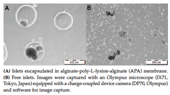
Figure 1. Pig Islets After 10 Years of Cryopreservation
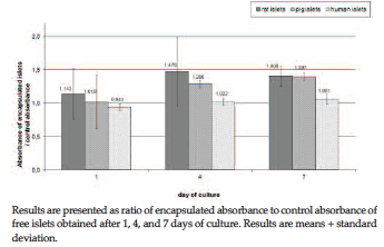
Figure 2. Mitochondrial Activity of Cryopreserved Islets

Table 1. Stimulation Index Results of Free and Encapsulated Islets After 10 Years of Cryopreservation